Journal of Nutrition & Food Sciences
Open Access
ISSN: 2155-9600
ISSN: 2155-9600
Commentary - (2015) Volume 5, Issue 3
Effect of post-hatch (PH) feed deprivation (FD) for 6, 12, 24 and 36 hrs., on the performance, gut development and differential expression of nutrient transporter genes in egg type chickens (crosses of White Leghorn) was studied. Significant decrease (P=0.001) in yolk utilization (in all the FD chicks) and relative weight of gizzard, intestine, liver and pancreas was observed in 24 and 36 hrs FD chicks. The 36 hrs FD chicks recorded lower (P=0.001) body weight, lower feed intake and inferior FCR, decreased serum glucose, but higher cholesterol and uric acid level than the control (immediate fed group) or 6 hrs FD chicks. Villi height in duodenum, jejunum and ileum decreased (P=0.001) but villi width increased (P=0.001) with increase in FD period and significant changes were observed particularly in 24 or 36 hrs FD chicks. Relative expression of Cdx gene decreased with age of the bird and the feed restriction period. Expression of SGLT, FABP gene was not associated with the feed deprivation period, while that of EAAT3 gene increased in 24 or 36 hrs FD chicks. No difference was observed in bursa, spleen and thymus weight. In vivo humoral and cellular immune response was significantly better in chicks FD for 6, 12 and 24 hrs than control and 36 hrs FD chicks. Expression of immunity related genes IL-6 and TLR-2 increased as the FD period increased. It may concluded that PH feed deprival for first 12 hrs did not affect growth performance, intestinal morphology and immune response but feed withdrawal for 24 or 36 hrs adversely affect the intestinal morphology and few nutrient transporter genes expression in egg type chickens.
Keywords: Feed deprivation; Performance; Intestine morphology; Gene expression; Egg type chickens
In commercial poultry production the newly hatched chicks do not have immediate access to feed and water as chicks hatched over a 12-24 hrs period are usually pulled out of the hatchers only when the majority of chicks have hatched. Besides, the hatchery operating procedures such as sex determination, vaccination, packaging and transportation to production facilities are also responsible for delay in access to feed [1]. The precocial group of birds usually starts foraging for feed immediately after hatching and begins to grow, but due to this delayed feeding chick remain fasted for 24-36 hrs and resulting in body weight loss [2].
Residual yolk sac forms the only source of energy before the utilization of feed is initiated. An increased post- hatch holding period has the detrimental effects on chicks due to potential dehydration and energy depletion [3]. Delayed feeding chicks reported to have poor response to vaccination, slow gastrointestinal tract and immune system development, poor resistance to diseases and pathogens and adverse effects on long-the term performance [4]. The gastrointestinal tract grows more rapidly than other body parts in the first few days following hatch and plays a critical role in the early stages of chick growth [5]. The development and function of the gastrointestinal tract (GI) of broiler chicks has been found to be negatively affected by delayed feeding [6-8]. Early access to feed and water is responsible for the weight gain, chick’s mortality reduction [4,7], immune system improvement and better resistance to disease challenges [9]. The early access to feed stimulates growth and development of the small intestine, while feed deprivation results in reduction of villi [10]. Nutrient transporters which are present in the small intestine are responsible for dietary nutrient absorption; so the expression of these nutrients transporters can influence overall nutrient status as well as the growth and development of the animal [11,12]. Geyra et al., [13] reported that starvation depressed the expression of both the cdxA and cdxB transcription factors, which appear to be involved in intestinal development and maintenance.
Many factors are responsible for early development of immune system in newly hatched chicks and out of this one is the early feed intake [9]. Feed provides nutrient which are essential for the growth and development of both primary and secondary lymphoid organs. The immune system of hatchling, particularly the mucosal immune system, also requires oral feed intake for rapid development [14]. Post hatch feed restriction harmful to the immune system [15] as well as growth [16]. Though nutrient requirements of juvenile is widely studied, but not much literature is available on early post-hatch feed withdrawal in egg type chicken (White Leghorn) i.e. slow growing chicken.
In the present study we have tried to evaluate its effect on post hatch performances, internal organ development, serum biochemical profiles, intestinal morphology and differential expression of few gut development and nutrient transporter genes.
Experimental design, birds and treatments
The experimental design was a completely randomized design (CRD) with five treatments, eight replications per treatment and 12 birds per replication having similar group mean weight. The main effect was variation in post-hatch (PH) feed placement. A total of 480 egg type chicks (cross of White Leghon) from single hatch were randomly assigned to 40 cages (45×60×37.5 cm each) in four-tier battery brooders housed in an environmentally controlled room. Average initial body weight of chicks was 33.54 ± 0.13 g. Each pen was equipped with a feeder and watered placed outside the cage. Initial brooding temperature was 32.0 °C, but this was gradually reduced to 24 ± 1°C at 21 d PH. Light period of 24 hrs was maintained throughout the trial period. Access to feed and water was provided ad libitum over the trial period. All birds were reared under uniform and standard management conditions and routine vaccination schedule was followed. All management and procedures in this study were carried out as per the code of practice approved by the Institute Animal Ethics Committee. The experimental treatments included access to feed at 0 (Control), 6, 12, 24, 36 hrs PH. Layer starter mash (20% crude protein, 2800 Kcal/ kg metabolisable energy as per the standard of Indian Council of Agricultural Research, New Delhi, 2013) were offered to the birds for a period of 42 d PH.
Intestinal organ development, yolk-sac utilization and serum bio-chemicals
At 6, 12, 24, 36 hrs, 7 d and 14 d PH, four birds (both the sex) were selected from each treatment at random, weighed and killed by cervical dislocation for measurement of organ weights. The proventriculus plus gizzard without contents, small intestine (the region from the distal end of gizzard to 1 cm above the ileo-caecal junction) without contents, pancreas, spleen, liver, heart and bursa of Fabricius were weighed. The relative yolk sac and intestinal organ weights were expressed in g/100 g of body weight.
Blood (at 36 hrs, 7 d and 14 d PH) was collected and allowed to clot at room temperature. Serum was separated and kept at –20C for blood biochemical analysis such as serum glucose (mg/dl) (Autospan liquid gold, SPAN diagnostic Ltd. India), serum protein(g/dl) (Autospan liquid gold, SPAN diagnostic Ltd., India), serum cholesterol (mg/dl) (Autospan liquid gold, SPAN diagnostic Ltd., India) and serum uric acid (mg/dl) (Cogent, SPAN diagnostic Ltd., India).
Response criteria
Individual body weight of chicks and the feed intake of all the birds in a pen were recorded at 7 d intervals throughout the experimental period. Feed conversion ratio was calculated for 0-14, 14-28, 28- 42 d and the overall period (0-42 d) by dividing feed intake to the corresponding weight gain.
In vivo immune response
To study the in vivo humoral immune response, (at 28 d PH) one ml, 1% suspension (v/v) of sheep red blood cells (SRBC) in phosphate buffer saline (PBS) was injected intravenously to a group of ten birds. The antibody response to SRBC was determined after five days post injection using a standard haemagglutination assay. The cellular immune response was assessed in another ten chicks (at 21 d PH) using the in vivo cutaneous basophilic hypersensitivity response to the lectin phytohaemagglutinin from Phaseolus vulgaris (PHA-P). The toe web thicknesses, between the third and fourth digits, of both left and right feet were measured using a micrometer. The detailed procedure for in vivo immune response has been described in the paper by Bhanja et al., [17].
Histopathology
For histo-pathology of the intestine, duodenum (portion extending from gizzard to end of duodenal loop), jejunum (portion from the end of the duodenal loop to Meckel’s diverticulum), and ileum (portion from Meckel’s diverticulum to the ileal-cecal junction) were collected aseptically after sacrificing four experimental birds (both the sex, identified through sex gonads) from each treatment group at 36 hrs and 7 d PH. After separation, segments were gently flushed with saline solution to remove the intestinal content. For morphological analysis, approximately 4-5 cm of the middle portion of the duodenum, jejunum and ileum was excised and fixed in 10% neutral buffer formalin at least for 24 hrs. Two cross sections of 10% neutral buffer formalin preserved segments were dehydrated in graded alcohol series, cleared in xylol and embedded in paraffin to obtain 5 μm-thick transversal histological cuts. Slides were stained by hematoxylin and eosin staining as described by Beçak and Paulete [18]. After staining, each stained cross section sample were observed at two to three villi section areas for villus height (VH) and villus width (VW). VH (from the tip of the villus to the lamina propria as the base) and VW were measured using the Image- Pro Plus microscope equipped with a video camera.
Tissue collection for gene expression studies
At 36 hr, 7 and 14 d PH, about 2 cm of the proximal portion of the jejunum [17] (where most of the digestive enzymes action takes place) and spleen were collected from four birds per treatment then opened and flushed with normal saline. The tissue was then homogenized using automated Kinematica TM Polytron TM homogenizer PT 1200 E (Thermo Fisher Scientific, Inc., Waltham, USA) and 50 mg of homogenized tissue was used for the total RNA isolation. For contingency uses about 150 mg of tissue was immersed in 500 μL RNA stabilizing solution (RNA Later, which fixes the total RNA and inhibits its degradation) and kept at 4°C overnight and then transferred to -80°C deep fridge.
RNA isolation and reverse transcription
Total cellular RNA from jejunum and spleen of each treatment group were isolated using RNAgents® Total RNA Isolation System (Promega, Madison, USA), purity and quantity were assessed by measuring the optical density of each sample at 260 versus 280 nm in a nanodrop. Any possible traces of genomic DNA were removed by treating 5 mg of each RNA sample with 5 U of RNase-Free DNase (Biogen Idec, Inc., Durham, USA) at 37°C for 1 h. The DNase was subsequently inactivated by incubation at 65°C for 10 min. Each DNase treated total RNA sample (2 μg) was reverse transcribed using the RevertAid First Strand cDNA Synthesis Kit (MBI Fermentas, Hanover, USA) according to the manufacturer’s instructions. The reverse transcription reaction was carried out in a final volume of 20 μl. The resultant cDNA was stored at −20°C for further use.
Standardization of primers for PCR
The primers for this study were designed using DNASTAR Lasergene software (Version 5.0, 1997). The oligonucleotide sequences of the primer have been presented in Table 1. The qPCR assays were evaluated by the generation of a standard curve. Calibration curves for each gene were done on an iQ5 cycler (Bio-Rad Laboratories, Hercules, USA) with five 10-fold serial dilutions (in triplicates) and were calculated by the Bio-Rad Optical System Software (Version 2.1, 2010) with the analysis mode “PCR base line subtracted”. Amplification efficiency (E) of qPCR reactions was calculated with the slope of the log linear portion of the calibration curve according to the equation: E = 10 (−1/slope) [19,20]. Expression of nutrient transporter and intestine developmental related genes like caudal type homeobox (Cdx), fatty acid binding protein (FABP), sodium dependent glucose transporter (SGLT) and excitatory amino acid transporter-3 (EAAT3) and immune related genes: interleukin-6 (IL-6) and tool like receptor-2 (TLR-2) were studied in real time PCR. The amplifications of genes were carried out using an iQ5 cycler (Bio-Rad Laboratories) in 25 μl volume containing 1X QuantiTect® SYBR® Green PCR Master Mix (SYBR® Green 1 dye, HotStartTaq DNA polymerase and dNTPs in optimized buffer components; Qiagen GmbH, Hilden, Germany), a 0.2 μM concentration of each gene-specific primer and 1 μl of cDNA template. PCR cycling conditions included initial denaturation at 95°C for 10 min, followed by 40 cycles of denaturation at 95° for 30 s, annealing for 30 s, and extension at 72° for 45 s. For each gene of interest, negative and positive controls were also included. Negative controls were samples in which cDNA was not added. A melting curve was performed for each sample after completion of amplification and analyzed in comparison to negative and positive controls to determine the specificity of PCR reaction.
| Sl No. | Gene | Primer (5I- 3I) | Annealing Temp. (oC) |
Size of Amplicon (bp) | Putative bio-logical role | Accession Number |
|---|---|---|---|---|---|---|
| 1 | EAAT | F-cagtgtttggaaccctaa | 56 | 139 | Nutrient transporter | XM_424930 |
| R-gatggctttgtagatggc | ||||||
| 2 | FABP | F-gcagaatgggaataagttc | 55.7 | 256 | Nutrient transporter | NM_204192.3 |
| R-cttgctaattctcttgtagg | ||||||
| 3 | SGLT | F-tattatcctgcttgctat | 46.1 | 173 | Nutrient transporter | AJ236903.1 |
| R-cattcatatacttctccat | ||||||
| 4 | CDX | F-ctcggacttcgccagctacc | 54 | 296 | Intestinal tract Dev. | AB046532 |
| R-tgcgcctcatccattcgtac | ||||||
| 5 | IL-6 | F-gaaatccctcctcgccaatctga | 57 | 281 | Humoral immunity related genes | NM001007079 |
| R-tgaaacggaacaacactgccatct | ||||||
| 6 | TLR-2 | F-gtggccatgtcgatcagcagaaac | 56 | 202 | Cellular immunity related genes | NM_204278.1 |
| R- tcagcggagagtcacagatgtagc | ||||||
| 7 | GAPDH | F-ccgtcctctctggcaaagtcc | 57.5 | 266 | House keeping | NM_204305 |
| R-agccccagccttctccatg |
Table 1: Oligonucleotide sequence of intestinal developmental and nutrient transporter gene primers.
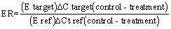
The relative expression ratio (ER) of a target gene is computed, based on its real-time PCR efficiencies (E) or a static efficiency of 2, and the cycle threshold (Ct) difference (Δ) of mean control versus each unknown sample (ΔCt control – treatment) as described below [21] using glyceraldehyde 3 phosphate dehydrogenase (GAPDH) as the reference housekeeping gene:
Statistical analysis
The statistical analysis and interpretation of data was done through SPSS software package Version 16.0 (2007). The body weight, organ weight, serum bio-chemicals and intestinal morphology were analyzed by one way ANOVA. mRNA expression levels (expression ratio) of intestinal, nutrient transporter and immune related genes were analyzed by REST 2009 software. Difference in mean values was considered as significant at the level of 95% (P < 0.05) and 99% (P < 0.01).
Body weight changes, yolk utilization and internal organ weight during first 36 hrs post hatch
After 6 hrs PH the control chicks (0 hr FD) gained 6.41 per cent weight correspond to their hatch weight, whereas 6 hrs FD chicks had 0.74 per cent weight loss. 6 hrs FD chicks had significantly higher (P=0.007) yolk weight than control chicks. There was significant decrease in pro-ventriculus (P=0.005), gizzard (P=0.032) and intestine weight (P=0.011) in 6 hrs FD chicks. However, liver (P=0.105) and pancreas weight (P=0.115) did not differ between 6 hrs FD and control chicks. After 12 hrs PH, 6 hrs FD chicks had significantly higher (P=0.001) weight gain than the control or 12 hrs FD chicks, whereas later had 1.14 per cent weight loos. Yolk utilization was significantly lower (P=0.001) in 6 hrs and 12 hrs FD chicks compared to control. There was no difference in intestinal organ weight except gizzard which was lower (P=0.011) in 12 hrs FD chicks (Table 2).
| Attributes* | Weight change | Yolk sac | Pro-ventriculus | Gizzard | Intestine | Liver | Pancreas |
|---|---|---|---|---|---|---|---|
| 6 hr post hatch | |||||||
| Control | 6.41b | 6.61a | 0.90b | 5.49b | 2.46b | 2.99 | 0.22 |
| 6hr FD | -0.74a | 8.22b | 0.77a | 3.56a | 1.56a | 2.54 | 0.16 |
| SEm | 1.38 | 0.36 | 0.02 | 0.48 | 0.21 | 0.14 | 0.02 |
| P-value | 0.001 | 0.007 | 0.005 | 0.032 | 0.011 | 0.105 | 0.115 |
| 12 hr post hatch | |||||||
| Control | 5.67b | 6.73a | 0.82 | 6.52b | 1.88 | 2.67ab | 0.19 |
| 6hr FD+ 6hr feeding | 8.09c | 7.68b | 0.78 | 6.50b | 2.16 | 3.01b | 0.21 |
| 12 hr FD | -1.14a | 8.79c | 0.8 | 4.74a | 1.78 | 2.54a | 0.18 |
| SEm | 1.24 | 0.27 | 0.08 | 0.6 | 0.23 | 0.09 | 0.02 |
| P-value | 0.001 | 0.001 | 0.933 | 0.011 | 0.093 | 0.053 | 0.865 |
| 24 hr post hatch | |||||||
| Control | 9.75c | 5.83a | 0.84 | 6.69bc | 2.64b | 3.22 | 0.3 |
| 6hr FD+ 18hr feeding | 6.50b | 7. 78b | 0.86 | 7.36c | 2.92b | 3.5 | 0.35 |
| 12hr FD+ 12hr feeding | 8.63bc | 7.72b | 0.84 | 6.04b | 2.6b | 3.11 | 0.25 |
| 24 Hr FD | -3.19a | 7.11b | 0.87 | 4.90a | 1.9a | 2.98 | 0.24 |
| SEm | 1.39 | 0.24 | 0.02 | 0.27 | 0.13 | 0.1 | 0.02 |
| P-value | 0.001 | 0.002 | 0.924 | 0.001 | 0.021 | 0.316 | 0.167 |
| At 36 hr post hatch | |||||||
| Control | 13.96c | 3.39a | 0.83 | 7.04b | 3.42c | 3.71b | 0.43 b |
| 6hr FD+ 30hr feeding | 21.46d | 4.30ab | 0.94 | 6.38b | 3.84c | 3.83b | 0.47 b |
| 12hr FD+ 24hr feeding | 19.08d | 4.02ab | 0.92 | 6.54b | 2.79b | 3.55b | 0.29a |
| 24 Hr FD+ 12hr feeding | 6.70b | 4.47b | 0.84 | 6.68b | 2.86b | 3. 87b | 0.18a |
| 36 Hr FD | -6.64a | 4.92b | 0.85 | 4.26a | 1.96a | 3.02a | 0.23a |
| SEm | 2.37 | 0.17 | 0.02 | 0.28 | 0.34 | 0.09 | 0.03 |
| P-value | 0.001 | 0.039 | 0.330 | 0.001 | 0.001 | 0.013 | 0.001 |
Table 2: Mean weight gain/loss, yolk weight and intestinal organ weight of chicks subjected to 0, 6, 12, 24 and 36 hrs delay in post hatch feed placement.
After 24 hrs post hatch, the control, 6 hr or 12 hrs FD chicks gained (P=0.001) 6.50 -9.75 % weight correspond to their hatch weight but 24 hrs FD chicks lose 3.19% body weight. There was no significant difference in the utilization of yolk in FD chicks but it was better (P=0.002) in control chicks. Weight of gizzard (P=0.001) and intestine (P=0.021) was lower in 24 hrs FD chicks than control and 6 hrs or 12 hrs FD chicks. Weight loss in 36 hrs FD chicks was 6.64%, however 6 hrs and 12 hrs FD chicks gained more (P=0.001) weight than control or 24 hrs FD chicks. There was significant reduction (P=0.001) in gizzard, intestine, liver and pancreas weight in 36 hrs FD chicks than the control or some of the FD chicks (Table 2).
Post hatch growth response
At 7 d PH there was no difference in the body weight (BW) of control chicks and those FD up to 12 hrs, but significantly decreased (P=0.001) in 24 and 36 hrs FD chicks. However, at 14 d PH only 36 hrs FD chicks recorded lower (P=0.001) body weight than the control or other FD chicks. At 28 and 42 d PH 36 hrs FD chicks had significantly lower (P<0.01) BW than the control and 6 hrs FD chicks but did not differ from the 12 or 24 hrs FD chicks BW. Feed intake (FI) of the control and FD chicks did not differ (P>0.05) during 8-14, 15-28 and 29-42 d PH but 24 and 36 hrs FD chicks recorded lower (P=0.001) FI during 0-7 d PH. The feed conversion ratio (FCR) of 36 hrs FD chicks was consistently poorer than the control or other FD chicks, and was only significant (P=0.001) during 0-14 d PH (Table 3).
| Attributes* | Delay in post hatch feed placement | SEM | P-value | ||||
|---|---|---|---|---|---|---|---|
| 0 hrs (Control) |
6 hrs | 12 hrs | 24 hrs | 36 hrs | |||
| BW (g) | |||||||
| Day old | 33.56 | 33.63 | 33.59 | 33.52 | 33.42 | 0.13 | 0.090 |
| 7 d | 64.44c | 64.69c | 62.72bc | 61.19b | 57.18a | 0.60 | 0.001 |
| 14 d | 109.92bc | 111.00c | 107.61bc | 104.64b | 96.03a | 1.27 | 0.001 |
| 28 d | 255.65bc | 263.61c | 248.24ab | 250.44ab | 237.89a | 2.33 | 0.003 |
| 42 d | 395.31c | 388.64bc | 370.64abc | 366.69ab | 346.08a | 4.66 | 0.002 |
| FI (g/b) | |||||||
| 0-7 d | 49.86b | 51.75b | 49.08b | 44.78a | 42.86a | 0.85 | 0.001 |
| 8-14d | 99.79 | 100.82 | 96.32 | 95.44 | 91.92 | 1.23 | 0.145 |
| 15-28 d | 383.29 | 384.81 | 381.61 | 375.75 | 363.07 | 2.92 | 0.106 |
| 29-42 d | 522.92 | 510.90 | 503.42 | 478.35 | 457.58 | 8.30 | 0.706 |
| FCR | |||||||
| 0-7 | 1.67 | 1.71 | 1.68 | 1.62 | 1.81 | 0.02 | 0.075 |
| 0-14 d | 1.99a | 1.99a | 1.96a | 1.97a | 2.17b | 0.02 | 0.038 |
| 0-28 d | 2.42 | 2.35 | 2.46 | 2.38 | 2.44 | 0.03 | 0.707 |
| 0-42 d | 2.93 | 2.97 | 3.06 | 2.99 | 3.06 | 0.03 | 0.443 |
Table 3: Mean body weight (BW), feed intake (FI) and feed conversion ratio (FCR) of birds subjected to 0, 6, 12, 24 and 36 hrs delay in post hatch feed placement.
Post hatch intestinal organ development
Mean weight of intestinal organs like pro-ventriculus, gizzard, intestine, liver and pancreas did not differ between control and FD chicks at 7 and 14 d PH (Table 4).
| Attributes* | Delay in post hatch feed placement | SEM | P-value | ||||
|---|---|---|---|---|---|---|---|
| 0 hrs (Control) |
6 hrs | 12 hrs | 24 hrs | 36 hrs | |||
| At 7 d post hatch | |||||||
| Pro-ventriculus | 0.95 | 1.11 | 1.05 | 1.11 | 1.03 | 0.03 | 0.605 |
| Gizzard | 9.24 | 10.18 | 9.67 | 9.73 | 8.74 | 0.25 | 0.492 |
| Intestine | 8.18 | 9.06 | 8.51 | 9.91 | 9.50 | 0.30 | 0.393 |
| Liver | 3.19 | 3.3 | 3.96 | 3.75 | 3.01 | 0.09 | 0.060 |
| Pancreas | 0.55 | 0.6 | 0.63 | 0.58 | 0.59 | 0.02 | 0.814 |
| At 14 d post hatch | |||||||
| Pro-ventriculus | 0.96 | 0.83 | 0.87 | 1.03 | 1.00 | 0.04 | 0.527 |
| Gizzard | 7.72 | 7.56 | 6.99 | 7.88 | 7.80 | 0.20 | 0.676 |
| Intestine | 8.69 | 7.87 | 7.73 | 8.24 | 8.05 | 0.22 | 0.130 |
| Liver | 3.17 | 3.12 | 2.91 | 2.91 | 2.84 | 0.07 | 0.404 |
| Pancreas | 0.50 | 0.52 | 0.58 | 0.55 | 0.62 | 0.02 | 0.351 |
Table 4: Mean intestinal organ weight of birds subjected to 0, 6, 12, 24 and 36 hrs delay in post hatch feed placement.
Serum bio-chemicals
When the serum samples of 36 h old chicks were compared, glucose level was similar in the control and those FD up to 24 hrs but in 36 hrs FD chicks it was significantly (P=0.048) lower. Serum protein level did not change due to feed withdrawal, but serum cholesterol and uric acid level was higher (P=0.001) in 36 hrs FD chicks than control and 6, 12 or 24 hrs FD chicks. The serum bio-chemical profiles at 7 and 14 d PH did not differ between control and FD chicks except the uric acid level of chicks at 14 d PH which had FD for 12 hrs or more (Table 5).
| Attributes* | Delay in post hatch feed placement | SEM | P-value | ||||
|---|---|---|---|---|---|---|---|
| 0 hrs (Control) |
6 hrs | 12 hrs | 24 hrs | 36 hrs | |||
| At 36 hrs (mg/dL) | |||||||
| Glucose | 248.29b | 243.90b | 244.51b | 254.57b | 195.12a | 6.33 | 0.048 |
| Total Protein | 4.32 | 4.42 | 4.46 | 4.32 | 4.24 | 0.08 | 0.924 |
| Cholesterol | 267.00ab | 225.50a | 286.50b | 262.50ab | 362.50c | 11.92 | 0.000 |
| Uric acid | 7.18a | 7.14a | 6.95a | 7.47b | 7.76c | 0.07 | 0.000 |
| At 7 day | |||||||
| Glucose | 136.59 | 135.98 | 136.95 | 159.15 | 165.24 | 4.86 | 0.128 |
| Total Protein | 3.91 | 3.87 | 4.23 | 3.79 | 4.28 | 0.10 | 0.442 |
| Cholesterol | 282.5 | 256.00 | 280.5 | 254 | 293.5 | 9.07 | 0.610 |
| Uric acid | 6.40 | 5.90 | 6.79 | 6.76 | 7.34 | 0.20 | 0.204 |
| At 14 day | |||||||
| Glucose | 160.98 | 167.68 | 152.44 | 156.59 | 186.59 | 5.22 | 0.170 |
| Total Protein | 4.13 | 4.07 | 4.67 | 4.54 | 4.50 | 0.09 | 0.081 |
| Cholesterol | 272.50 | 278.50 | 272.00 | 257.50 | 289.50 | 7.08 | 0.751 |
| Uric acid | 5.71a | 5.75a | 6.84b | 6.94b | 7.15b | 0.17 | 0.000 |
Table 5: Serum Profile of birds subjected to 0, 6, 12, 24 and 36 hrs delay in post hatch feed placement.
Intestinal morphology
Intestinal villi morphology of the birds subjected to 0, 6, 12, 24 and 36 hrs FD has been presented in Table 6. At 36 hrs of PH the duodenum villi height (VH) decreased (P=0.001) but duodenum villi width (VW) increased (P=0.001) with increase in FD period. However, duodenum VH and VW did not differ between control and 6 or 12 hrs FD chicks. The ratio of VH to VW was significantly reduced (P=0.001) in FD chicks as compared to control chicks. The FD chicks had significantly lower (P=0.001) VH in jejunum and ileum. Jejunum VW was higher (P=0.006) in 24 hrs FD chicks but ileum VW decreased in chicks FD for 12 hrs or more. At 7 d PH VH in duodenum, jejunum and ileum significantly decreased in chicks FD for 24 or 36 hrs compared to control or 6 hrs FD chicks. Jejunum and ileum VW also significantly deceased (P=0.001) in 24 or 36 hrs FD chicks, but the ratio of jejunum VH to VW increased (P=0.001).
| Attributes* | Delay in post hatch feed placement | SEM | P-value | ||||
|---|---|---|---|---|---|---|---|
| 0 hrs (Control) |
6 hrs | 12 hrs | 24 hrs | 36 hrs | |||
| At 36 hrs post hatch | |||||||
| Duodenum | |||||||
| Villi Height (VH) | 688.68c | 669.96c | 647.03c | 566.48b | 363.31a | 27.2 | 0.001 |
| Villi Width (VW) | 78.45a | 90.30a | 94.13a | 117.82b | 123.65b | 3.56 | 0.001 |
| Ratio VH:VW | 9.36d | 7.11c | 6.52c | 4.90b | 2.95a | 0.49 | 0.001 |
| Jejunum | |||||||
| Villi Height (VH) | 528.85b | 446.60a | 360.39a | 375.93a | 395.94a | 15.52 | 0.001 |
| Villi Width (VW) | 71.74a | 72.70a | 80.28ab | 97.28b | 81.09ab | 2.61 | 0.006 |
| Ratio VH:VW | 7.28b | 5.82ab | 4.56a | 4.24a | 4.70a | 0.30 | 0.001 |
| Ileum | |||||||
| Villi Height (VH) | 427.38b | 429.40b | 260.04a | 277.59a | 286.82a | 15.79 | 0.001 |
| Villi Width (VW) | 91.82b | 85.62b | 68.59a | 65.79a | 72.07a | 2.23 | 0.001 |
| Ratio VH:VW | 4.64 | 5.22 | 3.39 | 4.51 | 4.01 | 0.23 | 0.109 |
| At 7 d post hatch | |||||||
| Duodenum | |||||||
| Villi Height (VH) | 1034.53b | 1022.50b | 950.93ab | 820.33a | 810.70a | 30.52 | 0.018 |
| Villi Width (VW) | 126.76 | 128.24 | 115.03 | 107.25 | 100.98 | 4.08 | 0.136 |
| Ratio VH:VW | 8.4 | 8.49 | 8.26 | 9.5 | 8.04 | 0.41 | 0.871 |
| Jejunum | |||||||
| Villi Height (VH) | 799.08b | 777.73b | 779.18b | 741.10ab | 701.58a | 10.55 | 0.008 |
| Villi Width (VW) | 120.63b | 119.74b | 130.98b | 65.48a | 76.46a | 4.64 | 0.001 |
| Ratio VH:VW | 6.71a | 6.46a | 5.99a | 11.36c | 9.13b | 0.47 | 0.001 |
| Ileum | |||||||
| Villi Height (VH) | 699.68c | 657.79c | 622.34bc | 541.68ab | 468.58a | 19.87 | 0.001 |
| Villi Width (VW) | 131.66c | 104.45ab | 115.13bc | 87.70a | 85.38a | 3.51 | 0.001 |
| Ratio VH:VW | 5.5 | 6.35 | 5.58 | 6.04 | 5.49 | 0.22 | 0.660 |
Table 6: Intestinal villi morphology (in micro meter) of chicks subjected to 0, 6, 12, 24 and 36 hrs delay in post hatch feed placement.
Expression of nutrient transfer genes
At 36 hrs PH relative expression of Cdx gene was higher (P=0.008) in 6 hrs FD chicks while the expression did not differ in 12, 24 and 36 hrs FD chicks as compared to control chicks. FABP gene expression significantly increased (P<0.05) in 12, 24 and 36 hrs FD chicks as compared to control chicks. Expression of SGLT gene increased (P=0.002) in 6 hrs FD chicks but did not differ between control and other FD chicks. However, EAAT gene expression was higher only in 12 hrs FD chicks (P=0.052) compared to the control chicks (Figure 1).

Figure 1: Relative fold expression of Cdx, FABP, SGLT and EAAT gene at 36 hrs pH in the intestinal tissues of control and feed deprived chicks. Expression of Control group is taken as 1.0 * and **indicates significant expression at the level of 95% (P<0.05) and 99% (P<0.01), respectively.
At 7 d PH Cdx gene expression was lower but not significant in 6 and 12 hrs FD chicks however, increased (P<0.05) in 24 or 36 hrs FD chicks. Expression of FABP and SGLT genes did not differ between control and FD chicks. Expression of EAAT was lower in 6, 12 and 24 hrs FD chicks though significant in 6 hr FD chicks (P=0.029) but up regulated in 36 hrs FD chicks (P=0.065) compared to control chicks (Figure 2).

Figure 2: Relative fold expression of Cdx, FABP, SGLT and EAAT gene at 7 d pH in the intestinal tissues of control and feed deprived chicks. Expression of Control group taken as 1.0 * and *indicated significant expression at the level of 95% (P<0.005) and 99% (P<0.01), respectively.
At 14 d PH Cdx gene expression was lower in all the FD chicks but significantly down regulated (P<0.05) in 24 and 36 hrs FD chicks. Expression of FABP gene was up regulated (P<0.05) in most of the FD chicks except those FD for 12 hrs. No significant difference was observed in the expression of SGLT genes between FD chicks and control chicks. EAAT expression was not different in 6 and 12 hrs FD chicks but up regulated (P<0.05) in 24 and 36 hrs FD chicks compared to control chicks (Figure 3).

Figure 3: Relative fold expression of Cdx, FABP, SGLT and EAAT gene at 14 d pH in the intestinal tissues of control and feed deprived chicks. Expression of control group id taken as 1.0 *indicates significant expression at the level of 95% (P<0.05).
Immune organ development and in vivo immune response
The relative weight of organ as expressed as g/100 g of live weight at 36 hrs, 7 d and 14 d PH has been presented in Table 6. No significant difference was observed in the bursa, spleen and thymus weight of FD and control chicks. Humoral and cellular immune responses evaluated through SRBC titer and foot web index respectively have been presented in Table 7. The antibody titre to SRBCs was significantly higher (P=0.001) in 6, 12 and 24 hrs FD chicks compare to control chicks. Though 36 hr FD chicks had lower antibody titer than 6 and 12 hrs FD chicks, but did not differ from the control and 24 hrs FD chicks. Foot web index in response to PHAP injection was significantly higher (P=0.021) in chicks feed deprived for 6, 12 and 24 hrs than control and 36 hrs FD chicks.
| Attributes* | Delay in post hatch feed placement | SEm | P-value | ||||
|---|---|---|---|---|---|---|---|
| 0 hr (Control) |
6 hrs | 12 hrs | 24 hrs | 36 hrs | |||
| At 36 hrs | |||||||
| Bursa | 0.13 | 0.18 | 0.16 | 0.16 | 0.20 | 0.01 | 0.452 |
| Spleen | 0.05 | 0.06 | 0.03 | 0.05 | 0.04 | 0.00 | 0.063 |
| Thymus | 0.12 | 0.13 | 0.14 | 0.15 | 0.11 | 0.01 | 0.156 |
| At 7 day | |||||||
| Bursa | 0.27 | 0.25 | 0.26 | 0.21 | 0.25 | 0.01 | 0.768 |
| Spleen | 0.11 | 0.12 | 0.11 | 0.11 | 0.13 | 0.01 | 0.763 |
| Thymus | 0.22 | 0.02 | 0.02 | 0.02 | 0.02 | 0.01 | 0.716 |
| At 14 day | |||||||
| Bursa | 0.31 | 0.37 | 0.41 | 0.42 | 0.46 | 0.03 | 0.404 |
| Spleen | 0.13 | 0.12 | 0.15 | 0.14 | 0.14 | 0.01 | 0.567 |
| Thymus | 0.26 | 0.21 | 0.21 | 0.24 | 0.24 | 0.01 | 0.560 |
Table 7: Immune organ weight (expressed as g/100g) of chicks subjected to 0, 6, 12, 24 and 36 hrs delay in post hatch feed placement.
Expression of immune related genes
At 36 hrs PH relative expression of IL-6 and TLR-2 gene was similar in 6 and 12 hrs FD chicks but up regulated (P<0.05) in 24 and 36 hrs FD chicks compared to control chicks (Figure 4). However, at 14 d PH the expression of IL-6 and TLR-2 gene was up regulated (P<0.05) only in 36 hrs FD chicks compare to the control chicks (Figure 5).

Figure 4: Relative fold expression of IL-6 and TLR-2 gene at 36 hrs, pH in the spleen tissues of control and feed deprived chicks. Expression of the control group is taken as 1.0 *indicates significant expression at the level of 95% (P<0.05).

Figure 5: Relative fold expression of IL-6 and TLR-2 gene at 14 d, pH in the spleen tissues of the control and feed deprived chicks. Expression of control group is taken as 1.0 *indicates significant expression at the level of 95% (P<0.05).
Body weight changes, yolk utilization and internal organ weight during first 36 hrs post hatch
We observed significant BW loss as FD period increased however BW gain increased in 6 or 12 hrs FD chicks after they were provided with feed. Noy and Sklan, [22] and Bhanja et al., [23] had also reported that, chicks fed immediately after hatch gained 10.5% more weight than those FD for 48 hrs. This may be due to some mechanical stimulant in the gut due to feeding which is absent in FD chicks [22]. We have also found that there was significant decrease in yolk sac content in control than the FD chicks at all the period of study. Yolk sac content is used up by the chicks that had access to feed immediately after hatching [24]. The yolk contributes to maintenance of the young bird during the first few days after hatching [25]. Following hatch, yolk is transported both to the circulation through the vascular system and to the intestine through the yolk stalk [24]. Therefore, it is expected that yolk nutrients would be utilized more quickly by fasted chicks compared to the fed chicks, allowing young chicks to maintain their physiological functions. On the contrary yolk utilization was more rapid in the fed chicks than the fasted chicks, suggesting that transfer of yolk is facilitated by intestinal motility of fed chicks [23].
We observed significant (P<0.01) decrease in the gizzard, intestinal, pancreas and liver weight of the chicks as the FD period increased, but no difference was observed in pro-ventriculus weight. Our data is in concomitant with Bhanja et al., [23] who have reported that the development of gastrointestinal tract and internal organs is directly related to FI. Many researchers have recommended that there is the need to feed chicks immediately after hatch to ensure proper development of gastrointestinal tract, liver and pancreas [26-29]. The PH feed and water deprivation adversely affects the liver weight also [30,31].
Post hatch growth response
Body weight changes: The results of the present study revealed that at 7 d PH significant decrease in BW was observed in 24 and 36 hrs FD chicks than control or up to 12 hrs FD chicks. However, the effect of feed deprivation was more pronounced in 36 hrs FD chicks. Research in broiler chickens and turkey poults revealed decrease in BW of the birds which had no access to feed compared to those fed the starter diet immediately after hatching [16,32-34]. Panda et al., [14] have also reported that the body weight gain during 0-21 and 0-42 d were significantly lower in the chicks that had access to feed at 48 hrs PH in compared to the early fed chicks. Juul-madsen et al., [15] also observed the unfavorable effect of feed deprivation up to 48 hrs on growth, while FD for 24 hrs was acceptable for normal performance. In the present study FI during first 7 d PH was lower in 24 and 36 hrs FD chicks but did not affect subsequent feed consumption. Similar findings were also reported by Tabeidian et al., [35] and Pourreza et al., [36] wherein the short period of FD (12 hrs) could impact the growth performance, FI and FCR in broilers.
Serum bio-chemicals: In the present study serum glucose level did not differ between controls and up to 24 hrs FD chicks but significantly decreased in 36 hrs FD chicks. However, serum protein level did not change due to feed withdrawal. Similarly, lower glucose values have been reported under fasting in broiler chicken [37,38]. Warris et al., [38] have also reported higher plasma protein values in chicks that submitted to FD for 48 hours, which is not in agreement with our results. In another study, significantly decreased glucose level, increased cholesterol level and no difference in total protein in chicks fasted up to 48 hrs was reported [36]. At 36 hrs PH, serum cholesterol and uric acid level was higher in 36 hrs FD chicks than control or 6, 12 or 24 hrs FD chicks. But, the serum bio-chemical profiles at 7 and 14 d PH did not differ between control and FD chicks except the uric acid level of 12 hrs FD chicks at 14 d PH. Increased uric acid level at early age of chicks may be due to protein catabolism or due to gluconeogenesis from glucogenic amino acids derived from the yolk sac of chicks which had been subjected to prolonged fasting [39].
Intestinal morphology
We observed that at 36 hrs of PH the duodenum VH decreased and VW increased with increase in FD period and there was no difference in control and 6 or 12 hrs FD chicks. The FD chicks had also significantly lower VH in jejunum and ileum. The negative effect of fasting immediately after hatching on the small intestine villi height is possibly related to an increase in villi extrusion rate, leading to a reduction in height. Our data is in agreement with Franco et al., [40] who reported that the reduction in duodenum and jejunum villi height at 7 d PH with the increase in feed withdrawal period. There was significant reduction in villi height and in number of enterocytes per villi only after five to six days of feed deprivation [41]. It is also reported that the morphological changes of the intestinal villi in broilers are dependent on the presence of digested nutrients in the small intestinal lumen [42].
At 7 d PH VH in duodenum, jejunum and ileum significantly decreased in chicks FD for 24 or 36 hrs compared to control or 6 hrs FD chicks. Jejunum and ileum VW also deceased in 24 or 36 hrs FD chicks with increase in the ratio of jejunum VH to VW. Uni et al., [43] reported that the villi surface area in the duodenum is initially depressed in the absence of feed but reaches values close to those of fed chicks after 4 d, whereas the villus surface area in the jejunum was lowered throughout the first week post hatch, while no significant effects were found in the ileum. The early feeding have been found to stimulates growth and development of the small intestine, opposite to the feed deprivation which results in reduction of villi [10]. Only providing of water itself does not have a large impact on small intestine development unless it is served sufficiently with feed together [29].
Immune organ development and in vivo immune response
No significant difference was observed in the bursa, spleen and thymus weight of FD and control chicks at 36 hrs, 7 and 14 d PH. Posthatching bursa development may begin after 72 hrs of hatching, which could explain the absence of effects of fasting on this organ during the experimental period of 0-42 d PH [30]. No significant differences between experimental treatments in bursa, spleen, and heart weights were found at day 21 and 42 due to PH feed deprivation. However, they found heavier spleen in the fed than in fasted chicks (48-72 hrs) which is contrary to the present finding [30].
Humoral immune response (antibody titer to sheep RBCs antigen) was significantly higher in 6, 12 and 24 hrs FD chicks compare to control or 36 hrs FD chicks. In line with our results Panda et al., [14] reported that the humoral immune response was significantly higher in the chicks accessed to feed immediately or 12 hrs after placement compared to those FD for either 24 or 48 hrs. SRBC titre was higher in the chicks accessed to feed at 24 hrs after hatch compared to 48 hrs.
We found that, cellular immune response (foot web index in response to PHAP injection) was significantly better in chicks feed deprived for 6, 12 and 24 hrs than control or 36 hrs feed deprived. However, Panda et al., [14] reported no difference in the cell-mediated immune response to PHAP inoculation due to post hatch feed deprivations up to 48 hrs. But the lymphocyte proliferation ratio decreased significantly in birds feed deprived for 24 and 48 hrs compared to those had access to feed immediately or after 12 hrs of hatch [14]. Non availability of nutrients during early life hampers the immune system which is incomplete at the time of hatching [9]. Further, in fasting chicks there is secretion of corticosteroids, which inhibits the immune cell proliferation resulting in low immune response [9]. Feeding on the day of hatch may provide an early antigen stimulus and thus facilitate rapid differentiation of the humoral response [44]. In the present study also humoral and cellular immune response was lower in 36 hrs FD chicks. Juul-Madsen et al., [34] have also reported that feed withdrawal for more than 24 hrs affect immunological response.
Expression of nutrient transfer genes
At 36 hrs PH we found higher expression of Cdx gene in 6 hrs FD chicks while it did not differ in 12, 24 and 36 hrs FD chicks. At 14 d PH it was down regulated in 24 or 36 hrs FD chicks all the FD chicks. It is reported that starving from 0 to 48 hrs PH retarded growth and decreased enterocyte proliferation and expression of cdxA and cdxB [13]. After access to feed, expression of cdx genes was enhanced. Small intestinal expressions of the cdxA and cdxB homeobox genes increased during embryonic and PH development but were depressed by lack of access to feed, which decreased intestinal growth in the chick (Table 8). Noy and Sklan, [16] also reported that lack of access to feed for 48 hrs depresses intestinal development and growth through the time of marketing in chicks and poults.
| Attributes* | Delay in post hatch feed placement | SEm | Prob. | ||||
|---|---|---|---|---|---|---|---|
| 0 hr (Control) |
6 hrs | 12 hrs | 24 hrs | 36 hrs | |||
| Foot web index (mm) | 0.35a | 0.47b | 0.48b | 0.48b | 0.37a | 0.02 | 0.021 |
| sRBC titer (Log2) | 8.75a | 12.40d | 11.14cd | 10.38bc | 9.13ab | 0.29 | 0.001 |
Table 8: Immune response of chicks subjected to 0, 6, 12, 24 and 36 hrs delay in post hatch feed placement.
We observed that at 36 hrs FABP gene expression significantly (P<0.05) increased in 12, 24 and 36 hrs FD chicks as compared to control chicks while at 7 d PH its expression did not differ in FD chicks. At 14 d PH the expression increased in most of the FD chicks compared to control chicks. Very scanty information is available on the expression pattern of FABP gene during the fasting period. However, research in channel catfish (Ictalurus punctatus) revealed that expressions of several fatty-acid binding proteins (FABP) were perturbed by fasting, indicating changes in lipid metabolism [45]. We observed that expression of SGLT gene at 36 hrs PH increased in 6 hrs FD chicks but did not differ between control and other FD chicks. At 7 and 14 d expression of SGLT gene was similar in FD and control chicks. Our observation is in concomitant with Gilbert et al., [46] who observed little response in SGLT1 mRNA expression to feed restriction. Feed restriction in birds decreases the amount of SGLT1 protein [47] and also the activity of this transporter gene [48].
EAAT expression was either lower or similar in 6 and 12 hrs FD chicks compared to control but up regulated (P<0.05) in 24 and 36 hrs FD chicks. Susbilla et al., [49] observed increased activities of amino- and di-peptidases in the small intestines of broiler chicks that were feed restricted to 40% of the control intake from 5 to 11 d of age. Feed-restriction, fasting or other sources of malnutrition result in reduced intestinal absorptive surface area, which may explain why expression of nutrient transporters increased with age in response to feed restriction. Delayed access to feed for 36 h post hatch resulted in depressed villus height and crypt depth, and depressed growth in all intestinal segments. Feed restriction (30% of ad libitum) in male broiler chicks decreased the surface area of the tip of the enterocytes in the small intestine at 14 d PH [50]. Thus, the mechanism leading to a fasting or malnutrition-related increase in nutrient transport may be a combination of increased gene expression and ratio of transporting to non-transporting cells.
Expression of immune related genes
At 36 hrs PH relative expression of IL-6 and TLR-2 gene were up regulated in 24 and 36 hr FD chicks while at 14 d PH it was up regulated only in 36 hr FD chicks compared to control chicks. IL-6 is the proinflammatory cytokine which induces final maturation of B cells into antibody-secreting plasma cells [51] thereby increasing the secretion of immunoglobins [52]. In the present study in vivo immune response was higher when the birds were subjected to lesser feed deprivation. We did not found any correlation between in vivo immune response and immune gene expression. However, the 24 hrs FD chicks had higher in vivo immune response and immune gene expression. TLRs initiate adaptive immunity by activating antigen presenting cells for the production of pro-inflammatory cytokines (IL-1β, IL-6, chemokines, and IL-8) and up regulating co-stimulatory molecules [53]. In the present study also increase in TLR-2 expression was associated with increased expression of IL-6.
It may concluded that PH feed deprival affected the utilization yolk sac contents, however its effect on digestive organ development was prominent in 24 or 36 hrs FD chicks. Feed deprivation for first 12 hrs did not affect the growth performance and intestinal morphology and immune response. However, feed withdrawal for 24 or 36 hrs adversely affected intestinal morphology, the villi height. Relative expression of Cdx, gene decreased with age of the bird and the feed restriction period, but SGLT and FABP genes expression were not associated with the feed deprivation period. Feed deprivation for more than 24 hrs increased the expression of EAAT3 gene. In vivo humoral and cellular immune response was significantly better in chicks FD for 6, 12 and 24 hrs than control and 36 hrs FD chicks. Expression of immunity related genes IL-6 and TLR-2 increased as the FD period increased.