
Journal of Clinical Trials
Open Access
ISSN: 2167-0870
ISSN: 2167-0870
Case Report - (2026)Volume 16, Issue 5
Background: Spondylolisthesis is a frequent cause of low back pain but is seldomly associated with infectious causes and multisystem failure.
Case presentation: We present a patient who complained of severe low back pain and acute onset of neurological deficits that progressed to cardiovascular collapse and developed into a complex diagnostic and therapeutic scenario requiring intensive care management and surgical intervention.
Conclusion: The goal of our article is to elucidate the potential causes of catastrophic low back pain and to explore the potential treatments and their timing.
Spondylolisthesis, Tuberculosis, Global medicine, Neurosurgery, General medicine
Spondylolisthesis is defined as a vertebral pathology in the pars interarticularis where one vertebra slips anteriorly over the underling vertebra. This condition causes localized pain and may result in lower extremity deficits if not corrected. We present a case of sudden onset low back pain with lower extremity paresthesia and weakness that, unlike other cases, progressed to altered mental status, shock, and coma.
56 Years old Hispanic male, recent immigrant from South America, with Past Medical History (PMH) relevant of diabetes, gout, alcohol, and tobacco use who presented with progressive severe back pain over two months that was initially managed with Non-Steroidal Anti-Inflammatory Drugs (NSAIDS) and tramadol. Based on a review of his medical records, the Primary Care Provider (PCP) had obtained a lumbar X-ray that showed spondylolisthesis, but no additional blood work (Figure 1).
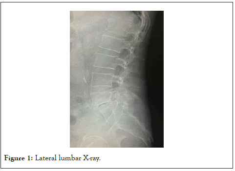
Figure 1: Lateral lumbar X-ray.
The patient developed paresthesia and leg weakness, and a Magnetic Resonance Imaging (MRI) confirmed the spondylolisthesis with spinal and neuroforaminal stenosis. He was prescribed pregabalin, but the pain remained unchanged.
During the third month, the patient developed unexplained tremors and altered mental status that progressed to unconsciousness, and he was admitted to the hospital. He was found to have acute hepatic and renal failure due to shock of unclear etiology and was admitted to the Intensive Care Unit (ICU).
His labs were relevant for thrombocytopenia, leukocytosis, elevated ammonia, and C-Reactive Protein (CRP). Blood cultures were positive for Staphylococcus aureus. His initial treatment consisted of IV antibiotics and pressors, and his clinical status improved.
During his hospital stay, he developed partial right lower extremity atrophy, continued to have altered mental status that progressed to coma, and underwent a lumbar puncture for Cerebrospinal Fluid (CSF) collection looking for any infectious or metabolic cause that failed to yield an etiological cause.
He continued to be bedridden and developed toxic megacolon due to C difficile infection, which responded to further antibiotics.
One month into his hospital admission, asymmetry and significant lower right extremity atrophy was noted and a repeat lumbar MRI with contrast showed paraspinal fluid collection, diskitis, and osteomyelitis (Figure 2). Tuberculosis blood and CSF cultures were negative. Further interventional testing, such as biopsy and aspiration, were precluded given his thrombocytopenia and coagulation disruption.
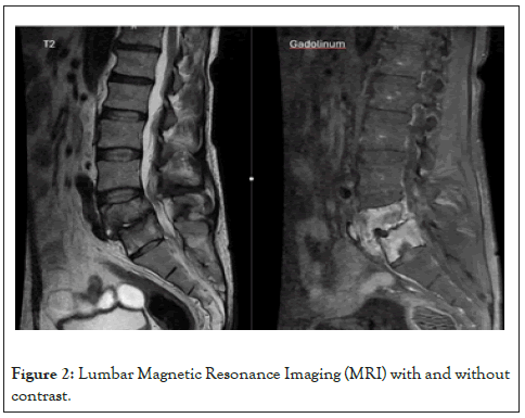
Figure 2: Lumbar Magnetic Resonance Imaging (MRI) with and without contrast.
The patient was kept in the hospital receiving broad spectrum antibiotics until his clinical condition improved, and he was scheduled for a lumbar fusion due to his evident neurological changes. During the surgery, the bone tissue showed caseating necrosis and was diagnosed as spinal tuberculosis after direct tissue collection.
Post-surgically, the patient started oral anti-tuberculous therapy and had slow but steady improvement. The patient continues to have pain but is ambulatory with a cane. Current pain medications include pregabalin, mirtazapine, and allopurinol.
We present this case due to the catastrophic series of events that lead to the proper etiologic cause of the pain. Initially, the vertebral damage was attributed to possible Staphylococcal osteomyelitis, but due to the critical state of the patient, interventional diagnostic procedures were precluded. In addition, conventional tuberculosis tests failed to elicit an early diagnosis without a clear explanation (e.g. immunosuppression). Furthermore, on retrospective inquiry, the patient did not admit contact with any infected individuals and was at the same risk of contagium as his peers.
Studies have shown various etiologies of spondylolisthesis, including congenital, degenerative, and trauma, to the posterior region of the spinal cord. The most frequent presentation of spinal tuberculosis is known as Pott’s disease in which the patient will experience back pain, stiffness, and muscle spasms, but seldomly neurological deficits. Furthermore, the patient will present with constitutional symptoms, including, but not limited to, weight loss, fever, and night sweats. The disease is more prevalent in underdeveloped countries and if found in an industrialized country is associated with other superimposed infections that may cause immunosuppression (e.g., Human Immunodeficiency Virus (HIV)).
One poorly understood cause of spondylolisthesis is an infectious process from spinal tuberculosis [1]. Tuberculosis infection can disseminate via the bloodstream and reach the vertebral column. Most cases involve the vertebral bodies and anterior regions of the spinal cord, which can lead to destruction of the intervertebral disc causing neurologic complications. The usual progression of spinal tuberculosis is not currently known to cause spondylolisthesis. However, there may be a correlation between either pre-existing or concomitant spinal tuberculosis and the development of spondylolisthesis.
As noted, spinal Tuberculosis (TB) affects the anterior aspects of the vertebral column with research estimating 95-98% of infections localizing to this region [2]. Tuberculosis involvement within the posterior segments of the vertebral column is rare but may lead to pathological fractures within the pars interarticularis. These fractures may lead to a secondary morbidity of spondylolisthesis. In addition, existing defects in the pars interarticularis may become symptomatic or exacerbated when associated with a tubercular infection. It is unclear whether these conditions are confounding factors of each other. However, clinical evidence has shown aggressive anti-TB treatment of patients diagnosed with TB with spondylolisthesis has led to significant symptomatic and radiologic improvement [2].
Regarding the cardiovascular collapse, the mortality in the United States due to sepsis is around 12.5%, the mortality from septic shock is around 35%, and the most common bacteria associated with septic shock include Escherichia coli (E. coli), Staphylococcus aureus (Staph), and Klebsiella pneumoniae [3]. Mycobacterium tuberculosis will present as a pulmonary infection but not as pneumonia. In 20% of the cases, it could present as disseminated tuberculosis without cardio vascular involvement and, in extremely rare cases, may cause hypotension when the adrenals glands are affected [4].
Mishra et al. reported 3 cases of septic shock associated with tuberculosis with a remarkably high rate of case fatality (3 out of 3) in which very broad-spectrum antibiotics failed to remedy the shock, and the diagnosis was made post mortem [5]. Unlike our patient, the confounding presence of the Staphylococcus species may have been the cause of his critical scenario, and, given his clinical response to the therapeutic treatment aimed at this specific bacterium, we can assume that this was the pathological agent.
Lastly, regarding the timing of the surgery, some advocate completing the full treatment of anti-tubercular antibiotics (9 months) prior to fusion while other experts believe that surgical treatment is paramount to stabilize the spine to prevent further neurological injury [6]. In our case, the surgeon reported significant issues with securing the pedicular screws due to the friability of the vertebral body and thus the screw position was not ideal (Figure 3). Based on these results, we recommend completing the full treatment of anti-tubercular antibiotics prior to fusion. Although the full treatment was not completed on this patient, it fortunately did not aggravate the patient’s clinical outcome.
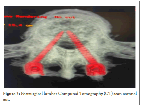
Figure 3: Postsurgical lumbar Computed Tomography (CT) scan coronal cut.
In conclusion, we believe the importance of this case lies in the fact that a catastrophic presentation could have been prevented with aggressive initial intervention. The discovery that this patient had evident inflammatory markers should have led to earlier admission and an attempt to diagnose the cause through interventional means before the onset of coagulopathy.
It is also a reminder that in a globalized world, diseases that are not usually considered initially should be explored based on the ethnic characteristics of each individual patient.
The patient provided written consent for the publication of this case report.
Citation: Penmetcha T, Subieta G. (2026). Tuberculosis Associated Spondylolisthesis and Radiculopathy . J Clin Trials. 16:650.
Received: 10-Jul-2026, Manuscript No. JCTR-26-43396; Editor assigned: 13-Jul-2026, Pre QC No. JCTR-26-43396; Reviewed: 27-Jul-2026, QC No. JCTR-26-43396; Revised: 03-Aug-2026, Manuscript No. JCTR-26-43396; Published: 10-Aug-2026 , DOI: 10.35248/2167-0870.26.16.651
Copyright: © 2026 Penmetcha T. This is an open-access article distributed under the terms of the Creative Commons Attribution License, which permits unrestricted use, distribution, and reproduction in any medium, provided the original author and source are credited.