Journal of Probiotics & Health
Open Access
ISSN: 2329-8901
ISSN: 2329-8901
Research Article - (2015) Volume 3, Issue 1
In the last few decades silver nanoparticles (Ag-NPs) have increasingly been employed thanks to their antimicrobial activity. With the use of the Transmission Electron Microscopy (TEM) we were able to evaluate the interaction between Ag-NPs and two Salmonella enterica strains (Enteritidis and Senftenberg) and to study morphological changes caused by the interaction with nanoparticles.
The Ag-NPs appeared to interact rapidly with the two Salmonella serovar, adhering mainly to the cell wall. The interaction with the Ag-NPs resulted to be time limited in the case of S. Senftenberg, while it was more long lasting for S. Enteritidis.
Cell responses to Ag-NPs morphologically differed in S. Enteritidis and S. Senftenberg. Ag-NPs were adsorbed to the cell membrane of S. Enteritidis and penetrated inside, thus modifying the cell structures. On the contrary, Ag- NPs were able to damage the cell wall of S. Senftenberg, but did not enter the cells. These results show that the two Salmonella strains display different sensitivity to silver, with S. Senftenberg exhibiting a resistant phenotype.
Keywords: Transmission Electron Microscopy (TEM); Salmonella; silver nanoparticles; silver resistance
Nanomaterials are a novel group of compounds with a wide range of possible uses in medicine, life sciences and biotechnology.
Different types of nanomaterials, such as copper, zinc, titanium, magnesium, gold, alginate and silver have been studied; however, silver nanoparticles (Ag-NPs) have proved to be most effective as they have good antimicrobial efficacy against a wide array of microbes, including activity against drug-resistant bacteria, viruses, and other eukaryotic microorganisms [1]. A study by Lara et al. [2] showed that Ag-NPs are bactericidal against multidrug-resistant P. aeruginosa, ampicillin resistant E. coli O157:H7, and erythromycin-resistant S. pyogenes. The bactericidal effect was the same whether tested in these drug resistant bacteria or in their drug-sensitive counterparts, showing that their drug-resistance proteins against antibiotics do not affect their sensitivity to Ag-NPs. In addition, Ag-NPs also increase the antibacterial effects of the antibiotics they are mixed with, including penicillin, amoxicillin, vancomycin, clindamycin and especially erythromycin, against S. aureus and E. coli [3].
Ag-NPs show a higher antibacterial action when compared to their bulk form and silver ions, given their increased surface area to volume ratio, their high catalytic capabilities and their ability to generate reactive oxygen species [4-6].
Various physicochemical properties of Ag-NPs have been demonstrated to affect their potential toxicity: concentration, shape, size, surface condition and synthesis methods [6].
The mechanism of the antibacterial activity of the Ag-NPs still remains unclear, although a plausible explanation has been outlined. The activity of nanoparticles against bacteria occurs mainly by the interaction with sulfur or phosphorous containing proteins present in the cell membrane, which is damaged by release of Ag+ ions or by generation of reactive oxygen species. Ag-NPs then penetrate inside the bacteria, inhibiting protein synthesis, DNA replication and respiratory chain [5,7,8].
The Transmission Electron Microscopy (TEM) is one of the most widely used methods for the morphological characterization of nanoparticles and a valuable tool to evaluate their interaction with microorganisms. Therefore, we analyzed two different Salmonella serovars (S. Enteritidis and S. Senftenberg), following the observation that these bacteria showed different sensitivity to Ag-NPs in vivo tests [9]. Briefly, Losasso and colleagues [9] demonstrated that the S. Senftenberg strain presented the Ag+ resistance determinant, while the S. Enteritidis strain was very sensitive to the Ag-NPs.
Thanks to the TEM, we were able to a) evaluate the interaction between the Ag-NPs and these two Salmonella serovars b) study the changes of cell morphology, c) analyze the structural damages and d) investigate whether Ag-NPs are capable of penetrating inside the bacteria cells, thus clarifying their antibacterial potential.
Nanoparticles
AgNPs suspensions (code NM-300K batch n. 6051) and their control dispersant medium were purchased from LGC Standards (UK). These nanoparticles are included in the Joint Research Center Nanomaterials Repository, are approved within the OECD WPMN (Working Party on Manufactured Nanomaterials) international testing program as selected representative reference nanomaterial (OECD Paris) and are delivered in aqueous solution containing 4% of Polyoxyethylene Glycerol Trioleate and Polyoxyethylene Sorbitan mono-Laurate (Tween 20).
Bacteria
Bacterial strains Salmonella enterica , subspecies Enteritidis and Senftenberg, were obtained from the collection of microorganisms at the OIE/National Reference Laboratory for Salmonellosis. The stock cultures were subcultured on Tryptone Agar (TA) slants at 4 °C, transferred in 15 mL of Mueller Hinton Broth (MHB) culture medium and incubated at 37°C.
Experimental design
Bare nanoparticles observation: TEM (Philips 208S) was utilized to determine the structure, shape and size of Ag-NPs and to confirm the manufacturer-identified characteristics. The sample was prepared by placing a drop of homogeneous suspension of Ag-NPs onto a formvarcoated copper mesh grid [10].
The same preparation was used to study, at 4 and 24 hours, the behavior of nanoparticles at different concentrations (50, 100, 200 mg/L) in MHB culture medium.
Interaction of nanoparticles with Salmonella (negative stain observations): To examine the bacterial growth and killing kinetics in the presence of silver nanoparticles, S. Enteritidis and S. Senftenberg (108 CFU/ml) were grown in MHB supplemented with 50 mg/L of Ag- NPs at 37°C as described by Losasso et al. [9].
The interaction between nanoparticles and bacteria was observed by TEM. Briefly, the cylindrical sample containers were placed horizontally on a shaker platform and agitated at 225 rpm. Samples of bacteria cultures (50 μl) were collected after 5, 10, 15, 30 min, 1, 2, and 4 h. The samples were prepared by placing a drop of homogeneous suspension on a formvar-coated copper mesh grid and observed by TEM.
A second experiment was performed to evaluate whether nanoparticles were truly attached to the surface of the cell wall or if they were inside the bacteria: samples of bacteria cultures (300 μl) were collected after 2, 4, 6 and 24 hours, centrifuged at 2100 x g for 5 min, washed with mili-Q water, centrifuged again at 2100 x g and resuspended in mili-Q water (7). Droplets of the sample were put on a formvar-coated copper mesh grid and observed by TEM.
Interaction of nanoparticles with Salmonella (ultrastructural observations): Samples from bacterial cultures, mixed with 50 mg/L of Ag-NPs, were collected at 1 and 4 hours and fixed overnight in 2.5% gluteraldehyde in 0.1M Cacodilate Buffer at 4°C; they were then washed three times in the same buffer and post-fixed for 2 h in 1% osmium tetroxide. The fixed samples were dehydrated through a graded acetone series (30–100%) and were embedded in Durcupan ACM resin. To be sure that the particles observed in bacterial cells were definitely Ag-NPs and not dye artefacts, we examined ultrathin sections without contrasting them by uranyl acetate and lead citrate. Ultrathin sections were then prepared with ultramicrotome Ultracut Richter Young and observed by TEM [10]. Micrographs were recorded on a side mounted Gatan CCD camera and the analysis of the images was performed using the analytical imaging software “Image Pro Express” (Media Cybernetics, USA).
Bare nanoparticles observation
The analysis by TEM revealed that the Ag-NPs had a spheroidal shape with dimensions ranging between 15 nm and 20 nm, as reported in the data sheet supplied by the manufacturer.
In addition, the TEM showed that Ag-NPs were both aggregated and dispersed, with the prevalence of the second form (Figure 1A).
Interaction of nanoparticles with Salmonella (negative stain observations): After 4 hours in MHB culture medium, the Ag-NPs (50 and 100 mg/L) appeared mainly in the dispersed form, whereas after 24 hours, they precipitated at the bottom of the test tube. By increasing the concentration of Ag-NPs up to 200 mg/L, the aggregates could already be observed after 4 hours (Figure 1B).
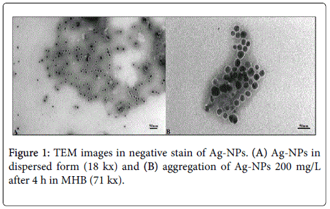
Figure 1: TEM images in negative stain of Ag-NPs. (A) Ag-NPs in dispersed form (18 kx) and (B) aggregation of Ag-NPs 200 mg/L after 4 h in MHB (71 kx).
The Ag-NPs mixed with the bacteria showed to bind rapidly to the cell wall of the two Salmonella enterica strains within the first 5 min (Figures 2A and 2B) and proved to adhere mainly to the cell wall of S. Enteritidis serovar (Figures 3A and 3B).
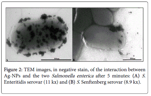
Figure 2: TEM images, in negative stain, of the interaction between Ag-NPs and the two Salmonella enterica after 5 minutes: (A) S. Enteritidis serovar (11 kx) and (B) S. Senftenberg serovar (8.9 kx).
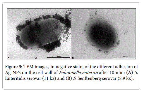
Figure 3: TEM images, in negative stain, of the different adhesion of Ag-NPs on the cell wall of Salmonella enterica after 10 min: (A) S. Enteritidis serovar (11 kx) and (B) S . Senftenberg serovar (8.9 kx).
The interaction between Ag-NPs and bacteria was evident even after repeated cycles of centrifugation and re-suspension; as well, the bond was visible up to 24 hours in the case of S. Enteritidis (Figure 4A), while it lasted only for 6 hours of incubation in the case of S. Senftenberg (Figure 4B).
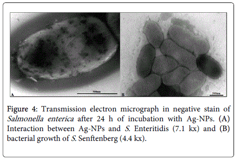
Figure 4: Transmission electron micrograph in negative stain of Salmonella enterica after 24 h of incubation with Ag-NPs. (A) Interaction between Ag-NPs and S. Enteritidis (7.1 kx) and (B) bacterial growth of S. Senftenberg (4.4 kx).
At this stage, there was no evidence of Ag-NPs entering inside the cells of Salmonella enterica .
Interaction of nanoparticles with Salmonella (ultrastructural observations): The response of S. Enteritidis to Ag-NPs after 1 hour included: disruption of the cell wall, lysis of the cell membrane, damage of the cytoplasm and cell deformation.
The interaction between Ag-NPs and the cell wall was characterized by the formation of “pits” (Figure 5A) and by their aggregation on the surface of the outer membrane (Figure 5B), thus determining an enhanced permeability of the bacterial membrane which allowed entry into the cell and, possibly, caused its death.
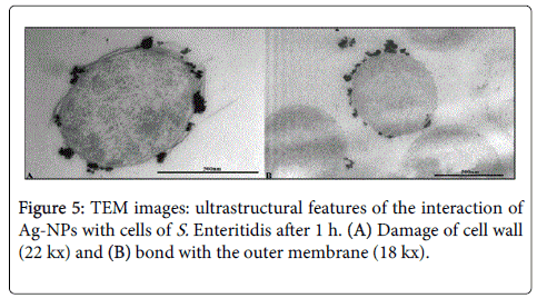
Figure 5: TEM images: ultrastructural features of the interaction of Ag-NPs with cells of S. Enteritidis after 1 h. (A) Damage of cell wall (22 kx) and (B) bond with the outer membrane (18 kx).
In addition, electron dense Ag-NPs were found in cytoplasm of S. Enteritidis (Figure 6A) and the damage of the cells displayed the formation of small electron lucent areas in cytoplasm. These regions appeared throughout the whole cell and aggregated in areas of high electron density located in the electron lucent cytoplasm (“empty cells”) (Figure 6B).

Figure 6: TEM images: ultrastructural analysis of S. Enteritidis after 1 h. (A) Ag-NPs in cytoplasm (28 kx) and (B) “empty cells” (14 kx).
Besides, when Ag-NPs were adsorbed, the cytoplasm became amorphous, in some cases spilling out of the cell (Figure 7A).
Another change induced by the adsorption of Ag-NPs was the widening of the periplasmic space, in which Ag-NPs had accumulated (Figure 7B).
Ultrathin sections of S. Enteritidis sometimes showed one or more membrane vesicles budding from the cells (Figure 7C).
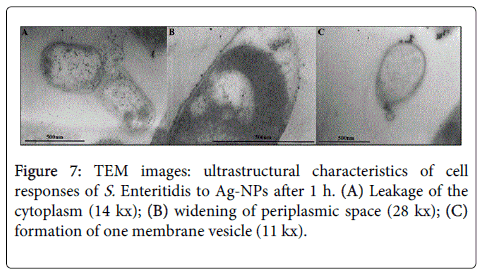
Figure 7: TEM images: ultrastructural characteristics of cell responses of S. Enteritidis to Ag-NPs after 1 h. (A) Leakage of the cytoplasm (14 kx); (B) widening of periplasmic space (28 kx); (C) formation of one membrane vesicle (11 kx).
With the concentration of Ag-NPs used, not all cells were impaired and the cultures showed that some cells had no visible signs of interaction with Ag-NPs, meaning no alterations.
After 4 hours of incubation, the same morphological damages were observed in the cultures of S. Enteritidis.
In the case of S. Senftenberg, the disruption of the cell wall was observed after 1 hour, although the Ag-NPs did not penetrate inside and we did not detect any damage to the cell shape (Figure 8A).
After 4 hours, the S. Senftenberg cells were mainly intact and maintained the same morphological structures as the control (Figure 8B).
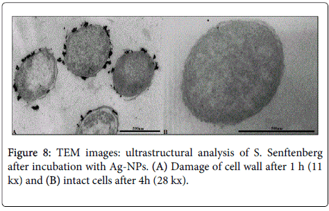
Figure 8: TEM images: ultrastructural analysis of S. Senftenberg after incubation with Ag-NPs. (A) Damage of cell wall after 1 h (11 kx) and (B) intact cells after 4h (28 kx).
Nanosized particles display specific physical and chemical properties and may be used in numerous biological, biomedical and pharmaceutical applications.
Considering the outbreaks of infectious diseases caused by different pathogenic bacteria and their ever increasing resistance to antibiotics, the pharmaceutical companies have been looking for new antibacterial agents. In the present scenario, the silver nanoparticles have proved to be the most suitable [11,12], although many aspects of their antibacterial activity are not yet fully understood.
The morphological characterization of nanoparticles can be performed by TEM, which undoubtedly is a very useful instrument to study the physical interactions between microorganisms and nanoparticles and may therefore be used to better understand the mechanisms of antibacterial activity of nanoparticles.
The results obtained by using the negative stain technique highlighted that in the first 5 minutes the Ag-NPs had interacted with both strains of Salmonella enterica . This may explain the rapid inhibition of bacteria growth observed in the assays of bactericidal activity, in which the number of dividing cells had significantly decreased after 30 min of incubation with Ag-NPs, differently from the control culture [9].
Remarkably, the interaction with the Ag-NPs was more noticeable and long lasting in the case of S. Enteritidis rather than in S. Senftenberg. This data could confirm that S. Enteritidis is more sensitive to the action of nanoparticles, as demonstrated by the in vivo test [9].
Furthermore, the ultrastructural analysis revealed some differences between S. Enteritidis and S. Seftenberg as regards their sensitivity to Ag-NPs.
The Ag-NPs attached to the cell membrane of both Salmonella enterica serovars, although they managed to penetrate only inside the cell of S. Enteritiditis. This observation is important to explain the antibacterial mechanism of nanoparticles: they interact with the bacterial membrane causing structural changes and cell death.
Notably, damages to the cell were observed in S. Enteritidis up to 4 hours, even if the in vivo test showed a resumed growth of bacterial cells already after 1 hour. This could be explained by the gradual decreasing concentration of the nanoparticles, caused by their interaction with the liquid medium and with the intracellular substances spilled out from the destroyed cells. As a matter of fact, the Ag-NPs have a limited use as bactericide because of their low functional stability: this suggests the necessity of repeated addition of Ag-NPs to control the microbial charge. As well, to use them combined with some classes of antibiotics (e.g.: beta-lactam) to enhance the antimicrobial effects and reduce resistance to drugs is a possibility to be taken into consideration.
On the contrary, the Ag-NPs, even if adhering to the S. Senftenberg cell wall, cannot enter within the bacteria, probably because of the presence of Ag+ resistance determinants [9].
In Salmonella , the silver resistance determinant present in some strains is encoded by genes located both on the plasmid and the chromosome. The silver determinant, studied on Salmonella plasmid pMG101, contains nine genes coding for one efflux ATPase (SilP), two metal-binding proteins (SilF and SilE), and one cation/proton antiporter (SilCBA). These proteins supposedly work in synergy: SilP releases Ag+ in the periplasmic space, SilF carries Ag+ from the periplasm to the inner membrane cation pump protein SilA, as a part of the SilCBA complex, which brings Ag+ out from the bacterial cell [13].
Finally, our results have demonstrated to what extent TEM observations are useful to evaluate the damages of cellular structures and the biological effects of nanomaterials.
By using negative stain, some authors [7,14-18] have described the cellular damage which can be observed in bacteria treated with Ag- NPs, such as the alteration of cell shape, the damage to the cell membrane and the gradual “efflux” of cytoplasm. However, we believe that further studies considering ultrathin sections are necessary to support these findings, because the negative stain alone was unable to visualize the fine structure of the cell.
The present study has demonstrated that Ag-NPs can be effective as an antimicrobial in the case of Salmonella , but its success is strongly strain-dependent, since differences in terms of time of action of Ag-NPs and sensitivity were observed for the two investigated serovars. This is probably due to genetic factors specifically intrinsic of each strain, including the presence of specific determinants of resistance, as demonstrated in the case of S. Senftenberg.
The ultrastructural observations, performed using TEM, attributed the different sensitivity of the tested bacteria to different morphological changes and structural damages in S. Enteritidis and S. Senftenberg following the interaction with Ag-NPs, thus confirming the utility of TEM to investigate the effect of nanoparticles on live bacteria.
Authors are grateful to Dr.ssa Antonia Ricci (OIE/National Reference Laboratory for Salmonellosis) for funding this experiment and study.
This work was supported financially by the Ministry of Health, IT (project code RC IZSVe 01/11 and RC IZSVe 04/12).
Authors acknowledge Mrs. Francesca Ellero for manuscript editing.
All authors have obtained permission from their employer or institution to publish and there are no conflicts of interest to report.