Journal of Fertilization: In Vitro - IVF-Worldwide, Reproductive Medicine, Genetics & Stem Cell Biol
Open Access
ISSN: 2375-4508
ISSN: 2375-4508
Research Article - (2015) Volume 3, Issue 4
Exposure of in vitro cultured human embryos to microvibrations can improve embryo development, but music as a source of mechanical vibrations has not yet been explored. To determine the effect of the exposure to music during in vitro culture, 967 oocytes (114 patients) were analyzed. Before insemination, oocytes from each patient were randomly assigned to two groups: embryo culture exposed to music (479 oocytes), and embryo culture without music (488 oocytes). Three different types of music were also tested: pop, heavy metal and classical. Fertilization rates and embryo quality (score, cleavage stage and multinucleation) were compared using a generalized linear mixed model (two levels were considered) and analyzed by means of Bayesian inferences using Integrated Nested Laplace (INLA). Results showed that fertilization rates were 4.82% higher when oocytes were exposed to music but no statistically significant differences were found regarding embryo quality on Day 2. Moreover, no statistically significant difference was observed between the different types of music played (pop, heavy metal and classical). As a conclusion, the routine use of music inside incubators during in vitro culture could be a useful tool to improve fertilization rates. The effect of music on embryo development up to Day 5 should be evaluated.
<Keywords: Microvibration, Music, In vitro development, Human embryo, Fertilization rate
Since the beginnings of assisted reproduction, in vitro culture conditions have always tried to mimic the in vivo situation, with developments focusing principally on temperature, light, carbon dioxide and oxygen levels, as well as ensuring that culture media contain the same nutrients as those found in the uterus. Embryos were initially cultured in large volumes of culture medium, but this volume has been reduced so that embryos are now cultured individually in microdrops (either flat or suspended). However, these are all static rather than dynamic cell cultures with one main limitation: toxic by-products, such as free radicals or ammonia, accumulate in the culture medium [1], and this can compromise embryo development. In natural circumstances, human embryos travel along the fallopian tubes to the uterus in conditions of constant cellular and peristaltic movement surrounded by ciliated secretory cells, thus allowing the interchange of metabolites [2]. This movement, apart from being necessary for the egg to reach the uterus, contributes to the dispersal of toxic metabolites generated by the oocyte, zygote or embryo and to the uptake of nutrients and molecules needed for further development [2-4]. Two different in vitro culture systems have been developed to mimic the natural environment effect: (i) movement applied directly to the culture media and (ii) dynamization of the embryo microculture [5]. Several groups have tried to apply a variety of methods, such as mechanical microvibrations to embryo culture plates [6-8], the use of dynamic fluids in embryo culture [9-16] or the use of a tilting embryo culture system [17]. An improvement in the quality of in vitro human blastocysts was noted when the tilting embryo culture system was used [18,19] and also when dynamic microfunnel culture conditions were applied [12]; in the latter study a higher rate of embryo implantation and subsequent pregnancy was also noted. Pregnancy rates were as well increased with the application of pulsatile mechanical microvibrations to the culture system (20 Hz over 5 seconds, once per hour) [6].
On the other hand, a number of studies have investigated the biological influence of sound, a physical agent that propagates through fluids as a mechanical wave producing pressure and displacement. In particular, research into infrasounds (10-4-20 Hz) [20] and ultrasounds (2 × 10⁴-1012 Hz) [21,22] have determined accurately their effects and applications, whereas knowledge about the biological impact of audible frequencies (20-2×10⁴ Hz) is scarce. Nevertheless, the effect of acoustic stimulation in the field of neuroscience is well-documented [23] and has also been observed during prenatal exposure [24].
In the search for a source of mechanical vibrations that might improve success rates, our group has been interested in the use of music during in vitro development prior to implantation. To our knowledge, this is an approach not yet described.
The objective of this study was to investigate the effects of sound pressure waves created by music on in vitro embryo culture.
Study population
Between December 2011 and August 2012, 114 patients were included in this study. Both recipients of egg donation and patients undergoing in vitro fecundation (IVF) with their own eggs were included. A minimum of 6 embryos were in culture for each patient. In total there were 967 oocytes. 912 were inseminated, of which 725 oocytes were fecundated: 378 from the music group and 347 from the non-music group (Table 1). After oocyte recovery and before decumulation, oocytes were divided into two groups (two different plates) independently of their morphology. Afterwards, they were inseminated and each plate was cultured in a separate incubator (with and without music, respectively).
| Total | Music Group | No Music Group | |
|---|---|---|---|
| Patients | 114 | 114 | 114 |
| Number of cycles | 114 | 114 | 114 |
| Number of oocytes | 967 | 479 | 488 |
| Number of inseminated oocytes (ICSI) | 912 | 466 | 446 |
| Number of fecundated oocytes | 725 | 378 (81.1%) Classical: 109 (82.5%) Pop: 140 (78.6%) Heavy: 129 (82.6%) |
347 (77.8%) |
| Number of scored embryos | 645 | 336 (67.6%) | 309 (63.3%) |
| Embryos with score >7 | 468 | 242 (48.7%) | 226 (46.3%) |
| Embryos with score >5 | 575 | 309 (62.2%) | 266 (54.5%) |
Table 1: Oocyte characteristics.
All women included gave written informed consent. Data were accessed and retrieved only by authorized healthcare professionals and anonymity was ensured for subsequent analysis, according to the Spanish Law 15/1999 on Personal Data Protection Act (LOPD) and the principles of the Declaration of Helsinki.
Experimental procedures
Following hormonal stimulation using ultrasound monitoring and estradiol analysis, the egg donors and the patients undergoing IVF with their own eggs completed ultrasound-guided egg collection 36 hours after the ovulation trigger. All culture media were from Vitrolife Sweden AB; Gotheborg, Sweden. G5-Series™ PLUS culture medium was used for both the micromanipulation of oocytes and embryo culture, and G-MOPS™ PLUS medium was used for egg collection. Oocytes were maintained in G-IVF™ PLUS culture medium in Labotect C200 incubators (Labotect Labor-Technik-Göttingen GmbH; Rosdorf, Germany) until the moment of decumulation.
Decumulation occurred approximately 3 hours after egg collection using a Hyase-10X™ and G-MOPS™ PLUS. Subsequently, those in metaphase II were kept in microdrops of IVF™ PLUS covered with OVOIL™ oil on NUNC™ 35 × 10 plates, until the moment of insemination. All the oocytes were inseminated using intracytoplasmic microinjection (ICSI) 4-5 hours post collection. Once microinjected, the embryos were kept in microdrops of G1™ PLUS, covered with oil, in their respective incubators.
Six incubators (Labotect C200) were assigned to this project: three with music (one for each music genre) and three without music. The oocytes recovered from the egg retrieval were 967, divided in two groups from each patient: half of them were cultured in Labotect 112 C200 incubators with music (n=479) and the other half in Labotect C200 without music (n=488). Prior to insemination oocytes were denuded, inseminated and placed back into incubators with music (n=466) and without music (n=446) (Figure 1). The source of music was a commercially available MP3 player (iPod, Apple Inc., California, USA) placed inside each incubator and played constantly throughout embryo culture. Embryos were located at the same distance from the speaker, and sound measurements (by Soundlab, Laboratorio de Mediciones Acústicas, Barcelona, Spain) guaranteed that all embryos in a given incubator were exposed to the same sound waves. Each MP3 player had been preprogramed with one of three music genres: classical music, pop music or heavy metal (Figure 1). This way, three styles of music were established in order to evaluate the effect of each of the different music genre on the embryo culture. Sound pressure levels created by the music were monitored.
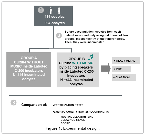
Figure 1: Experimental design.
In order to guarantee similar culture conditions in all the incubators, daily temperature and CO2 levels were measured, ensuring the values were 37°C and 6.0-6.4% of CO2, respectively. pH was recorded weekly in the culture media to ensure this was also correct (pH = 7.2-7.4). To avoid possible variation between different incubators, all the incubators were randomly used (exchanged at different time points of the study) to provide music to embryos in the Music Group, and incubators containing embryos cultured with and without music were switched at half of the culture time.
Fertilization and developmental morphology follow up was determined following Istanbul consensus workshop [25]. Sixteen to 19 hours post-insemination, the fertilization rate of the oocytes was evaluated. Correctly fertilized oocytes (2pN-2PB) were transferred to a new culture dish containing G1™ PLUS culture medium and then returned to their allocated incubator.
Embryo morphology was assessed 44 ± 1 h post-insemination (i.e., Day 2) according to the following criteria: number of cells, cell symmetry [26], percentage of cell fragmentation, presence of multinucleation and cytoplasm appearance. Embryos were classified on Day 2 according to an internal score consisting of 1 to 10 points, where 1 is the lowest quality and 10 the best quality. From an initial score of 10 points, 0 to 4 points could be subtracted for each criteria depending upon its quality, according to different morphological parameters (Table 2). Embryos scoring ≥ 7 were considered first choice for a potential transfer, which was done on Day 3. The fertilization rate and the embryo quality (embryo score) on Day 2 of embryo development were calculated.
| Day 2 (points) | |
|---|---|
| Number of cells | |
| 4 cells | 0 |
| 4 cells | 0 |
| 4 cells | 0 |
| 4 cells | 0 |
| 5 cells | -1 |
| 2 cells | -4 |
| 2 cells | -4 |
| 2 cells | -4 |
| 2 cells | -4 |
| 3 cells | -2 |
| ≥ 6 cells | -4 |
| Asymmetry (*) | |
| + (>20% asymmetry) | -1 |
| ++/+++ (>50% asymmetry) | -2 |
| Multinucleated blastomere | |
| Embryo discarded | |
| Percentage of fragmentation | |
| 0% | 0 |
| <10% of fragments | 0 |
| >10-20% of fragments | -1 |
| 21-35% fragments | -2 |
| 35-50% fragments | -3 |
| >50% fragments | Embryo discarded |
| Fragmentation appearance | |
| small, associated to one blastomere, in small amount | 0 |
| fragment filling the space | 0 |
| small fragments at random at all levels or at the edges | -1 |
| All of them are big fragments | -2 |
| necrotic fragments | Embryo discarded |
(*) Embryos that present cellular asymmetry at 3 or 5 cells-stage are not downgraded.
Using this procedure, fertilized oocytes initially start with a score of 10 points.
Table 2: Chart to calculate the embryo score, showing the morphologic parameters assessed and their weight on the final score.
Measurement of sound
Independent sound technicians (Soundlab) analyzed in their own laboratory the equipment used in reproductive medicine and the musical spectrum of the music chosen. A pure frequency spectrum for the music was obtained in the sound lab, with a class 1 soundlevel meter model 01dB-Solo (ACOEM, Limonest, France). This incorporated a noise spectrum and reverberation time analyzer with an MCE-212 microphone and preamplifier PRE21-S. To monitor the sound inside the incubators (Figure 2), an integrated sound level meter CESVA SC310 (Cesva Instruments S.L.U., Barcelona, Spain) with a C-130 microphone and PA-13 preamplifier with an extension cable was used. A sound calibrator CESVA CB-5 was also used. Background noise in the incubators was measured to determine possible interference or influence with the music. In all cases, the volume of the music played was the maximum possible for the piece of equipment used. Equivalent continuous sound pressure levels (Leq) were measured in thirds of octave spectral distribution.
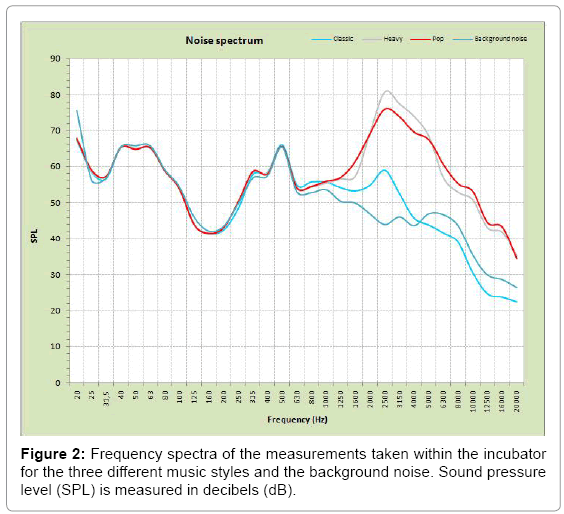
Figure 2: Frequency spectra of the measurements taken within the incubator for the three different music styles and the background noise. Sound pressure level (SPL) is measured in decibels (dB).
Statistical analysis
The main objective was to evaluate the effect of music (the independent variable) on the two dependent variables: fertilization rate and quality of the embryos on Day 2. Both the general effect of the music and that of the three individual types of music used were analyzed: classical, pop and heavy metal.
Descriptive analysis of the variables in question was performed. Mean and standard deviation were calculated for quantitative variables (e.g. number of cells and embryo score) in each condition: presence or absence of music in general, and presence of one of the three different types of music (classical, heavy or pop). Qualitative variables were expressed as percentages and frequencies or categorized, and the different categories were compared in each scenario.
Generalized linear mixed models were estimated for both dependent variables, since music might affect each parameter assessed on more than one level. We considered that the effect of music on the fertilization rate and the embryo score might not be the same for all patients (first level) and that the music might affect the fertilization rate in one patient differently in two separate cycles of ovarian stimulation (second level).
The generalized linear mixed models were chosen according to the nature of the dependent variable [27]. A Poisson model was used when the dependent variable was quantitative and discrete (e.g., quality of the embryos as measured by the number of cells or by a 1 to 10 score) whereas a binomial distribution was used when the variable was dichotomous and qualitative, e.g. fertilization, multinucleation (classified as multinucleated versus not multinucleated or binucleated) or embryo quality ranges (score ≥ 7, ≥ 5). All the explicative variables were qualitative and dichotomous (presence or absence of music in general, and presence or absence of classical, pop or heavy metal music).
A Bayesian focus was used to make all the inferences by using Integrated Nested Laplace approximations (INLA) [28,29]. All the analyses were completed using the R-Project for statistical computing, version 2.15.2 [29] and the R-INLA package (The R-INLA project, http://r-inla.org/home).
Sound measurements
The integrated sound levels inside the incubators where music was played, and the background noise where no music was played, are shown in Table 3.
| Type of music | LeqA [dBA] |
|---|---|
| Classical | 67.3 |
| Heavy metal | 84.5 |
| Pop | 80.7 |
| Background noise | 64.6 |
LeqA – A-weighted equivalent continuous noise level
Table 3: Equivalent sound values produced within the incubator in the treatment cycles where music was used, as well as the background noise, shown in A-weighted decibels (dBA).
Heavy metal music produced the highest sound levels due to its musical characteristics (Figure 2). Both heavy and pop music produced levels well above those produced by classical music. Classical music produced low sound levels but, even so, it produced an increase of 3 dBA above the background noise. Figure 2 compares the frequency spectra of the measurements taken within the incubator, from 20Hz to 20 kHz. As can be seen, the levels obtained from the different types of music and the background noise remained almost the same until about 630 Hz.
Embryological and clinical results after IVF
The results of the descriptive analyses showed that fertilization rates were significantly higher (p<0.05) in the group exposed to music when compared with those not exposed to music (81.1% vs. 77.8% respectively). There was no overlap of the 95% confidence intervals between the group with music (80.7% - 83.3%) and the group without music (76.3% and 79.3%).
The same results were obtained when multivariate models were used. Specifically it was possible to observe an increase in the fertilization rate of 4.82% (95% confidence intervals 3.44%-6.21%) in the group exposed to music (Table 4).
| Music | Percentage of variation in fertilization rate | Percentage of variation in score (≥7) | Percentage of variation in score (≥5) |
|---|---|---|---|
| Any type | 4.82% (3.44%-6.21%) | 0.0035% (-2.17%, 2.19%) | 0.049% (-2.02%, 2.29%) |
| Classical | 2.82% (1.79%-5.47%) | 0.0061% (-2.14%, 2.17%) | 0.013% (-2.11%, 2.18%) |
| Pop | 5.34% (3.21%-7.48%) | -0.0240% (-2.22%, 2.09%) | 0.006% (-2.14%, 2.17%) |
| Heavy | 5.94% (3.47%-8-42%) | 0.0280% (-2.11%, 2.27%) | 0.030% (-2.08%, 2.24%) |
Table 4: Results of the estimation of the multivariate models (mean percentage of variation with 95% credible intervals).
However, when different types of music were analyzed, the 95% confidence intervals overlapped: classical (79.6%-82.9%), pop (76.4%- 79.9%) and heavy metal (79.0-82.7%), indicating that there is no statistically significant difference between their effects. These results were corroborated when multivariate models were used (data not shown).
Similarly, using either descriptive analysis or multivariate models, neither the presence of music nor the specific three types of music were statistically associated with either the embryo score or with some of the quality variables used to calculate that score (number of cells and percentage of cellular fragmentation).
When two groups of embryos were established based on their quality (embryos of first choice for potential transfer with a score ≥ 7 and <7), no significant differences were observed in the group of embryos exposed to music when compared with those not exposed (p=0.539). A further estimate established a group of transferable embryos (score ≥ 5 independently of whether or not they would have been first choice for potential transfer) versus embryos that were not transferable. This was done to take into account all embryos of an appropriate quality for potential freezing and possible use in a subsequent cycle. No statistically significant differences were found between these two groups either (p=0.884) (Table 4).
To our knowledge, this is the first study to demonstrate that direct exposure of human embryos to music during the IVF process can positively affect fertilization rates, with an increase of 4.82%.
It is known that some cellular processes require mechanotransduction, that is, the chemomechanical coupling of reactions through a process of interacting vibrational information networks [30,31]. Vibration seems to play a role in some processes related to embryo culturing, where it increases fertilization rates, although the exact reason for this effect is unknown [6,12,19]. Indeed, mechanical stimulation has been shown to activate DNA synthesis and gene transcription in endothelial and bone cells [32], an effect thought to be a direct result of the microvibrations on cytoplasmic maturation.
It has been demonstrated that cells other than auditory hair cells could respond to audible sound [33]. Several studies have proved the effects of single frequencies or pure sounds in cell growth [34-39] either increasing or decreasing the proliferation rate. Furthermore, more recently, music has shown to alter the cell cycle and morphofunctional parameters of human cell lines [33] with an increase in the percentage of cells in S phase, the one devoted to DNA replication. Music has also been shown to produce specific changes in the expression of particular molecules during the process of neurogenesis related to neural cell behavior [40] and to affect the peripheral immune response, upregulating anti-inflammatory cytokines [41].
Nevertheless, not all sound pressure has the same effect. Different wave frequencies, exposure times or the harmonic organization of the sound (features that differentiate music from simple noise) have different consequences. In fact, noise can cause growth retardation and decreased neurogenesis when applied to developing rats, the opposite to the outcomes of music [42].
In our study, we noted a statistically significant increase in the fertilization rates of the group of embryos exposed to music during embryo culture when compared with embryos from the same patients not exposed to music, indicating that music could smooth the process of fertilization. The exact biological mechanism possibly induced by music is still unknown and additional studies are needed. One possibility is that music could promote changes affecting DNA synthesis, facilitating the meiosis II spermatozoid activation inside the oocyte, although this study was not designed to explore such a possibility. Additionally, vibrations triggered by music could aid dissipating toxic by-products.
Regarding the different types of music chosen, as Figure 2 shows, there are some differences in intensities and frequency ranges (between 4000-6000 Hz) between classical music on one hand and pop and rock music on the other, that could account for the non-statistically differences observed among them (Table 4). Sound waves travel through fluids displacing the particles of the medium through oscillations that are proportional to the wave energy. Thus, without considering the influence of harmony, more acute frequencies and higher intensities may be related to better fertilization rates. Nonetheless, the small difference in intensity between classical music and background noise (64.6 versus 67.3 dBA) (Table 3) rules out the possibility of sound volume being the whole explanation for this effect (Figure 2).
This study was designed to evaluate embryo quality until Day 2 of development but we were unable to detect statistically significant differences using this parameter. Further studies need to be done to discriminate whether music exerts an exclusive effect on the earliest stages of fecundation, resulting in higher fertilization rates, or if it also affects subsequent stages of embryo development, not revealed currently due to specific limitations of this study, such as the short period of time under evaluation. In this respect, it will be necessary to evaluate whether or not music can affect embryo quality up to Day 5 of evolution, just prior to embryo transfer.
According to several reports, dynamization of cell culture seems to improve results in assisted reproduction, whether by means of dynamic microfluids, the application of microvibrations or, as shown here, by exposure to music. However, the costs in terms of installation, maintenance and implementation all need to be taken into account. Equipment intended to be used routinely should be easy to install and manipulate. Dependence on specific apparatus is not desirable, while adaptability for use in all incubators is an advantage. In this regard, the system described here, only requiring basic sound equipment, is very simple, and a small music player can be left permanently inside the incubator with minimum maintenance.
We can conclude that this is the first study to show that the use of music in the in vitro setting has a statistically significant, positive effect on fertilization rates in human embryos prior to implantation. In contrast to other methods of dynamizing culture media, this technique is easy to apply in assisted reproduction laboratories.