
Journal of Bone Research
Open Access
ISSN: 2572-4916
ISSN: 2572-4916
Research Article - (2025)Volume 13, Issue 5
Background: Bone Marrow-derived Mesenchymal Stem Cells (BMMSCs) have been extensively studied for their therapeutic role in regenerative medicine. However, their effectiveness in BMMSCs-based therapies is constrained due to the hypoxic microenvironment encountered at injured sites. This study aims to investigate the cellular mechanisms that regulate mitochondrial function through mitophagy in hypoxic BMMSCs to improve their therapeutic potential with minimal adverse effects. Material and Methods: C57BL/6 mouse BMMSCs were subjected to a simulated ischemic environment at hypoxia with 1% O2 for 48 hours. Mitophagy markers, including LC3B and BNIP3, were assessed using quantitative real-time PCR, and Western blotting. The hypoxic response was further characterized by evaluating HIF-1α expression and its role in the transcriptional regulation of mitophagy-related genes. Angiogenic factors, such as VEGF, were also quantified to assess the regenerative potential of hypoxia- preconditioned BMMSCs. Results: Hypoxia significantly increased LC3B and BNIP3 expression, suggesting that hypoxia promotes mitophagy in BMMSCs. HIF-1α level was also elevated in these cells and drove BNIP3 transcription. Furthermore, VEGF expression was upregulated in hypoxic BMMSCs, indicative of enhanced reparative functionality. Conclusion: Hypoxic preconditioning promotes mitophagy and survival and reparative capacity of BMMSCs through the upregulation of BNIP3, among other regulators of key mitophagy
BMMSCs; Hypoxic microenvironment; Mitophagy; HIF-1α; BNIP3; LC3
Mitochondrial quality control is important for cellular function and survival. Deteriorated mitochondria are constantly renewed through mitochondrial biogenesis and autophagy, particularly mitophagy, which removes dysfunctional mitochondria to maintain cellular homeostasis and prevent harmful cellular consequences such as oxidative stress, inflammation, and apoptosis [1-3]. Mitophagy is a highly regulated process that ensures mitochondrial dynamics, including fission, fusion, and turnover, are preserved. Under cellular stress conditions, such as hypoxia, mitophagy is critical for maintaining mitochondrial integrity [1,2].
Hypoxic conditions, those defined by low oxygen availability, activate hypoxia- inducible factors, notably HIF-1α, which controls a wide array of target genes to enable the cell to adapt to low oxygen stress. One of the important target genes induced by HIF is VEGF, considered to be a critical regulator of angiogenesis [4]. The role of HIF has already been established for the monitoring it gives to the cell's hypoxic response; however, hypoxia and mitochondrial quality control, particularly mitophagy, are still incompletely understood [5]. Mesenchymal Stem Cells (MSCs), including bone marrow-derived MSCs (BMMSCs), have been recognized for their regenerative capacity, making them promising candidates for cell-based therapies aimed at treating a variety of diseases [6-8]. BMMSCs are pluripotent, with the potential to differentiate into various cell types, and they possess the ability to promote tissue repair through paracrine signaling, thereby contributing to the restoration of damaged tissues. While MSCs have garnered interest in regenerative applications, their efficiency in vivo is often limited due to harsh microenvironments such as ischemia and hypoxia, which can lead to cell apoptosis and hinder the therapeutic potential of transplanted cells [6-8].
It has been shown that hypoxic preconditioning, which includes exposing MSCs to low oxygen conditions before transplantation, improves the cells' survival and proliferation and enhances their therapeutic efficacy. Of note, this preconditioning can affect the metabolic pathways of the stem cells and improve functional regulation, increasing their therapeutic potential in regenerative medicine [1-3,9]. Despite the numerous studies focused on the impact of hypoxia on the behavior of stem cells, the related mechanisms, especially concerning mitochondrial quality control and mitophagy, are poorly investigated [10,11].
The present study aimed to elucidate the involvement of hypoxia in the regulation of mitophagy in BMMSCs, focusing on the molecular mechanisms by which hypoxic preconditioning enhances mitochondrial turnover and function. Elucidation of these processes could be of great importance for optimizing stem cell-based therapies for clinical applications, especially for diseases where mitochondrial dysfunction and subsequent tissue damage occur [12-14]. Furthermore, this study has also explored how mitophagy regulators such as BNIP3 contribute to the hypoxic induction of a mitophagic response in the stem cells [15-17]. Thus, the purpose of this review is to provide an explanation of how hypoxic preconditioning affects mitochondrial quality control and metabolic regulation within BMMSCs for the development of more advanced stem cell treatments in regenerative medicine [18,19].
Cell culture
C57BL/6 mouse mesenchymal stem cells at passage 6 from Cyagen of China were plated in mouse MSC growth medium of Cyagen origin, 10% Fetal Bovine Serum, Penicillin, 100 U/ml, Streptomycin 100 μg/ml. MSCs were kept at 37°C in 5% CO2 in a humidified environment and sub-cultured upon reaching 70-80% confluency. To induce mitophagy, cells were incubated under hypoxic conditions of 1% O2, 5% CO2 at 37°C for 48 hours with an AnaeroPack system Mitsubishi Gas Chemical Co., Inc. Tokyo, Japan. For MitoTracker staining, a working solution of 500 nM was prepared from a 1 mM stock solution of MitoTracker M7512, Thermo Fisher Scientific, Waltham, MA, USA, in prewarmed medium.
RNA extraction and quantitative real-time polymerase chain reaction
Total RNA was isolated from cells (n=3 per group) using an RNeasy Mini Kit (Qiagen, Tokyo, Japan). The RNA preparations were further treated with a TURBO DNA-free™ Kit (Thermo Fisher Scientific, Waltham, MA) to remove contaminating DNA. First-strand cDNAs were reverse-transcribed using a High-Capacity cDNA Archive Kit (Thermo Fisher Scientific). The relative expressions of the target mRNAs to β-actin RNA were determined by real-time PCR. TaqMan MGB probes (Mm00468869_m1 for HIF-1α, Mm01275600_g1 for BNIP3, Mm00786306_s1 for BNIP3L, Mm00458724_m1 for LC3A, Mm00782868_sH for LC3B, Mm00437306_m1 for VEGF and Mm02619580_g1 for β-actin, Thermo Fisher Scientific) and a QuantStudio 7 Real Time PCR System (Applied Biosystems) were utilized. Thermal cycling conditions followed the TaqMan Fast Universal PCR protocol.
Western blot analysis
Cells were lysed in RIPA lysis buffer (Santa Cruz Biotechnology), and protein concentrations were measured using a BCA Protein Assay Kit (Pierce Biotechnology). Twenty micrograms of protein from each sample were loaded onto the gel for electrophoresis, followed by transfer to PVDF membranes. Then, membranes were incubated with the following primary antibodies: HIF-1α (1:1000, 20960-1-AP; Proteintech Group, Inc, Rosemont, IL, USA), BNIP3 (1:1000, ab109362; Abcam, Cambridge, UK), BNIP3L (1:1000, ab109414; Abcam), LC3-A/B (1:1000, 12741; Cell Signaling Technology), VEGFA (1:500, ab46154; Abcam), β-actin (1:1000, 4970; Cell Signaling Technology), and GAPDH (1:1000, 2118; Cell Signaling Technology). The membranes were then incubated with horseradish peroxidase-conjugated anti-mouse or anti-rabbit IgG (1:2000; Cell Signaling Technology) for one hour at room temperature. Protein bands were visualized using an ECL Plus Western Blotting Detection System (GE Healthcare), and the images were captured and analyzed using Luminescent Image Quant LAS 4000 Mini (GE Healthcare) and ImageJ software (National Institutes of Health).
Statistics analysis
Data are presented as means ± standard deviation. Student’s t-test was used for pairwise comparisons between normoxic and hypoxic groups. One-way ANOVA followed by post hoc Tukey’s test was used when comparing more than two groups. A p-value < 0.05 was considered statistically significant.
Mitotracker analysis shows increased mitochondrial activity and mitophagy in hypoxic BMMSCs
MitoTracker staining was used to assess mitochondrial activity and morphology in BMMSCs under hypoxic conditions. Hypoxia exposure led to a noticeable increase in mitochondrial mass, as indicated by the enhanced MitoTracker fluorescence intensity (Figure 1). Indeed, the BMMSCs cultured under low oxygen showed a far stronger mitochondrial signal compared to their normoxic controls, which reflects the compensatory increase in mitochondrial activity under hypoxic stress (Figure 1). This result supports that hypoxic preconditioning can provide improved mitochondrial function and homeostasis.
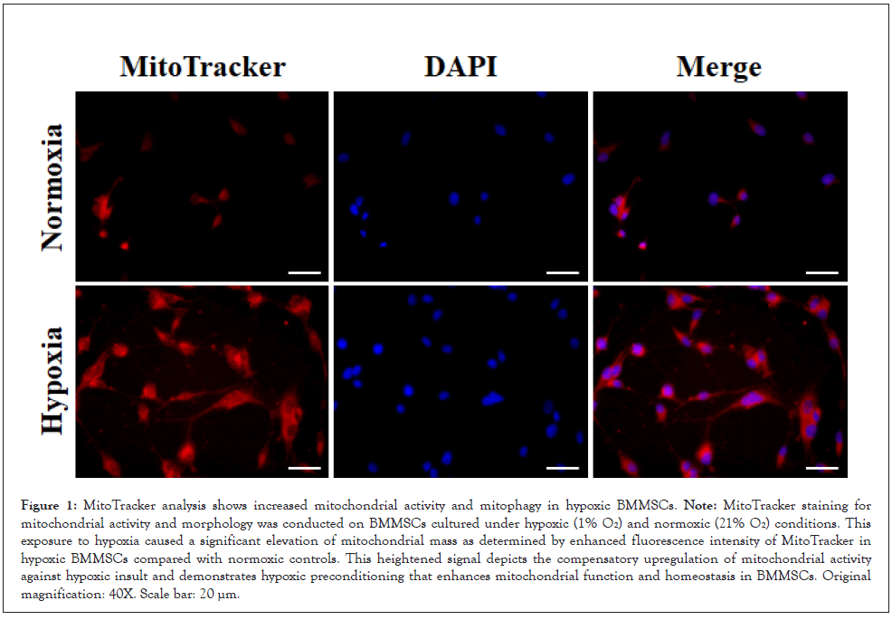
Figure 1: MitoTracker analysis shows increased mitochondrial activity and mitophagy in hypoxic BMMSCs. Note: MitoTracker staining for mitochondrial activity and morphology was conducted on BMMSCs cultured under hypoxic (1% Oâ) and normoxic (21% Oâ) conditions. This exposure to hypoxia caused a significant elevation of mitochondrial mass as determined by enhanced fluorescence intensity of MitoTracker in hypoxic BMMSCs compared with normoxic controls. This heightened signal depicts the compensatory upregulation of mitochondrial activity against hypoxic insult and demonstrates hypoxic preconditioning that enhances mitochondrial function and homeostasis in BMMSCs. Original magnification: 40X. Scale bar: 20 µm.
Gene expression profiling further presented that mitophagy is higher in hypoxic BMMSCs
To explore the expression of crucial genes involved in mitophagy and hypoxic signaling pathways, the relative expression was detected by quantitative real-time PCR in BMMSCs. The expressions of HIF-1α, BNIP3, BNIP3L, LC3A, LC3B, and VEGF genes were significantly upregulated by approximately 2.1-, 2.9-, 5.4-, 3.0-, 6.0- , and 1.6-fold, respectively, in hypoxic BMMSCs compared with normoxic controls; β-actin is used as an internal reference gene to normalize the gene expressions (Figure 2). Precisely, hypoxia caused a significant elevation of HIF-1α expression, showing the turning on of hypoxic pathways. BNIP3 and LC3B expressions were also drastically upregulated; both are critical regulators of mitophagy. This supports our hypothesis that hypoxic preconditioning increases the mitophagic activity in BMMSCs. VEGF expression was significantly increased, suggesting the possible role of hypoxia on affecting angiogenesis-related pathways as well (Figure 2). These results reinforce the role played by both the mitophagic and hypoxic signaling pathways in the maintenance of mitochondrial homeostasis and survival of cells under stress conditions.
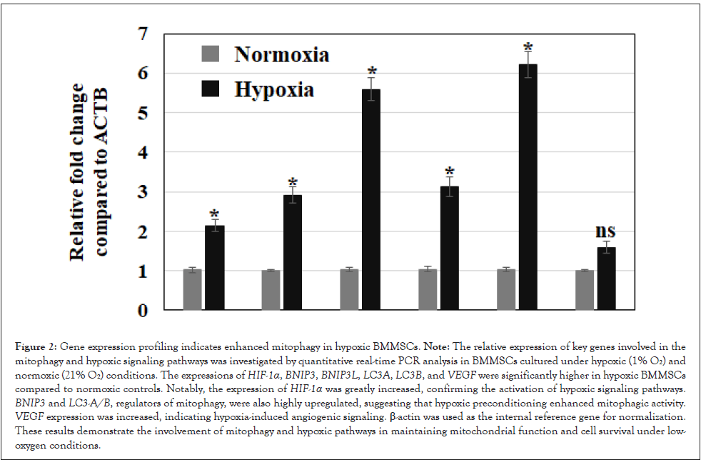
Figure 2: Gene expression profiling indicates enhanced mitophagy in hypoxic BMMSCs. Note: The relative expression of key genes involved in the mitophagy and hypoxic signaling pathways was investigated by quantitative real-time PCR analysis in BMMSCs cultured under hypoxic (1% Oâ) and normoxic (21% Oâ) conditions. The expressions of HIF-1α, BNIP3, BNIP3L, LC3A, LC3B, and VEGF were significantly higher in hypoxic BMMSCs compared to normoxic controls. Notably, the expression of HIF-1α was greatly increased, confirming the activation of hypoxic signaling pathways. BNIP3 and LC3-A/B, regulators of mitophagy, were also highly upregulated, suggesting that hypoxic preconditioning enhanced mitophagic activity. VEGF expression was increased, indicating hypoxia-induced angiogenic signaling. β-actin was used as the internal reference gene for normalization. These results demonstrate the involvement of mitophagy and hypoxic pathways in maintaining mitochondrial function and cell survival under lowoxygen conditions.
Western blot analysis shows enhanced mitophagy and hypoxic pathway proteins in hypoxic BMMSCs.
Next, the protein expression levels of major mitophagy and hypoxic pathway markers were analyzed in BMMSCs under hypoxic conditions by Western blot analysis. The results indicated that proteins HIF-1α, BNIP3, and LC3-A/B were significantly upregulated in hypoxic cells compared to normoxic controls, which corroborated the results obtained from RNA expression analysis (Figure 3). Protein levels showed a corresponding increase with HIF-1α, BNIP3, BNIP3L, LC3-A/B, and VEGF elevated by approximately 2.5-, 1.8-, 2.0-, 2.2-, and 3.5-fold, respectively (Fig. 3b). HIF-1α was highly upregulated, indicating the activation of hypoxic signaling in response to low oxygen levels (Figure 3). Accordingly, BNIP3, a regulator of mitophagy, significantly increased its protein expression under hypoxia, reflecting enhanced mitophagic activity. Similarly, LC3-A/B implicated in the formation of autophagosomes was also increased, which further supported the hypothesis of increased autophagic flux in response to hypoxic stress (Figure 3). Consistent with the role of hypoxia in promoting angiogenesis, VEGF expression was also increased in hypoxic BMMSCs (Figure 3). The expressions of β-actin and GAPDH were used as the loading controls. These findings provide further confirmation that hypoxic preconditioning evokes key regulatory proteins, associated with mitophagy and low oxygen adaptation, thereby supporting BMMSCs for possible therapeutic applications in regenerative medicine
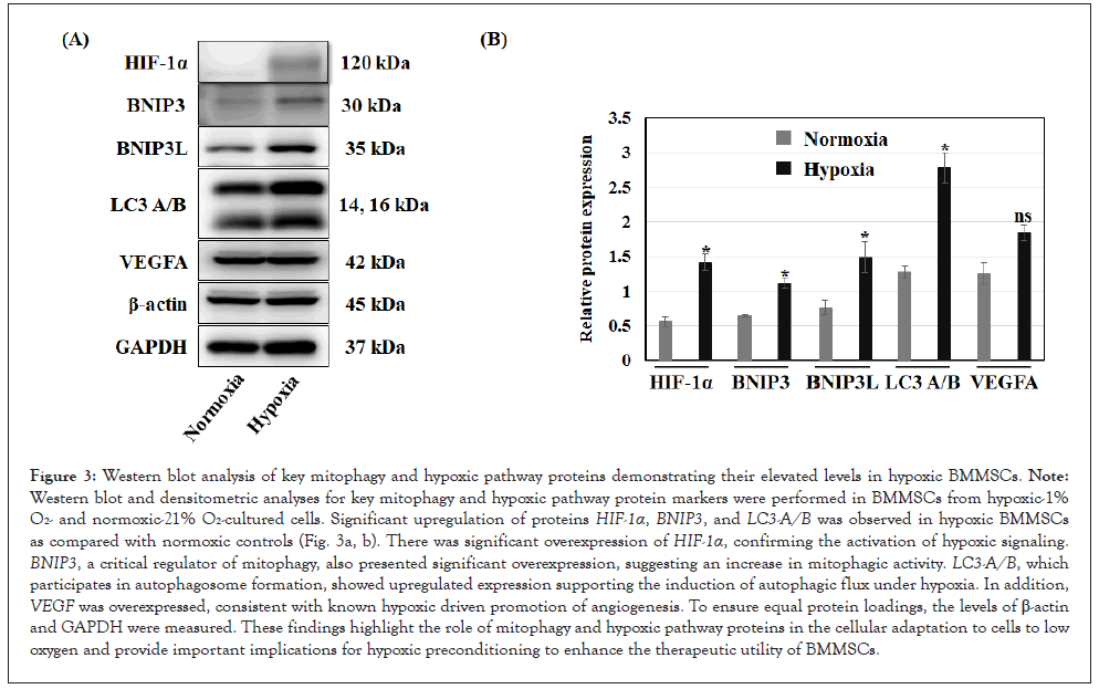
Figure 3: Western blot analysis of key mitophagy and hypoxic pathway proteins demonstrating their elevated levels in hypoxic BMMSCs. Note: Western blot and densitometric analyses for key mitophagy and hypoxic pathway protein markers were performed in BMMSCs from hypoxic-1% Oâ- and normoxic-21% Oâ-cultured cells. Significant upregulation of proteins HIF-1α, BNIP3, and LC3-A/B was observed in hypoxic BMMSCs as compared with normoxic controls (Fig. 3a, b). There was significant overexpression of HIF-1α, confirming the activation of hypoxic signaling. BNIP3, a critical regulator of mitophagy, also presented significant overexpression, suggesting an increase in mitophagic activity. LC3-A/B, which participates in autophagosome formation, showed upregulated expression supporting the induction of autophagic flux under hypoxia. In addition, VEGF was overexpressed, consistent with known hypoxic driven promotion of angiogenesis. To ensure equal protein loadings, the levels of β-actin and GAPDH were measured. These findings highlight the role of mitophagy and hypoxic pathway proteins in the cellular adaptation to cells to low oxygen and provide important implications for hypoxic preconditioning to enhance the therapeutic utility of BMMSCs.
The study focused on the cellular mechanisms of mitochondrial function and mitophagy in bone marrow-derived mesenchymal stem cells under hypoxic conditions for their reparative role and evasion of side effects in stem cell therapy. This review will further analyze how these stress responses may promote the reparative function of the cells through mitophagy pathways in BMMSCs after being treated with hypoxia and VEGF signaling. In clinical applications, BMMSCs often encounter a hostile ischemic microenvironment characterized by low oxygen and nutrient deprivation, which severely limits their survival and therapeutic efficacy. Enhancing mitochondrial quality control mechanisms such as mitophagy may therefore be crucial for improving BMMSC viability post-transplantation.
Maintaining mitochondrial homeostasis is importantly dependent on the process of mitophagy under stress conditions. Impaired mitophagy contributes to the accumulation of dysfunctional mitochondria, consequently leading to cell damage and progressive disease, including CVDs [20]. Our findings presented that hypoxia markedly induced the expression of LC3B, an autophagic marker, which has an important role in the formation of autophagosomes and the removal of damaged mitochondria. Upregulation of LC3B would therefore suggest that hypoxic BMMSCs have an enhanced mitophagic response. In other cell types, LC3B has also been demonstrated as essential in the autophagic/mitophagic mechanisms of several cell types and in those of coronary artery disease [21]. These results can be considered a part of hypoxia-driven cellular defense mechanisms, particularly for stem cell therapy.
BNIP3 was significantly upregulated in hypoxic BMMSCs. BNIP3 is a receptor of mitophagy, which facilitates the removal of injured mitochondria by interacting with LC3B [22]. Increased expression of BNIP3 in hypoxia supports the involvement of the HIF-1α- BNIP3 axis, which was shown to be involved in the regulation of mitochondrial quality control in low-oxygen conditions [23]. These observations agree with earlier reports that BNIP3 induction plays an important role in mitochondrial maintenance for cell survival under hypoxic conditions [24]. On the contrary, BNIP3 is a pro- apoptotic factor and protective factor, indicating that mitophagic regulation under hypoxic stress is complex [25].
HIF-1α, the master transcription factor of the hypoxic response, was also upregulated in hypoxic BMMSCs, further supporting hypoxic signaling in the regulation of mitophagy. According to previous studies, HIF-1α not only modulates the gene expression involved in mitochondrial functions but also in cell survival under low oxygen tension conditions [26]. Our research data showed that hypoxia triggered HIF-1α induction, which in turn influenced BNIP3 transcription. Potential upstream regulators of the HIF- 1α-BNIP3 axis include signaling pathways such as PI3K/Akt and AMPK, which have been reported to modulate hypoxic responses and mitophagy in other cell types [27-29]. Investigating these pathways in BMMSCs under hypoxia warrants further study. It is concluded that HIF-1α may be an important regulatory factor for mitochondrial function by mitophagy in stressed stem cells [30].
The potential of BMMSCs for tissue repair and regeneration has been widely recognized, with studies demonstrating their efficacy in models of myocardial infarction, ischemic stroke, and other diseases [31-33]. However, the success of BMMSC-based therapies largely depends on the survival and functionality of transplanted cells in the target tissue. One major challenge is the harsh hypoxic microenvironment at the site of injury, which limits the effectiveness of BMMSC transplantation. Our findings show that hypoxic preconditioning induces mitophagy and mitochondrial quality control, events that are important for cell survival under ischemic conditions and thus promote the survival of BMMSCs. Increased expression of vascular endothelial growth factor in hypoxic BMMSCs further supports the reparative potential of these cells, especially in promoting angiogenesis and tissue regeneration [34]. These findings therefore point to the therapeutic potential of hypoxic-preconditioned BMMSCs in regenerative medicine.
There are, however, several limitations that need to be pointed out. Although this study has highlighted the role of mitophagy in relation to hypoxia, it cannot be denied that autophagy and apoptosis may also be contributing to the development of BMMSCs. This study’s findings are based on in vitro experiments, which may not fully replicate the complex in vivo microenvironment where BMMSCs encounter multiple systemic factors. Future in vivo studies are essential to validate mitophagy-related mechanisms and their therapeutic relevance. In addition, investigation of crosstalk between these pathways and functional implications on BMMSCs' therapeutic potential is required. Moreover, although BNIP3 and LC3B were identified as key regulators of mitophagy, the exact molecular mechanisms involved in their regulation in BMMSCs remain unclear. The involvement of post-translational modifications in regulating the activity of these proteins under hypoxic conditions should be explored in future studies.
Clinical translation of hypoxic preconditioning in BMMSCs: Translating hypoxic preconditioning into BMMSCs into the clinic requires further studies. Although our study demonstrates the potential benefits of hypoxic preconditioning for enhancing BMMSC survival and function, optimization of the conditions for hypoxia exposure will be required to maximize therapeutic outcomes, such as oxygen concentration and duration. It is prudent to ascertain the underlying benefits of this phenomenon and the avenues for uneventful clinical translation. Several studies have shown that hypoxic preconditioning enhances the function of BMMSCs in ischemic models, but the long-term safety and efficacy of such treatments need to be assessed in preclinical and clinical settings [28]. Moreover, apart from these, the effect of hypoxia on other types of stem cells and their interaction with the immune system remains a field that requires intensive research to understand, in detail, the potential of hypoxic-preconditioned stem cells for clinical applications.
In summary, this study defines the sophisticated regulation of mitochondrial function and angiogenesis pathways in hypoxic BMMSCs and points out that BNIP3 and mitophagy are key regulators in modulating the activity of these cells. Our study provides a rationale for the therapeutic application of hypoxia- preconditioned BMMSCs for improvement in tissue repair and thus holds great promise for diseases related to mitochondrial dysfunction. While further studies are needed to address long-term effects and clinical applications, the current study has set a basis for the development of better stem cell therapies by targeting mitophagy to promote cell survival and function in hypoxic conditions.
This work was supported by a research grant from Honam University, Korea
The authors declare no conflict of interest.
Citation: Kim S, Bhawal UK (2025) Hypoxic Preconditioning Enhances Mitophagy in BMMSCs. J Bone Res. 13:324.
Received: 30-May-2025, Manuscript No. BMRJ-25-37506; Editor assigned: 02-Jun-2025, Pre QC No. BMRJ-25-37506; Reviewed: 16-Jun-2025, QC No. BMRJ-25-37506; Revised: 23-Jun-2025, Manuscript No. BMRJ-25-37506; Published: 30-Sep-2025 , DOI: 10.35841/2572-4916.25.13.324
Copyright: ©2025 Kim S, et al. This is an open-access article distributed under the terms of the Creative Commons Attribution License, which permits unrestricted use, distribution, and reproduction in any medium, provided the original author and source are credited.
Sources of funding : This work was supported by a research grant from Honam University, Korea