Journal of Bone Research
Open Access
ISSN: 2572-4916
ISSN: 2572-4916
Case Report - (2022)Volume 10, Issue 6
Osteomas are a benign neoplasm. In facial region are very rare and just some few articles relating this type of tumour are found in the scientific literature. Most osteomas of the maxillofacial region occur in the mandible. Our article presents two rare cases of osteomas, one case located in the Temporomandibular Joint (TMJ) and the other one in the zygomatic arch. These two cases are very important for diagnosis and treatment planning of bone tumours, because it shows the variable characteristics and the possible auto limited development of the tumour. Thus, the treatment can be, in some cases, totally conservative if there is no compromise of important structures, function or facial aesthetic.
Osteomas; Skull; Mandible; Bone tumors; Temporomandibular joint
Osteoma is a benign neoplasm resulting from the continuous formation of both cortical and cancellous bone. Most osteomas of the maxillofacial region occur in the mandible [1-4]. Histologically they have normal membranous bone varying from insignificant thickening to large masses affecting the skeletal system largely [5].
Osteomas originate from membranous bones of the skull and face, from the cartilage or periosteum or embryonal periosteum, and structurally they are divided into cortical (ivory) osteoma, cancellous osteoma or a combination of both. Cortical type is more common in males whereas women have a high incidence of cancellous type. The mandible is more affected, especially in the posterior aspect of the mandible, lingual side of the ramus or inferior border below the molars. Mandibular lesions may be exophytic [6,7].
The first reported case of osteoma of the condylar process was described by Ivy in 1927 [2,7]. Since then, according to Ostrofsky et al. [7], there are only 22 cases reported in the English literature. The pathogenesis of osteomas are unknown, but they are mostly referred to as developmental anomalies, true neoplasms or reactive lesions triggered by trauma, muscle traction or infection [1,3,7,8]. However, according Ostrofsky et al. [7], and Saikrishna et al. [5], the cause of osteoma in the maxillofacial region is reported to be reactive bone hyperplasia or advanced bone ossification.
Coronoid osteomas are largely asymptomatic and are non-tender until their size and position incommodes with functioning [5,7]. Generally, the most common presenting complaints include facial asymmetry, trismus, malocclusion, swelling in TMJ area and dull pain. Only two cases were ever reported to have severe pain in the TMJ. Contrary to osteoid osteomas where pain may be the chief complaint, patients with osteomas rarely have pain as a main complaint [1,5,7].
The growth of osteomas occurring in the condyle or condylar process may result in morphologic and functional disturbances, including facial asymmetry and temporomandibular joint (TMJ) dysfunction [2]. Benign tumors of the TMJ tend to present less frequently than malignant lesions. Although they can occur at any age, they are most commonly found in patients over 35 years of age and the lesion tends to occur more commonly in males than females [7].
First case report
A 57 years old male patient sought Odontologic treatment at December 2019. A panoramic radiographic examination performed for other purposes, showed an abnormal growth next to the coronoid process, and it was unknown by the patient until that. There was no pain, no swelling, no trism or malocclusion caused by the lesion or moving limitation of the TMJ.
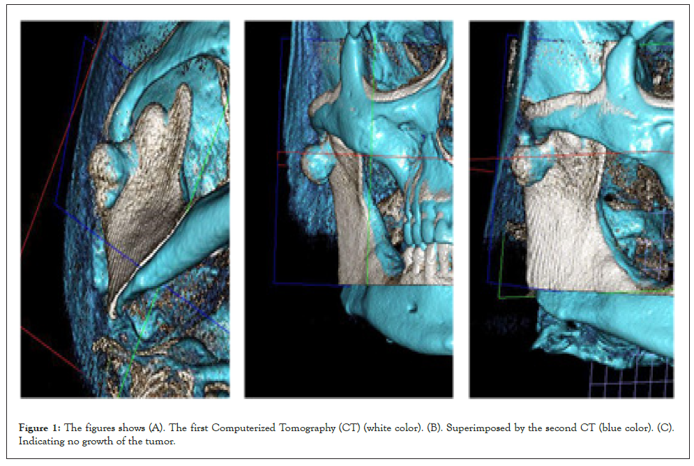
A Computerized Tomography (CT) was requested and it shows bone growth at the coronoid process; it seems like 2 spheres of cortical bone joined together, without another structure involved. The patient was accompanied for about 2 years and, after comparing a new CT analysis with the initial, we observed no increase in the dimensions and no one different aspect of the lesion (Figures 1A-1C).

Figure 1: The figures shows (A). The first Computerized Tomography (CT) (white color). (B). Superimposed by the second CT (blue color). (C). Indicating no growth of the tumor.
The clinical and radiographic examination was compatible with osteoma. And, because of a grave cardiological disease discovered by the patient, we took the decision of don't remove the osteoma, after all, this was no causing problems to esthetic or function to the patient.
Second case report
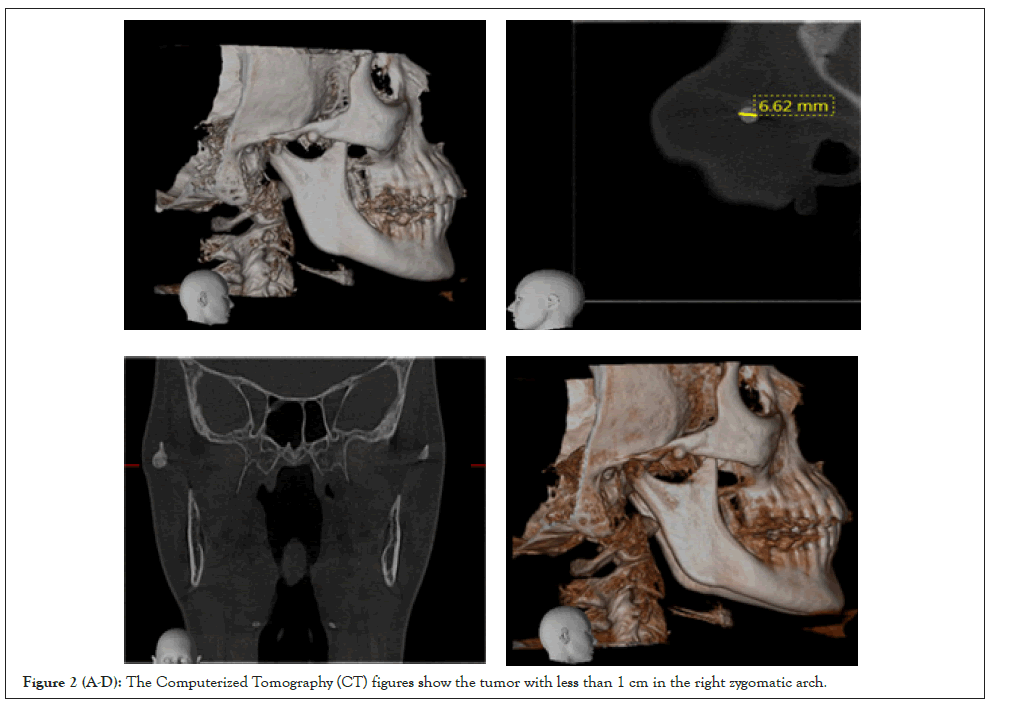
A 60 year old female patient sought care after noticing a nodule on the right side of the face. She reported having no pain or noticeable size change. The patient history has no trauma, infection or another fact that could be related. Clinically, it was possible to notice a nodular volume increase palpable, fixed in the zygomatic arch, as part of it, on the right side of the face, without other alterations. The CT shows a nodule with cortical bone aspect 6.62 mm diameter, approximately. No one anatomic structure was related with the lesion (Figures 2A-2D).

Figure 2 (A-D): The Computerized Tomography (CT) figures show the tumor with less than 1 cm in the right zygomatic arch.
The treatment chosen by the patient was conservative, because there are no aesthetic deficits or functional alteration. So, surgical treatment was no option for her. The patient will be followed up periodically to observe the evolution of the tumor.
As we found in the literature review, the osteomas may vary from insignificant thickening to large masses affecting the skeletal system largely. The cases shown in this study present a significant difference in their sizes, reaffirming that was said by Saikrishna, et al. [5]. According to Ostrofsky, et al. [7], osteoma is more commonly found in female patients, but we could not confirm this because the first case was male patient and the second case was female. And, as reported by de Souza et al. [1], Kondoh et al. [2], Valente et al. [3], and Yonezu, et. al. [4], most maxillofacial osteomas occur in the mandible, but in the second case where the osteoma is located in the zygomatic arch is even rarer.
Bhatt et al. [8], de Souza, et al. [1], Ostrofsky et al. [7], and Valente, et al. [3], said that the pathogenesis of osteomas is unknown, but they are mostly referred to as developmental anomalies, true neoplasms, or reactive lesions triggered by trauma, muscle traction or infection. In both cases, we couldn't find the etiology of the osteomas. There was no prior medical history, infection, or other sign of symptom associate with the tumors.
Despite being described in the literature for many years, the osteomas have no precise etiology. The articles mentioned in our literature review, referred to the osteomas as developmental anomalies, true neoplasms or reactive lesions triggered by trauma, muscle traction or infection, reactive bone hyperplasia or advanced bone ossification. In our present cases, no one trigger was associated; sustaining the possibility of osteoma being a true neoplasm and the biggest differential diagnostic was pain absence, eliminating the hypothesis of osteoma osteoid. Another conclusion that we believe is that the tumor is self-limiting, because no growth was observed in the two cases presented by us. Possibly, many other cases of osteoma may occur without the patient knowledge, because of the pain absence or no trigger associated. Other fact about the low incidence of these tumors may be unfamiliarity with the osteoma by doctors, not being diagnosticated.
Although being considered rare and of unknown etiology, osteomas do not show harm to patients when they are small in size and do not cause aesthetic or functional problems. Thus, the treatment of choice may be simple periodic follow-up and patient guidance. The most important is evaluate all characteristics of the tumor, facts relates by patients and especially ask about pain before choose a treatment plane.
[Crossref] [Google Scholar] [PubMed]
[Crossref] [Google Scholar] [PubMed]
[Crossref] [Google Scholar] [PubMed]
[Crossref] [Google Scholar] [PubMed]
[Crossref] [Google Scholar] [PubMed]
[Crossref] [Google Scholar] [PubMed]
[Crossref] [Google Scholar] [PubMed]
[Crossref] [Google Scholar] [PubMed]
Citation: Ciaramicolo ND, Junior OF, Bisson GB, Yaedú RYF, Silveira IT (2022) Two Rare Cases of Facial Osteoma and Literature Review. J Bone Res. 10:181.
Received: 08-Jul-2022, Manuscript No. BMRJ-22-18554; Editor assigned: 11-Jul-2022, Pre QC No. BMRJ-22-18554 (PQ); Reviewed: 25-Jul-2022, QC No. BMRJ-22-18554; Revised: 01-Aug-2022, Manuscript No. BMRJ-22-18554 (R); Published: 08-Aug-2022 , DOI: 10.35248/2572-4916.22.10.181
Copyright: © 2022 Ciaramicolo ND, et al. This is an open-access article distributed under the terms of the Creative Commons Attribution License, which permits unrestricted use, distribution, and reproduction in any medium, provided the original author and source are credited.