Journal of Hematology & Thromboembolic Diseases
Open Access
ISSN: 2329-8790
ISSN: 2329-8790
Short Communication - (2016) Volume 4, Issue 3
Objective: Several Western European countries and Canada have introduced white blood cell reduction as a universal standard to prevent potential adverse effects of allogeneic blood transfusions. However, there is an ongoing controversy regarding its appropriateness in other countries such as North America. We were interested in the whereabouts of leukocytes following whole blood transfusion in mice to evaluate their potential sites of (adverse) action.
Materials and methods: Wildtype C57BL/6 were transfused with non-leukoreduced blood from enhanced green fluorescent protein transgenic C57BL/6-Tg(CAGEGFP) 1 Osb/J mice. Cells were tracked on day 3 and day 5 after transfusion by flow cytometry.
Results: Enhanced green fluorescent protein-labeled leukocytes were found in the blood circulation, the myocardium, bone marrow and lung tissue up until day 5, but not in the kidney, the liver or the spleen. Myeloperoxidase activity in the lungs was significantly higher on day 5 when compared to control animals (0.09 ± 0.01% vs. 0.26 ± 0.08%, p=0.047). There was no increase in cytokines.
Conclusion: Syngeneic white blood cells survive in the recipient of non-leukoreduced red blood cell units. They circulate in the blood stream and can be found in the bone marrow and the lungs up to five days after transfusion.
Keywords: Blood transfusion; Leukoreduction; Cell tracking
Red Blood Cell (RBC) transfusions are used worldwide as a lifesaving procedure in many health care settings. Several Western European countries and Canada have introduced white blood cell (WBC) reduction as a universal standard to prevent potential adverse effects of allogeneic blood transfusions. There is an ongoing controversy regarding its appropriateness in other countries such as North America [1]. WBC reduction has been shown to reduce febrile, non-hemolytic transfusion reactions [2], alloimmunization to leukocyte antigens [3] and transmission of cytomegalovirus [4]. Potential, but not established, benefits of leukoreduction include reduction of transfusion associated immunomodulation, bacterial overgrowth, reperfusion injury following cardiopulmonary bypass and RBC storage lesion. Additionally, the potential to reduce infection by pathogens that are primarily associated with WBCs may extend beyond cytomegalovirus. The U.S. Food and Drug Administration (FDA) supports the use of leukocytes reduced blood [5], but leukoreduction has not been fully implemented by all blood establishments. This in vivo study examines where syngeneic leukocytes can be found in the recipient after transfusion of nonleukoreduced blood in a time-dependent murine model in order to provide further insights into potential mechanisms of WBC-associated adverse events.
Animals
All animal experiments received institutional approval by the Regierungspräsidium Darmstadt, Germany. 12-14 weeks old, male enhanced green fluorescent protein (EGFP) transgenic C57BL/6- Tg(CAG-EGFP)1 Osb/J mice (kindly provided by Dr. Johannes Holfeld and Prof. Michael Grimm, Innsbruck, Department of cardiothoracic surgery, University Hospital Innsbruck, Austria), which express EGFP in all blood cells with the exception of erythrocytes, were used as blood donors [6]. 10-12 weeks old C57BL/6 wildtype mice (Janvier Laboratory, St Berthevin Cedex, France) were used as blood recipients.
Blood collection and transfusion
Animal handling and surgery were performed according to the European guidelines. Blood donor mice were anesthetized via intraperitoneal application of ketamine/xylazine (100/10 mg/kg) and anesthesia was maintained with isoflurane per inhalationem. Blood was retrieved via an intra-arterial catheter, anticoagulated with 14% citrate-phosphatedextrose-adenine (CPDA)-1 (Sigma-Aldrich, Munich, Germany) and pooled. It was then centrifuged for 15 min at 600 x g, adjusted to a hematocrit of 70% to 75% by removing plasma, and stored in Eppendorf tubes at 4°C until transfusion the next day. C57BL/6 wildtype mice were randomized into a control group (n=3) and a transfusion group (n=6). Each mouse of the latter group was transfused with 110 μl of EGFP-labeled blood via a tail vein. Control mice received the same volume of 0.9% sodium chloride balanced electrolyte solution. Blood and organs of three transfused mice and one control mouse were harvested on day 3 and day 5 after transfusion, respectively. For the extraction of bone marrow, the femur was explanted, the epiphyses were cut off and the medullary cavity flushed with PBS.
Flow cytometry
For FACS analysis, tissue was placed in PBS, homogenised and filtered through a 70 μm filter in order to produce a single cell suspension. These suspensions and the withdrawn blood were stained with anti-CD45-PerCP and erythrocytes were lysed using BD Lysing solution (BD Biosciences, Heidelberg, Germany). Analysis was performed using BD FACS Canto II and BD FACS Diva Software.
Myeloperoxidase assay
In order to assess the extent of neutrophil activity in the lungs, myeloperoxidase (MPO) activity was quantified. Tissue was homogenized and lysed with 10% Triton X-100. After centrifugation, the supernatant was transferred to a new tube and incubated for 10 min with the same volume of resuspension buffer (1 M citric acid, 1 M sodium citrate, pH 4.2) and again centrifuged. MPO activity was finally determined using a spectraphotometric reaction with 2,2’- azino-bis(3-ethylbenzothiazoline-6-sulphonic-acid) at 405 nm. Absorption was measured at 405 nm and normalized to tissue protein content.
Statistics
Data are expressed as mean ± SD. They were analyzed using unpaired Student’s t-test. *p<0.05.
ResultsTo measure the amount of EGFP+ leukocytes on day 3 and 5 after transfusion, blood and tissue single cell suspensions were stained with CD45, and flow cytometric analysis was performed. Detected events with low forward and side scatter were considered as detritus and only cells within P1 were considered for further analysis (Figure 1A). In the blood of the transfused mice, 0.17% ( ± 0.07%) of all CD45+ cells in the blood sample were EGFP+ on day 3 and 0.16% ( ± 0.02%) on day 5 (Figure 1B). Analyzing myocardium, bone marrow and lungs, we found a non-significant increase of EGFP+/CD45+ cells in the myocardium on day 3 (Figure 1C) and a non-significant elevation in the bone marrow on day 5 after transfusion (Figure 1D), compared to control animals. We observed increasing numbers of EGFP+ leukocytes within the lungs over time with a significantly higher amount of EGFP+ leukocytes on day 5 (0.04 ± 0.02% vs. 0.19 ± 0.02%, p=0.01) when compared to control animals (Figure 1E). MPO activity in the lungs was significantly higher on day 5 after transfusion compared to control animals (0.09 ± 0.01% vs . 0.26 ± 0.08%, p=0.047; Figure 1F). We could not detect EGFP+/CD45+ cells in spleen, kidney or liver. There was also no rise in WBC-derived interleukin-6 and interleukin-1β, determined by ELISA and RT-PCR in lung tissue.
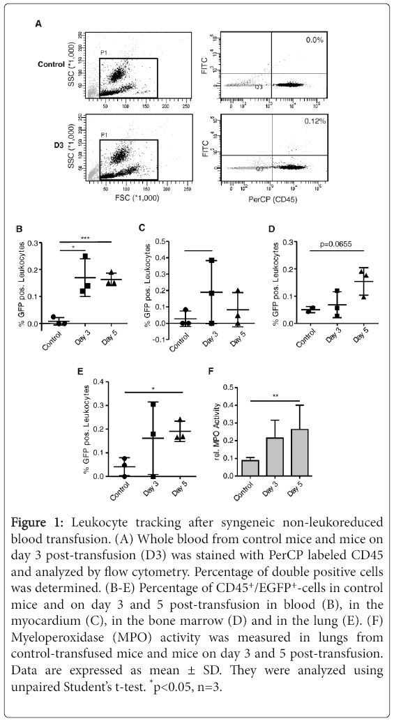
Figure 1: Leukocyte tracking after syngeneic non-leukoreduced blood transfusion. (A) Whole blood from control mice and mice on day 3 post-transfusion (D3) was stained with PerCP labeled CD45 and analyzed by flow cytometry. Percentage of double positive cells was determined. (B-E) Percentage of CD45+/EGFP+-cells in control mice and on day 3 and 5 post-transfusion in blood (B), in the myocardium (C), in the bone marrow (D) and in the lung (E). (F) Myeloperoxidase (MPO) activity was measured in lungs from control-transfused mice and mice on day 3 and 5 post-transfusion. Data are expressed as mean ± SD. They were analyzed using unpaired Student’s t-test. *p<0.05, n=3.
Syngeneic leukocytes were found in the blood stream up to five days after transfusion. The ones detected in the myocardium on day 3 could stem from the coronary arteries rather than the muscle tissue, which is something that cannot be distinguished in single cell suspensions. Very interesting, however, is the non-significant increase of EGFP+ leukocytes in the bone marrow, which might either show homing or even cell reproduction. However, the recipient’s immune systems probably tolerated the syngeneic WBCs much better than they would have done with allogeneic blood WBCs and therefore might be clinically irrelevant.
Furthermore, the significant increase of EGFP+ leukocytes in the lungs is very interesting, especially as the MPO activity is also elevated.
Transfusion with leuko-reduced blood would be an ideal control to determine the effects of WBC on the MPO activity in lungs with nonleuko- reduced blood. Our observation so far might either show that the syngeneic WBCs determined in the lung tissue are mainly neutrophils or that autologous WBCs were activated by the transfusion. Analysing further markers of WBCs cells would be helpful in determining the different subtypes. We furthermore detected fluorescent events at the spectral range emitted by EGFP in specimens of the control group, which represents auto fluorescence. This phenomenon is regularly observed in this spectral range as it is close to the spectrum emitted by hemoglobin.
In conclusion, syngeneic WBCs survive in the recipient of nonleukoreduced RBC units. They circulate in the blood stream and can be found in the bone marrow and the lungs up to five days after transfusion. This observation might add credence to the practice of pre-storage leukoreduction.
Source of support: The project was funded by the B. Braun Foundation.
The authors declare that they have no conflicts of interest relevant to the manuscript other than those stated above.