Journal of Thermodynamics & Catalysis
Open Access
ISSN: 2157-7544
ISSN: 2157-7544
Research Article - (2015) Volume 6, Issue 3
Meniscus degeneration is a common problem in the human knee. Meniscal diseases can significantly alter the quality of life. Thermal analysis of human meniscus extracted during surgery can provide insight into the degenerative process. The aim of present study was to characterise the water loss from the investigated biological samples caused by altered metabolism, and compare the thermogravimetric properties of normal, moderately, and severely degenerated meniscus samples. Although total water content was similar in normal, moderately and severely degenerated meniscal cartilages (70.26, 75.51 and 76.29% respectively), there was a significant difference in activation energy (44.92, 51.44 and 62.04 kJ M-1). This can be attributed to the difference in chemical composition of meniscus fibrocartilagenous matrix between the healthy and degenerated samples.
Keywords: Activation energy; Human meniscus; Osteoarthritis; Thermogravimetry
In present study the authors demonstrate thermogravimetric analysis of healthy and degenerated human knee joint menisci. Meniscus mainly consists of robust fibrocartilage, it is an important shock absorber in the knee. It protects the hyaline cartilage of the joint, shielding it from physical damage and therefore contributing to prevention of early osteoarthritis [1]. A large proportion of body weight is distributed through the meniscus during walking, running, and jumping. Menisci contribute to the stability of the knee by helping the shape of the femur to conform to the tibia. Several factors can cause damage to the menisci. They can tear or rip from force, or get pinched between the femur and the tibia [2,3]. It does not always require a major fall or twist to cause a meniscal tear. Degenerative changes of knee menisci frequently result from trauma to the joint or associate with joint diseases; in other cases they are caused as a result of chronic overload, and in some cases even with no obvious reason. Degeneration of human knee joint menisci has been the subject of numerous morphological studies [4,5] but detailed studies on the chemical composition and the metabolism of menisci are still limited.
Several thermoanalytical techniques can be applied to measure the change of physical or chemical properties of a sample as a function of temperature. Thermogravimetric analysis (TG) is one of the oldest thermal analytical procedures; it has been used extensively in the study of polymeric systems and similarly in case of biological samples. The technique involves monitoring the mass loss of the sample in a specific atmosphere (usually nitrogen or air) as a function of temperature. The applied equipment – among others – is suitable for simultaneous recording of thermogravimetric, derivative thermogravimetric (DTG) and differential thermoanalytical (DTA) curves [6]. The usefulness of TG for analyzing complex systems was greatly enhanced by the introduction of the ability to record the first derivative of the mass loss simultaneously. The ability of TG to generate fundamental quantitative data from almost any class of materials, has led to its widespread use in virtually every scientific field. Compositional analysis is a key application: by careful choice of temperature programming and gaseous environment, complex materials or mixtures can be analyzed by selectively decomposing them, or removing their components. This approach is regularly used to analyze moisture content of many substances. TG is inherently quantitative technique, and it does not give direct chemical information. The ability to analyze the volatile products during a mass loss is also a valuable possibility, when available [7-10]. Previously differential scanning calorimetry (DSC) was used for the investigation of normal and degenerative human meniscus cartilage, and water content has not been measured [11-14]. A significant paper from this field was the study of Than et al. [15], further studies measured the difference between primary osteoarthritis and septic arthritis [16]. Most of the known changes in the extracellular matrix in OA come from animal models, because human samples are only available for experiment to a limited extent. Thermogravimetric study of human hyaline cartilage has been carried out previously [17], although meniscus has not been studied extensively. The purpose of this investigation was to gain insight on the role of water content in contributing to progression of fibrocartilage degeneration, and to reveal the kinetic character of water loss effect of heating on menisci in good health and in different stages of degeneration.
Materials
The human knee joint meniscus specimens were received from the Orthopaedic Department, University of Szeged. After surgical removal, the menisci were dissected free of their attachments and a 5 mm thick disc was produced under sterile conditions from the middle segment of each meniscus. The disc was first washed in sterile saline, and then stored in 20 ml saline for transportation at room temperature. Mean storage time was 6 hours (Minimum: 1 hour, maximum: 26 hours), 10 samples out of 11 were studied within 6 h of preparation. One sample was stored over-night at 5°C. Pre-emptive control examinations did not show any change in the calorimetric and thermogravimetric properties after storage for 26 hours at 5°C. All tissues were managed in accordance to legal regulation, international ethical concerns, and patients’ consent. Diagnosis of the patient was established on basis of the medical history, clinical signs and radiological findings preoperatively. In order to conduct the thermoanalytical study, 11 samples were collected. The state of the joint degeneration, and the meniscus was determined intraoperatively. All patients in the moderately osteoarthritic group (mean age: 60.47 years) were considered to be Osteoarthritis Research Society International (OARSI) [18] grade 1-3 articular degeneration. Severely osteoarthritic group (mean age: 70.88) were considered to be OARSI grade 4-6. Normal samples were extracted during arthroscopic surgeries of patients (mean age: 40.94 years) with knee injury, when ruptured but otherwise healthy meniscus had to be sacrificed for the partial meniscectomy procedure. The gender distribution was the same in both osteoarthritic groups.
Methods
We used a new protocol established by Sohár et al. [17] for sample extraction during live surgery using simple saline solution instead of the previously used phosphate buffer. This new method proved to be suitable for the thermoanalytical investigations. Thermal analysis was performed with a MOM Derivatograph-C (MOM, Budapest, Hungary), and the TG, DTG, and DTA curves were determined. TG allows the determination of the mass as a function of temperature. This thermal technique provides information concerning the thermal stability and composition of the sample and of any intermediate compound which may be formed. The heating was linear from 25°C to 150°C and the rate of heating was 5 K minute-1. Platinum sample holders were used, Al2O3 was used as reference material, the purge gas was air. In the first step, the total water loss and kinetic parameters were calculated. The kinetic parameters calculated by the software are the following: the reaction order (n), the activation energy (Eact) and the pre-exponential factor (A). The value of n (reaction order) is allocated by the Kissinger method [19] and it is the first kinetic parameter calculated by the computer:
 (1)
(1)
where S is the form factor which presents the absolute value of the gradients of DTG curves in the points of min/max. The activation energy (Eact) is determined according to the natural logarithmic form of the Arrhenius-equation
 (2)
(2)
which is widely used in the literature [20,21]. The results of the investigations were statistically evaluated by Microsoft Excel. A Paired t-test was used to compare mean water loss of normal and degenerative meniscus during thermal analysis. The linear range of the first step of TG curve in water loss was analyzed to establish its slope. The determination of linear parts of TG curves and fitting linear straight line on the curve was performed by the statistical software SPSS.
TG curves of healthy meniscus, samples with no degeneration, and degenerated menisci are presented on Figure 1. The results show, that the total water content of healthy fibrocartilage was 70.26%, to remove the water content 44.92 kJ M-1 was needed. Fibrocartilage obtained from the moderately degenerated meniscus samples had a 75.51% water content, which differed significantly from the healthy tissue.
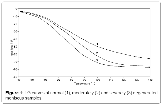
Figure 1: TG curves of normal (1), moderately (2) and severely (3) degenerated meniscus samples.
Extraction of water content needed 51.44 kJ M-1 energy on average. Fibrocartilage obtained from the severely degenerated meniscus samples had a 76.29% water content, which did not differ significantly from the moderately degenerated samples, but differed significantly from results of normal tissue. Extraction of water content needed 62.04 kJ M-1 energy on average. Loss of water content in all groups is presented with a sharp step on the TG curve, starting on average temperature of 44.65°C and ending at 137.2°C. Linear part of the TG curve begins at 63.53°C for normal meniscus, 63.73°C for moderately degenerated meniscus, and 62.65°C for severely degenerated meniscus; while ends at 96.73°C for normal meniscus, 97.20 °C for moderately degenerated meniscus, and 96.55°C for severely degenerated meniscus. A straight line was fitted to the curve in this section and the slope of this curve was calculated which represents the rate of the water content loss (Table 1). The slope of the linear region correlated in both groups. In case of the normal fibrocartilage 1.0% °C-1 mass loss was detected. In case of moderately degenerated samples 1.5% °C-1 decrease in mass was observed. In the severely degenerated samples also 1.5% °C-1 mass reduction was observed. The resulting amount of mass loss in the linear region was recounted from these results (Table 1).
| Sample group | Sample number | TG step linear region/°C | Mass loss/% | Reaction order/nSD | Slope of linearregion | ||
|---|---|---|---|---|---|---|---|
| Normal | 3 | 63.53 | – | 96.73 | 37.51 | 1.00 | 0.22 |
| 0.44 | |||||||
| Moderate | 4 | 63.73 | – | 97.20 | 45.45 | 1.00 | 0.23 |
| 0.18 | |||||||
| Severe | 4 | 62.65 | – | 96.55 | 46.88 | 1.13 | 0.21 |
| 0.26 | |||||||
Table 1: Reaction kinetic parameters of meniscus samples with no degeneration, moderate, and severe degree of degeneration.
The fibrocartilage of knee menisci is a functionally specialized tissue that plays a role in weight bearing, nutrition, and lubrication of the articular cartilage and in joint stability. Past biomechanical studies done under static loading have shown that the menisci, regardless of their condition, contribute to force [5] and stress distribution [1]. Meniscus degeneration in the knee is frequently a result from trauma to the joint or it is associated with joint diseases; in other cases it arises after chronic overload, or even spontaneously [4]. Our study has several potential limitations. First of all, the sample size was not large enough to draw definitive conclusions. Although the mass loss change of the process initiated by the temperature change showed marked difference between the normal and pathological groups (Table 2), statistical analysis did not show significant alteration at p ≤ 0.05. Consequently, further measurements are needed to confirm the results of our study. Previous thermo analytical studies used cadaver samples for the investigation as normal human meniscus. All samples that were extracted for our studies were obtained during live surgeries and were macroscopically intact. Only full thickness meniscus was included in the analysis of normal group. More information will be needed on the events leading to meniscus degeneration and the interactions between different pathologic effects in order to discover new therapeutic targets. Several potential disease modifying agents are studied currently that may significantly contribute to changes in the treatment approach; therefore sensitive tools are needed to study biological matter in clinical trials. Meniscus pathology plays an important role in osteoarthritis pathophysiology. However, whether meniscus damage or cartilage degeneration is the first factor is unknown. Meniscal rupture seems to be a strong risk factor in the development and progression of knee osteoarthritis. Meniscectomy increases knee osteoarthritis risk twofold [1,22]. Modifiable risk factors are natural target for clinical efforts in managing knee degeneration. At present, there is a number of modifying therapeutic options that may be used to alter the rate of disease progression. While these therapies are currently underutilized, they might play more prominent role when modification of disease progression has been demonstrated. However, the role of these therapies is unclear, given the paucity of long-term, well-designed controlled trials [23]. In summary, we examined the thermal properties of human meniscus tissue of normal origin in adults and in patients with moderate and severe stages of degeneration. A newly established thermogravimetric protocol was used for our experiments. This method proved to be suitable for thermogravimetric analysis of normal and degenerative human meniscus samples.
| Sample group | Sample number | TG step /°C | Total mass loss /%SD | Eact / kJ M-1SD | ||
|---|---|---|---|---|---|---|
| Normal | 3 | 30.50 | – | 130.20 | 70.26 | 44.92 |
| 7.44 | 10.50 | |||||
| Moderate | 4 | 27.88 | – | 133.05 | 75.51 | 51.44 |
| 3.66 | 12.41 | |||||
| ssSevere | 4 | 27.23 | – | 129.45 | 76.29 | 62.04 |
| 4.29 | 17.12 | |||||
Table 2: Mass loss and activation energy of normal, moderately, and severely degenerated meniscus samples.
Grant: This study was supported by OTKA T-047166.
No conflicts of interest to declare.