Translational Medicine
Open Access
ISSN: 2161-1025
ISSN: 2161-1025
Research Article - (2025)Volume 15, Issue 1
Background: In 2018, the World Health Organization introduced cancer as one of the deadliest types of disease worldwide. Although there are several unique advantages for cancer therapy, in recent years, problems such as poor targeting efficacy, increased tumor hypoxia, severe coronary syndromes, excessive ventricular conduction, druginduced drug resistance and increased risk of tumor metastasis have hindered their potential use in clinically limited.
Objectives: Based on current research, we are looking to identify natural medicinal compounds, including compounds found in sage plant extract, as anti-angiogenic compounds to prevent tumor growth, which have the most effectiveness and least side effects in the treatment of cancers.
Methods: 24 embryonated eggs (specific pathogen free) were randomly distributed into 4 equal groups, treated with a solution of hydroalcoholic Salvia officinalis extract and after 8 day, the experimental groups were treated with 150 μg/ml, 250 μg/ml and 350 μg/ml.
Results: On the 12th day, the Chorioallantoic Membrane (CAM) of all samples was photographed, the number and diameter of vascular branches were measured using Image J software and the resulting data were analyzed using SPSS test (P=0.01).
Conclusion: According to the studies conducted in this research, S. officinalis compounds play an important role in reducing the number of branches and the diameter of vessels in the CAM membrane of chick embryos and opening the door to a new era in the use of natural products as medicines.
Angiogenesis; Chorioallantoic membrane; Salvia; Cancer; VEGF
CAM: Chorioallantoic Membrane; Sa-A: Density 150 μg/ml; Sa-B: Density 250 μg/ml; Sa-C: Density 350 μg/ml; Ctr: Control group; VEGF: Vascular Endothelial Growth Factors; bFGF: Epithelial Neutrophil-Activating Peptide; EGF: Epidermal Growth Factor; IL: Interleukin; INF-γ: Interferon gamma; pIGF: Placental Growth Factor; TIMP: Tissue Inhibitor of Metalloproteinases 1-2; NF-κB: Nuclear Factor kappa-light-chain-enhancer of activated B cells; PI3K: Phosphoinositide 3-kinases; ERK: Extracellular Signal-Regulated Kinase 1/2
In 2018, the World Health Organization identified cancer as one of the major causes of death worldwide which is responsible for the deaths of 9.6 million people in 2018 [1,2]. In Iran, according to the statistics of the Global Cancer Observatory (GCO), the International Cancer Research Agency of the World Health Organization, 79,136 people have died by 2020. Most tumour treatment include a combination of surgery, chemotherapy or radiation therapy. However, there are various challenges in classical chemotherapy, including harmful side effects [3-6]. Therefore, in the last decade, new methods have emerged in the treatment of cancer; among them are treatments according to the histological sub-group and influence micro-tumours [7]. One of the alternative methods of tumour treatment is treatment through the vascular system based on the increase in tumour size must be coordinated with the increase in blood supply [8]. Proliferating tumours, due to the increase in the distance between cells and vessels, the need for oxygen and the supply of oxygen in them have exceeded, which prevents the diffusion of oxygen and creates a more hypoxic environment [9,10]. New blood vessels increase the amount of oxygen and the nutrients needed based on the cell volume bring the cancer mass to the tumour cells and most importantly, they facilitate the metastasis of cancer cells to other places [11]. Accordingly, angiogenesis, by supplying oxygen, nutrients, growth factors, proteolytic enzymes, coagulation factors and fibrinolytic factors support tumour growth [12]. Folkman et al. identified the main factors effective in tumour angiogenesis and by inhibiting those factors, were able to use the strategy of tumour starvation and its death in the treatment of the disease [13-17]. In the last 10 years, many potential anti-angiogenic targets had discovered with successive discovery of factors such as fibroblast growth factor, matrix metalloproteinase, tumour-related stromal cells and adhesion molecules [18].
Vascular Endothelial Growth Factor (VEGF)
VEGFs play a key role in the regulation of angiogenesis [19,20]. Scientists have stated that the regulation of VEGF levels caused by hypoxia is the main driving force of angiogenesis in the path of tumour progression. VEGF increases vascular permeability and cause the increase in vascular permeability leads to the release of plasma proteins and their entry into the interstitial space, which leads to the migration and proliferation of endothelial and angiogenic cells. VEGF family includes subtypes VEGFC, VEGFB, VEGFA, VEGFF, VEGFE, VEGFD and placental growth factor, but it is thought that VEGFA is the key regulator of angiogenesis during homeostasis and disease. VEGFA increases the secretion of Matrix Metalloproteinase (MMP) and proliferation of endothelial cells. VEGFC and VEGFD are key lymphangiogenic factors during development. VEGFE, which is structurally almost identical to VEGFA. Another family member, VEGFB, does not appear to display angiogenic activity, but is a key regulator of fatty acid metabolism.
Angiopoietins (Angs)
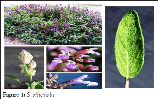
The family of angiopoietin (Ang) is an angiogenic stimulating factor, including Ang-1, Ang-2, Ang-3 and Ang-4. These molecules bind to an endothelial receptor tyrosine kinase, Tie-2, to promote angiogenesis. Angs control the homeostasis of endothelial cells by modulating maturation and vascular stability and cell survival. VEGFs are activated in the early stages of angiogenesis, while Ang/Tie-2 systems are activated in the later stages, control vessel assembly and maturation of the fatal vascular system as well as vascular homeostasis of the adult vascular system. Ang-2 acts with VEGF to initiate angiogenesis. In addition, Platelet-Derived Growth Factor (PDGF) and TGF-β other angiogenic factors stabilize new vessels. One of the main ways to reduce angiogenesis in tumour tissue is the use of food and supplements food. Notably, several components of natural resources have been reported to have anticancer activities on cancer cells through different mechanisms, including induction of apoptosis and necrosis and inhibition angiogenesis is possible. Since angiogenesis plays a prominent role in tumour growth and metastasis, inhibition of angiogenesis is considered as an important strategy for cancer treatment [14]. In addition, inhibition of angiogenesis before initiation, which is known as angioma prevention, has the potential to prevent the spread of hyperplastic foci and subsequent tumour development in the premalignant stage. Salvia officinalis (Figure 1) is one of the largest genera of the Lamiaceae family, a perennial herb that can grow about 30 to 60 cm. Several species produce woody stems and grow more like a shrub. Based on the ability of this plant to grow in different geographical areas, this plant can be identified in regions with different climates. All over the world, this species is used in traditional medicine as antibacterial, antioxidant, anti-diabetic, anti-tumour, herbal tea and food seasoning. Salvia plant essential oils exhibit broad-spectrum pharmacological activities and represent great interest for food preservation as potential natural products. The phenolic compounds of methanolic extracts of Salvia pomifera and Salvia fruticosa were identified by liquid chromatography tandem mass spectrometry. Carnosic acid and its metabolite carnosol were the most abundant terpene phenolic compounds of S. fruticosa, while they were completely absent in S. pomifera.

Figure 1: S. officinalis.
The analysis of the hydroalcoholic extract of S. officinalis showed it contain various flavonoids and phenolic compounds. The most important flavonoids include quercetin, kaempferol, luteolin (soluble in water), luteolin-7-glucoside, apigenin-7- glucoside, myristin and murine with strong antioxidant activity. They are analgesic and anti-inflammatory, in addition, it contains various other compounds such as steroids, saponins, tannins, phenols and polypeptides, carnosol, rosmarinic acid and camosic acid. In recent years, the potential use of salvia as a new anticancer agent has been recognized. Therefore, in this article, the anticancer property of phytochemicals is investigated.
Objectives
Previous studies have shown that the angiogenic system is involved in the growth of tumors whose size is estimated to be 2 mm. Also, no studies have been conducted to find medicinal compounds on the capillary branches that are moving from the main vessel to the tumor mass. Therefore, the purpose of this study is to investigate the natural medicinal compounds, including the compounds found in the hydroalcoholic extract of sage plant.
These compounds as anti-angiogenic compounds to prevent tumor growth and reduce the number of branching vessels towards the tumor mass, without affecting the main vessel, Based on this, it has the most effectiveness and the least side effects in the treatment of cancers.
Isolation and analysis of the essential oils: S. officinalis was collected from Bageran mountain in Southern Khorasan (Iran) in July 2022 (Table 1). The aerial parts dried in a cool and dark place. 200 gr of dried powder put in a percolator with 70% alcohol for 72 hours. The obtained extract was placed on a bainmarie and finally the crystalline structure of the extract kept in the refrigerator.
Shimadzu RID-10A HPLC was used to identify the polyphenolic compounds of the ethanolic extract of S. officinalis. Extraction was done by 8 × 10⁻⁴ RIU or higher detector at 339 nm wavelength for 2 hours.
| Location | Herbarium number | Geographical attributes | ASL (m) | Annual temperature (°C) | Average rainfall (mm) | Average humidity |
| Southern Khorasan-Birjand | 2912 | 32°52'16.9"N 59°10' 47.1"E | 1491 | 16°C(24°C-8°C) | 120 | 36% |
Table 1: Geographical features of collection place and herbarium number of studied species of Iranian S. officinalis.
The CAM assay: 24 specific pathogens free embryonated eggs were obtained from the Razi vaccine and serum center in Shiraz in September 2022. The eggs were placed in the hatcher machine with a temperature of 37°C and humidity 65% to 70%. On third and seventh day the embryos were checked and on the eighth day under a laminar hood with sterile forceps made a hole (1 × 1 cm) on the wide part of the egg. The experiment carried with four groups; (1) control group the eggs kept in the same position without moving in the incubation until the twelfth day. The second group treated with 250 μg/ml, the third group treated with 250 μg/ml and the last group treated with 350 μg/ml aqueous extract of S. officinalis. Then the opened part covered with paraffin and placed inside the incubator until the twelfth day.
The chorioallantoic membrane photographed (64X) with stereomicroscope (Ziess) and in order to improve the number of pixels, the data were analysis by Image J Software. The measurement of the index of the number of vessels and the diameter of the branches was carried out in squares with dimensions of 2 × 2 cm. SPSS software with t-test at a significance level of P ≤ 0.01 and ANOVA statistical test was also used to confirm the obtained results.
Based on HPLC results, three flavonoids identified; Quercetin, kaempferol and Isorhamnetin with separation times of 14:41, 19:37 for Quercetin and 29:68, 32:64 for kaempferol and Isorhamnetin. These times for the standard sample are 14:12, 19:87 for quercetin and 28:210, 31:620 for kaempferol and isorhemantin, respectively (Figure 2).
Figure 2: HPLC chromatography, measured at 339 nm. The retention time for quercetin, kaempferol and isorhemantin was 14:12, 14:41, 19:37, 19:87, 28:21, 27:66, 31:62 and 32:64 minutes, respectively.
The vessel diameter (25.7 ± 2.19) in experimental Sa-A group compared to the Ctr group showed a significant decrease (p=0.004). Also, the decrease in the average diameter of blood vessel branches in experimental Sa-B group (19.38 ± 2.77) compared to the Ctr group was significant (p=0.002) and compared to the average total diameter of blood vessels in experimental Sa-C group (8.73 ± 1.51) with the Ctr group, decrease was significant (p=0.001) (Figure 3).
Examination of vessel branching in experimental Sa-A group compared to Ctr group (10.2 ± 1.17) showed a significant decrease (p=0.005). The average difference in the number of vascular branches in experimental Sa-B group (7.85 ± 1.80) compared to the Ctr group showed a significant decrease (p=0.006). The average number of branches of blood vessels in experimental Sa-C group (3.92 ± 0.97) compared to the Ctr had a significant decrease (p=0.000) (Figure 3).
Figure 3: The graph of average values of the diameter and the number of branches of the vessel in the groups under the test drawn using SPSS software.
In total, the extraction at the concentrations of 150 and 250 μg/ml had a mild inhibitory effect and 350 μg/ml completely inhibited the growth of buds (Figure 4). The average number of capillary formation of three independent experiments showed S. officinalis extract has an inhibitory effect on angiogenesis in vivo in the CAM model.
Anti-tumorigenic effects of this species proved on breast cancer. Privitera et al. revealed this species as an important source of substances with anti-proliferative activity and could improve the treatment of this devastating disease.
Figure 4: Images taken by stereomicroscope of (CAM). A) Ctr group, B) Sa-A, C) Sa-B and D) Sa-C.
Meanwhile, with the decrease in the diameter of the main vessel and the decrease in the number of branches in the fetus, no weight loss disorders, dwarfism and abnormalities observed. The larger the diameter of the vessels, the colour of the vessels in the images will be closer to the colour spectrum of 120-150 (Figure 5).
Figure 5: Intensity diagram of vessel diameter according to the amount of pixels of the photographs prepared from A) Ctr, B) Sa-A, C) Sa-B and D) Sa-C experimental groups.
Figure 6 the three-dimensional representation of the vessels of the measured area of the chorioallantoic membrane of the chicken embryo shows. As shown in the image, the larger the diameter of the vessels, the more pixels there are in that area and accordingly, the color of the vessels in the images will be closer to the color spectrum of 120-150 pixels.
Figure 6: Three-dimensional representation of the diameter of blood vessels in a specific area of the chorioallantoic membrane, A) Ctr, B) Sa-A, C) Sa-B and D) Sa-C.
Bin Zhu et al. cancer cells treated with S. officinalis. It was found that the treatment leads to differential secretion of angiogenic cytokines in PC-3 and DU-145 cells. Cytokine levels of angiogenin, PDGF-BB, MCP-1, LEPTIN, RANTES and VEGFD decreased significantly in both prostate cancer cell lines. Significant decrease in ENA-78, bFGF and EGF levels in DU-cells 145. Also, PC-3 cells were more sensitive than DU-145 cells to exposure to S. officinalis, which resulted in increased levels of GRO, IL-6, IL-8, IFN-γ, PIGF, TIMP-1 TIMP-2, thrombopoietin and VEGF decreased significantly.
Ahmed as well proved the importance of S. officinalis as strong anti-angiogenic effects. The extract of this plant significantly inhibited the growth of blood vessels in CAM. This mechanism may be related to the regulation of the expression of VEGF, Matrix Metalloproteinases (MMPs), EGFR and inhibition of NF-κB, PI3K/Akt, ERK1/2 signaling pathways.
The results in the present study showed a decrease in the number or diameter of vascular branches in the concentrations of 150, 250 and 350 μg/ml of the hydroalcoholic extract of arial parts of S. officinalis. It should be noted that the percentage of fetal mortality in none of the used concentrations showed a significant difference compared to the control. Considering the importance of angiogenesis and its inhibitory factors for the treatment of various diseases, including tumors, angiogenesis inhibition methods are considered a promising way to treat angiogenesis-related diseases. Further research can be effective in knowing the possibility of practical use of S. officinalis as medicine or diet to treat diseases including cancer.
In summary, the hydroalcoholic extract of Salvia officinalis demonstrates a notable influence on the angiogenesis rate in the chorioallantoic membrane of chicken embryos. The findings suggest that the bioactive compounds within the extract may play a pivotal role in modulating vascular development, potentially offering insights for future research on natural angiogenesis regulators. These results underscore the importance of exploring herbal extracts as viable therapeutic agents in vascular-related conditions, paving the way for innovative approaches in both pharmacology and regenerative medicine.
No funding was received for conducting this study.
The authors declare that there is no conflict of interest.
[Crossref] [Google Scholar] [PubMed]
[Crossref] [Google Scholar] [PubMed]
[Crossref] [Google Scholar] [PubMed]
[Crossref] [Google Scholar] [PubMed]
[Crossref] [Google Scholar] [PubMed]
[Crossref] [Google Scholar] [PubMed]
[Crossref] [Google Scholar] [PubMed]
[Crossref] [Google Scholar] [PubMed]
[Crossref] [Google Scholar] [PubMed]
[Crossref] [Google Scholar] [PubMed]
[Crossref] [Google Scholar] [PubMed]
[Crossref] [Google Scholar] [PubMed]
[Crossref] [Google Scholar] [PubMed]
[Crossref] [Google Scholar] [PubMed]
[Crossref] [Google Scholar] [PubMed]
[Crossref] [Google Scholar] [PubMed]
[Google Scholar] [PubMed]
Citation: Rouygar F, Golab NG, Ghollasimood S, Tanideh N (2025) The Effect Hydroalcoholic Extract of Salvia officinalis on the Angiogenesis Rate in the Chorioallantoic Membrane (CAM) of Chicken Embryo. Trans Med. 15:342.
Received: 09-Mar-2024, Manuscript No. TMCR-24-30080; Editor assigned: 11-Mar-2024, Pre QC No. TMCR-24-30080 (PQ); Reviewed: 25-Mar-2024, QC No. TMCR-24-30080; Revised: 04-Mar-2025, Manuscript No. TMCR-24-30080 (R); Published: 11-Mar-2025 , DOI: 10.35248/2161-1025.25.15.342
Copyright: © 2025 Rouygar F, et al. This is an open-access article distributed under the terms of the Creative Commons Attribution License, which permits unrestricted use, distribution, and reproduction in any medium, provided the original author and source are credited.