Medicinal & Aromatic Plants
Open Access
ISSN: 2167-0412
ISSN: 2167-0412
Research Article - (2016) Volume 5, Issue 2
Transverse sections of the leaf blade and petiole of three species of the genus Chrysophyllum namely C. albidum, C. subnudum and C. cainito were investigated to establish taxonomic relationship among them. The result obtained revealed that the shape of vascular bundle of the leaf in C. albidum and C. subnudum are semi-circular while in C. cainito, it is V-shaped. The vascular bundle is bicollateral in the three taxa investigated. The shape of petiole is semicircular in C. albidum and C. cainito but rounded in C. subundum. C. cainito and C. subundum have well developed Vascular bundle, about 6-7 arranged to form an arc with distinct xylem and phloem cells while in C. albidum, they are 3-7, arranged to form an arc with in the cortex. The result further strengthened the inter-specific relationship existing among them
Keywords: Taxonomic; Relationship; Chrysophyllum; Anatomy; Leaf; Petiole
Chrysophyllum is derived from Greek, meaning “golden leaf ” from the color of the hairs of some species. It is a tropical tree growing rapidly to 10-20 m or more in height. Chrysophyllum has about 70-80 species with seven species occurring in Nigeria [1]. The fruits of the genus Chrysophyllum are large berry containing five large flattened seeds or sometimes fewer by abortion [2]. Keay [1], gave a very good account of the Nigeria species.
Chrysophyllum albidum G. Don. and Chrysophyllum subnudum Baker., were among the seven species identified by Keay [1] while Chrysophyllum cainito Linn., was recorded to have originated from the West Indies and Central America. It is frequently planted in the tropics for its edible fruit. All the species in Nigeria are tall or medium sized trees except C. welnitschii Engl. which is a climbing shrub.
Many species of the genus Chrysophyllum are cultivated for their edible fruits and for ornamental purposes. These include C. albidum, C. submidum, C. cainito and C. giganteum.C. cainito has the potential to improve nutrition and boost food security. Its ripe fruit fresh spooned can be mixed with that of mango, citrus, pineapple, other fruits and coconut water and serve as Jamaica fruit salad ice. C. cainito is second most valuable timber tree in Asia. C.albidum wood brownish white and soft often is used in houses and hut buildings, though perishable, in contact with ground and is also used for household articles and tool handles. The copious latex found in Chrysophyllum wood has been utilized as an adulterant of gutta-percha and chewing gum. The seeds of C. albidum are strung together and worn as anklets or girdles which also serve as rattles during dancing [3]. Medically, a ripe fruit of C. cainito, because of its mucilaginous character, is eaten to sooth inflammation in laryngitis and pneumonia. It is given as a treatment for diabetes mellitus and as a decoction in gargled to relieve angina. A decoction of the tannin-rich, astringent bark is drunk as a tonic and stimulant, and is taken to halt diarrhea, dysentery and hemorrhages and as a treatment for gonorrhea and “catarrh of the bladder”. Some of these species such as C. cainito and C.albidum are planted for ornamental purposes.
Plant anatomy traditionally focused mainly on the characterization and the mode of development of the various organs of the plant body. The increasing importance of plant anatomy has led to the development of not only numerous techniques but also to more and better equipment for the study. Anatomical characters are now generally believed to be just as valuable as morphological characters. They are also widely used in other aspects of taxonomy and have been applied to the elucidation of “phylogenetic” relationship in determining relationships between different genera, families, orders and other taxonomic categories; where the anatomical characters has been found most useful. Anatomical character such as the type of vascular bundles, rays, type, size, shape, wall sculpture and pattern of wood cells, stelar patterns, ground tissue and parenchyma, epidermal and mesophyll tissue stomata, trichomes, sclereids, node, phloem cells, obtained from various parts of the plant – stem, root, petiole node, leaves, flower, and fruit, has been found taxonomically valuable. In Dioscoreaceae, certain anatomical features were used in characterization of Dioscorea alata L. and D. Smilacifolia L. including other species of Dioscora [4]. Raganna and Ramakrishuan, reported the significance of anatomical features in the genus Camellia. Metcalfe [5] in several genera of cyperaceae has used several characters of leaf anatomy in differentiating species. The distribution pattern of Sclerenchyma in Carex and Festuca has been used in distinguishing species. Metcalfe and Chalk [6] and Howard have suggested that the petiole is also of taxonomic significance. Mbagwu and Nwachukwu used petiolar anatomy of eight Vigna species in Nigeria to establish interspecies relationship among the investigated taxa. Edeoga et al. [4] distinguished between species of the genus Phyllathus using anatomical features. Nwachukwu and Mbagwu in Indegofera, and Mbagwu et al. in genus Viscum L. applied the anatomical features of these genera to distinguish between various species they studied.
Due to close morphological resemblance of C. albidum and . subnudum, some authors have placed C. subnudum as variety of C. albidum. Also owing to limited taxonomical work on this genus in Nigeria, there is needed to carry out this study in order to agree or disagree with this placement with others around the world. This study is therefore based on the leaf and petiole anatomy of three species of the genus Chrysophyllum.
Specimen collection
Specimens of the three Chrysophyllum species (namely C. cainito, C. albidum and C. subnudum) were collected from three villages in Aboh Mbaise Local Government Area of Imo State. The villages are Ngali Obibi, Umuayara and Oboama Enyiogugu. C. cainito were collected from Oboama Enyeogugu while C. albidum and . subnudum were collected from both Ngali Obibi and Umuayara [7].
Specimen identification
The specimens were identified by a taxonomist, Professor S.E. Okeke and were confirmed at the Forest Herberum (FHI), Ibadan, Oyo State. Voucher specimens were deposited in Imo State University Herberium (IMSUH) Owerri [8].
Anatomical studies
Mature and fresh parts of the leaves and petioles of the three species were collected and sectioned. The cut sections were done at various portions. For the leaves, cutting was done from the middle portion while that of the petiole was at 0.5 cm from the node. Before sectioning commenced, the specimens were made to undergo a pretreatment process referred to as Killing and Fixing. The aim of this process is to terminate suddenly and permanently all life processes within the specimens and preserve the cells composing the materials as close to their original condition as possible [9]. For the pretreatment, the specimens were treated using Formalin-Acetic Alcohol (FAA) for 48 hours and then washed thoroughly in distilled water.
The specimens were washed in two changes of 30% ethanol and are dehydrated in a graded series of ethanol (30%-50%-70%-95%) for at least 15 minutes in each grade.
To infiltrate wax into the specimens, they were covered for 3 hours in each of the following solutions containing a ratio of absolute alcohol to pure chloroform (v/v: 3:1, 1:1, 1:3), then pure chloroform. At the stage of pure chloroform, wax pellets (60°C melting point) were added and the wax is changed periodically. The specimen containers were transferred to an oven for 2-7 days to remove the chloroform. To embed in wax, the contents of the vial were carefully transferred into moulds. The specimens were arranged using a flamed (hot) mounting needle and then transferred to a cold water bath where it remains until the wax is sufficiently solid and was later stored in a refrigerator for two days. A very thin section of 10-20 μ were made using a Leica rotary microtome. Ribbon cut sections obtained from the microtome sectioning were placed on clean slides covered with a thin film of Haupt’s albumen and allowed to dry. Three or four drops of distilled water were added before mounting. The slides were placed on a hot plate at 40ºC for few minutes to allow the ribbons to expand and were kept overnight.
The slides were immersed in pure xylene for 2-5 minutes in a solution of xylene and absolute alcohol with a ratio of 1:1 (v/v). They were later transferred to another solution of xylene and alcohol graded series (95%, 90%, 70% and 50%) in the ratio of 3:3 (v/v) for few minutes. Drops of safranin were used to stain the section for 5 minutes, washed off with distilled water and then counter stained with alcian blue for 2 minutes and then dehydrate in 50% alcohol, 70%, 80% and 90% xylene/ alcohol solution respectively and pure xylene at intervals. Mounting medium used is the D.P.X. mountant. Drops of D.P.X. were introduced onto the slides enough to cover the length of the sectioned material and then covered with cover slip. The slides containing the mounted sections were placed on a hot plate at 30°C to dry. Photographs of materials sectioned were taken using a Leitz Wetzler Ortholux digital microscope [10,11].
Leaf Anatomy
Leaf anatomy features of the three species of Chrysophyllum studied are summarized in Table 1 and illustrated in Plates 1a-f. The cuticle are thick on the upper lamina of the three species considered (Plates 1b, d and f). Epidermal cells are uniseriate in C. cainito but multiseriate in both C. albidum and . subnudum (Plates 1b, d and f). Single layered palisade cell in C. albidum while the layer is double-layered in C. cainito and . subnudum. The vascular bundle is bicollateral in the three taxa forming a semi-circular shape in C. albidum and . subnudum but V-shape in C. cainito (Plates 1a, c and e). Number of Vascular bundles for C. albidum and . subnudum range from 3-5 while it ranges from 4-5 in C. cainito (Table 1). Sclerechyma cells are present the three taxa studied.
| Character | C. albidum | C. cainito | C. subnudum |
|---|---|---|---|
| Cuticle | Thick | Thick | Thick |
| Epidermal cells | Multiseriate | Uniseriate | Multi seriate |
| Shape of epidermal cells | Rectangular-Oval | Rectangular | Oval-rectangular |
| Palisade layer | 1 | 2 | 2 |
| Spongy layer | Present | Absent | Absent |
| Shape of midrib | Semi-circular | Semi-circular | Semi-circular |
| Shape of Vascular bundle | Semi-circular | V-shaped | Semi-circular |
| Number of Vascular Bundle | 3-5 | 4-5 | 3-5 |
| Nature of vascular Bundle | Bicollateral | Bicollateral | Bicollateral |
| Sclerenchyma cell | Present | Present | Present |
| Bundle sheath | Present | Present | Present |
Table 1: Leaf anatomical features of the three species of Chrysophyllum.
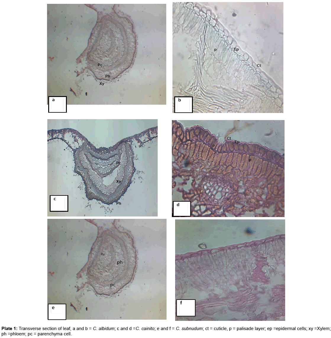
Plate 1: Transverse section of leaf, a and b = C. albidum; c and d =C. cainito; e and f = C. subnudum; ct = cuticle, p = palisade layer; ep =epidermal cells; xy =Xylem; ph =phloem; pc = parenchyma cell.
Petiole anatomy
The results showed that the shape of petiole is semicircular in C. albidum and C. cainito but rounded in C. subundum (Plate 2). Cuticle structure and bundle sheath are all present in the three taxa studied. Epidermal cells are uniseriate in all the taxa considered. Also the vascular bundle arrangements form an arc with invagination that curves inwards at the ends in the three taxa (Plates 2a-c). Trichomes are absent. On the other hand, C. cainito and C. subundum have well developed Vascular bundle about 6-7 arranged to form an arc with distinct xylem and phloem cells. While in C. albidum they are 3-7 arranged to form an arc with in the cortex (Table 2). The shape of epidermal cells is rectangular in C. albidum and C. cainito while it varies from rectangular to pentagonal in C. subundum.
| Characters | C. albidum | C. cainito | C. subnudum |
|---|---|---|---|
| Petiole shape | Semi-circular | Semi circular | Rounded |
| Cuticle structure | Present | Present | Present |
| Epidermal cells | Uniseriate | Uniseriate | Uniseriated |
| Shape of epidermal cells | Rectangular | Rectangular | Rectangular -Pentagonal |
| Number of vascular Bundle | 3-7 | 6-7 | 6-7 |
| Nature of vascular bundle | arc with invaginated ends | arc with invaginated ends | arc with invaginated ends |
| Bundle sheath | Present | Present | Present |
| Trichomes | Absent | Absent | Absent |
Table 2: Petiole anatomical features of the three Chrysophyllum species.
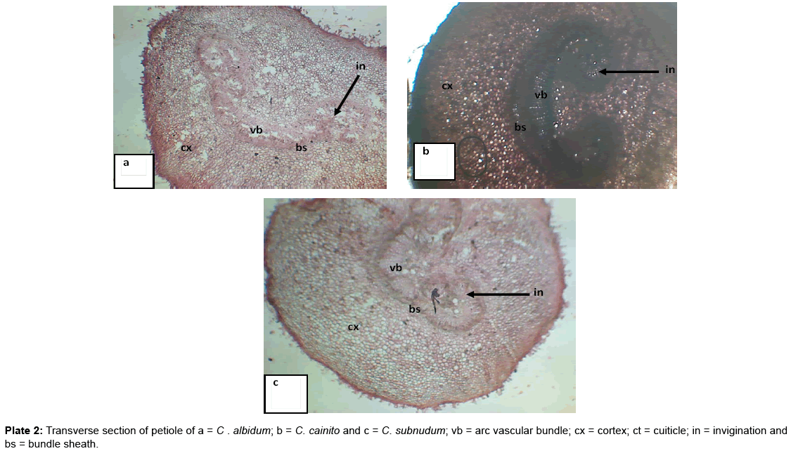
Plate 2: Transverse section of petiole of a = C . albidum; b = C. cainito and c = C. subnudum; vb = arc vascular bundle; cx = cortex; ct = cuiticle; in = invigination and bs = bundle sheath.
The leaf anatomical studies revealed the upper and the lower epidermis of the three Chrysophyllum investigated is covered with a thick layer of cuticle and can be considered for taxonomic purpose at the genus level. Rejanna and Ramakrishnan observed same thick cuticle in the twelve clones of genus Cemellia. The shape of epidermal cells could be used as taxonomic purpose at the species level. The flat epidermal cells in C. cainito delimit it from the other two taxa investigated. Also, the double layered epidermal cells observed in the two taxa C. albidum and . subnudum separated the two species from C. cainito with single layered epidermal cell. Semicircular shape of the mid rib observed in the three taxa is not strange as same feature has been reported by Makbul in genus Epilobuim L.
The numbers of palisade mesophyll observed in the species of Chrysophyllum studied are found to be diagonistic. The mesophyll consists of palisade and spongy layers in C. albidum while C. cainito and . subnudum contained only palisade mesophyll. C. cainito and . subnudum have double layered palisade mesophyll while C. albidum has single layered palisade mesophyll. The difference in number of palisade mesophyll separated C. albidum from the other two taxa investigated. Also Sponge mesophyll was absent in both C. cainito and . subnudum but present in C. albidum. Number of mesophyll could be used to delimitate the three taxa. Edeoga et al. [4] used number of palisade mesophyll to distinguish three taxa of Phyllanthus species. Whereas P. niruroides has 1 layer of palisade mesophyll; P. amarus and P. urinaria has 2 layers. Also, Metcalf and Chalk [6] used similar parameter in taxonomic conclusion of the Phyllanthus species. Semicircular shape of the midrib was observed to be common in the three taxa studied, also common among the taxa are the presence of Sclerenchyma cells, and bundle sheath. These could be of taxonomic value in the genera or species level. C. albidum could be separated from the other two taxa studied base on leaf anatomical features. Shape of vascular bundle in C. cainito was observed to be V-shaped but semi-circular in C. albidum and . subnudum. Similarly, the minimum number of vascular bundle when considered, per species, showed that C. albidum and . subnudum have a minimum of 3 vascular bundle each while C. cainito has minimum of 4. This value is of no taxonomical significant. Based on leaf anatomical analysis, C. albidum and . subnudum have strong closer affinity than C. cainito [12-14].
The pattern of vascular bundle observed on the transverse section of the petiole is diagnostic. The pattern of vascular bundle in the three taxa studied showed that it forms an arc with invaginated ends in the three taxa investigated. This pattern could reflect the intraspecific affinity among the taxa studied. Epidermal cell of the petiole among the species were found to be uniserate; bundle sheath was also recorded and no trichomes were present in the taxa, strongly affirm the intraspecific relationship among the species investigated. The number of contex in the taxa studied showed that C. cainito and . subnudum have closer affinity than C. albinudum. Also, number of vascular bundle separated C. albidum from other two taxa studied. Epidermals cell shape of the three taxa showed that C. albidum and C. cainito have rectangularshaped epidermal cells on the petiole; . subnudum has epidermal cells of various shapes; rectangular to pentagonal. Similarities observed in the petiole anatomy are believed to be of taxonomic value to show the intraspecific affinity among the three taxa investigated while differences could explain why the taxa stand as different species.
The leaf and petiole anatomic studies carried out showed strong intraspecific relationship among the three taxa studied, hence reasons for their placement under the same genus Chrysophyllum, more so there are numerous variations among the features studied indicating taxonomic reasons for the three taxa to exist as distinct species.