Journal of Glycomics & Lipidomics
Open Access
ISSN: 2153-0637
ISSN: 2153-0637
Research Article - (2012) Volume 0, Issue 0
Na+/K+-ATPase is a membrane glycoprotein composed of α, β, and γ subunits, generating ion gradients across plasma membranes. Ion pumping is mainly accomplished by the α subunit, while the glycosylated β subunit binds tightly to the α subunit to assemble the pump and plays an essential role in the stabilization and maturation of Na+/K+-ATPase. Accumulating evidence suggests that the N-glycans of the β subunit contribute to cell-cell interaction and the tightness of cell contacts is modulated by N-glycan branching. However, N-glycan function is not fully understood due to a lack of detailed information on the oligosaccharide structure. We, here, perform glycosylation profi ling of the N-glycans attached to pig kidney Na+/K+-ATPase in order to better understand the mechanism of Na+/K+-ATPase-mediated cell adhesion.
We purifi ed Na+/K+-ATPase from pig kidney outer medulla to homogeneity and solubilized it with the detergent C12E8. The enzyme thus obtained was identifi ed as α1β1 subtype by LC-MS/MS analysis of tryptic digests. During the course of MS analysis, we found that Lys456 of the α subunit was partially modifi ed with 4-hydroxynonenal, an aldehydric lipid peroxidation product. Three N-glycosylation sites on the β1 subunit were confi rmed to be fully occupied by time course analysis of enzymatic deglycosylation monitored by SDS-PAGE. HPLC profi ling of pyridylaminated oligosaccharides derived from Na+/K+-ATPase showed that high-mannose type oligosaccharides predominate while most of the less abundant complex-type oligosaccharides are capped with galactose residues. No glycans could be detected on the four consensus N-glycosylation sites on the α1 subunit. We briefl y discuss the possibility that oligomerization of Na+/K+- ATPase via β-β interactions assembles the N-glycans and promotes cell-cell adhesion.
<Keywords: N-glycan; Na+/K+-ATPase; HPLC; MS/MS; 4-hydroxynonenal
Na+/K+-ATPase is an integral membrane protein that is responsible for the ATP-dependent transport of Na+ and K+ across the cell membrane [1]. This transport generates the electrochemical gradients that are required for electrical excitability, cellular uptake of ions, nutrients and neurotransmitters, and regulation of cell volume and intracellular pH. Na+/K+-ATPase consists of three non-covalently linked subunits, designated as α, β and γ. The catalytic α subunit, with a molecular weight of approximately 113 kDa, contains the binding sites for Na+, K+, ATP, phosphate, and specific cardiac glycoside inhibitors. The β subunit (35~55 kDa) is composed of a short N-terminal cytoplasmic tail, a transmembrane segment and a large extracellular C terminal domain [2,3]. Recent reports on crystal structures of Na+/K+-ATPase reveal the overall topology at 2.4~4.6 Å resolution [4-7]. The role of the β subunit is less well defined but direct and indirect evidence suggests a role in maturation, intracellular transport to the plasma membrane and stabilization of the K+-occluded intermediate [8,9]. The small γ subunit (7 kDa), also called FXYD protein, regulates activity and also stabilizes the enzyme [10,11]. Na+/K+-ATPase is expressed in several isozymes, and in mammalian cells, four α isoforms (α1, α2, α3 and α4) and three β isoforms (β1, β2 and β3) have been identified [12-14]. Both α and β isoforms of Na+/K+-ATPase exhibit tissue-specific expression patterns. The α1 isoform in association with the β1 subunit is found in almost every tissue and α1β1 is the principal isozyme of the kidney, a tissue often used as a source of Na+/K+-ATPase [12].
The β subunit of Na+/K+-ATPase also plays a role in cell-cell adhesion [15]. The β2 subunit was first described as an adhesion molecule on glia (AMOG) in rat brain [16]. Na+/K+-ATPase plays a crucial role in the intercellular junction formation in Drosophila tracheal epithelium in an isoform-specific manner [17-19]. The β1 subunit of Na+/K+-ATPase is necessary for normal intercellular adhesion in MDCK cells, a cell line with distil renal tubule characteristics [20,21]. These functions of the β subunit may be partly related to the fact that it is N-glycosylated. Basigin (HAb18G/CD147), important in neuron-glia interactions [22], binds to the oligomannosidic carbohydrates expressed on the β2 subunit of mouse brain Na+/K+-ATPase (AOMG) [23]. MDCK cells expressing an unglycosylated mutant of the Na+/K+-ATPase β1 subunit form cellcell contacts more slowly compared with non-transfected MDCK cells [20]. A decrease of N-glycan branching on the β1 subunit in MDCK cells increases paracellular permeability of MDCK cell monolayers [24], suggesting that changes in the structure of the N-glycans affects intercellular adhesion. Kitamura et al. [25] report that mouse Na+/ K+-ATPase β1-subunit is a potassium-dependent lectin that binds GlcNAc-terminating oligosaccharides, which may be involved in neural cell interactions. N-glycosylation on the β1 subunit of mouse Na+/K+-ATPase is required for binding to GlcNAc-agarose [25].
Information on carbohydrate structure of Na+/K+-ATPase comes mostly from lectin-binding analyses. Na+/K+-ATPases from dog, lamb and rat kidney bind to wheat germ agglutinin (WGA) but not to Concanavalin A (ConA), lectins specific for N-acetylglucosamine residues and high mannose type glycans, respectively [26-28]. Tissuespecific glycosylation was reported for Na+/K+-ATPases from brain (human, rabbit, and rat) and kidney (human, rabbit, rat, pig and dog) by use of Datura stramonium aggulutinin (DSA) and Galanthus nivalis aggulutinin (GNA), which bind lactosamine and terminal mannose groups, respectively [29,30]. It is well-established that N-glycans are attached to the β-subunit, while the α-subunit is considered to be non-glycosylated. Some groups, however, maintain that there is glycosylation of the α-subunit [29-35], but the issue remains controversial on account of uncertainty regarding the purity of the various preparations. Sialylated glycans are reported to be present on the β subunit of lamb [33], dog [26,36,37] and rabbit kidney [38]. Mass spectrometric analysis of the major oligosaccharides at each of the three N-glycosylation sites in the β subunit of lamb and dog kidney Na+/K+-ATPase [28] indicates that most glycans are tetra-antennary structures with or without repeating N-acetyllactosamine units on a single antenna or on multiple antennae. AMOG (ß2 subunit of Na+/K+- ATPase) is about 80% modified with oligomannosidic glycans according to an anti-oligomannose antibody study [39]. The oligomannose series of glycans are most abundant in a rat brain membrane preparation enriched in Na+/K+-ATPase while tetra-antennary glycans with up to four lactosaminyl units predominate in the oligosaccharides from a Na+/K+-ATPase-enriched kidney membrane preparation [40]. To date, however, a detailed structural analysis of N-glycans of highly purified Na+/K+-ATPase has not been performed. We therefore purified Na+/K+- ATPase from pig kidney to homogeneity and analysed the N-glycans. The oligosaccharide structures are briefly discussed in terms of cell-cell interaction.
Enzymes
Peptide:N-glycosidase F (PNGase F) from Flavobacterium meningosepticum was purchased from New England BioLabs, Inc. Mortierella vinacea α-galactosidase, jack bean β-galacosidase and β-N-acetylhexosaminidase were purchased from Seikagaku Kogyo Co. Bovine kidney α-L-fucosidase was purchased from Sigma-Aldrich. Recombinant human soluble GnT-V (delta 73, Ala74-Leu741) with C-terminal hexahistidine tag was expressed in a baculovirus-insect cell system and purified by Ni2+-chelating affinity chromatography as described previously [41,42].
Reference oligosaccharides
Pyridylamino derivatives of α-1,6 glucose oligomers DP=3-22 (indicating the degree of polymerization of glucose residues) and reference PA-oligosaccharides were purchased from Takara Bio Inc. and Masuda Chemical Industries Co., Ltd. A reference oligosaccharide (Code no. 310.11) was enzymatically prepared from PA-oligosaccharide (Code no. 210.1) as follows. Substrate PA-oligosaccharide (1 μmol/mL) was dissolved in 20 mM MES buffer, pH 6.3 in the presence of human soluble GnT-V (130 pg/mL) and 4.2 mM UDP-GlcNAc and incubated for 16 h at 37°C. The product was purified with an ODS column and used as a reference standard.
Other chemicals
C12E8 (octaethyleneglycol n-dodecylether) was obtained from Nikko Chemical Co. HEPES and free acid of EDTA were products of Dojindo Laboratories. Borane dimethylamine complex was from Sigma-Aldrich. Acetonitrile, 2-aminopyridine, 2,5-hihydroxybenzoic acid, orcin and UDP-GlcNAc were from Wako Pure Chemical Industries, Ltd. Trifluoroacetic acid was from Nakalai Tesque. All the other chemicals (special grade) were of analytical grade and used without further purification.
Preparation of detergent-solubilized Na+/K+-ATPase
Na+/K+-ATPase was purified from microsomes of the outer medulla of frozen pig kidneys according to the method of Jørgensen [43] with some modifications [44]. Briefly, the microsomes were treated with SDS at final microsomal protein and SDS concentration of 1.4 mg/ mL and 0.6 mg/mL, respectively. The sample was subjected to sucrose density gradient centrifugation using a Beckmann Ti-15 zonal rotor to obtain the Na+/K+-ATPase-enriched membrane fragments. The membrane fragments thus obtained were washed to remove Na+ and K+ contamination as described previously [45], and finally suspended in 20% (w/v) glycerol, 12 mM imidazole, 28 mM HEPES and pH 7.1. The suspension of membrane-bound enzyme was frozen and stored at -80°C until use. For solubilization, the membrane-bound enzyme was incubated in the presence of various concentrations of KCl and/or NaCl with C12E8 at pH 7.0 and 0°C for 5 min [44]. The final composition was 2 mg/mL protein, 6 mg/mL C12E8, 1 mM EDTA, 10% (w/v) glycerol, 14 mM imidazole and 22 mM HEPES. The solution was centrifuged at 436,000 g for 5 min at 2°C and the supernatant used within 12 h as the solubilized Na+/K+-ATPase enzyme. The protein concentration of the solubilized enzyme was determined from the absorbance at 280 nm using an absorption coefficient of 1.22 mg-1ml cm-1 [45]. The specific ATPase activity of the membrane-bound enzyme was determined at 37°C under optimal conditions as described previously [44].
LC-MS/MS analysis of tryptic peptides of Na+/K+-ATPase
The membrane-bound Na+/K+-ATPase was dissolved at a concentration of 10 mg/mL in 6 M guanidinium chloride, pH 8.5 and incubated at 60°C for 15 min. After addition of 0.5 mM TCEP and 0.1 mM iodoacetamide, the reaction mixture was incubated at room temperature for 15 min in the dark. The sample solution was diluted ten-fold with 100 mM NH4HCO3 buffer and incubated with 5 μg/mL of modified trypsin (Promega) at 37°C overnight. The solution of tryptic peptides was diluted ten-fold with 0.1% (v/v) TFA and subjected to nanoLC-Ion Trap MS system. The solution of peptide mixture was loaded onto Ultimate 3000 Bio-compatible nanoLC system (DIONEX Co.) equipped with two Pepmap100 C18 columns (guard column; 0.3×1 mm, 5 μm, analytical column; 0.075×150 mm, 3 mm, DIONEX Co.). Tandemly connected C18 columns were equilibrated with 0.1% (v/v) formic acid and the trypticpeptides eluted with a gradient of acetonitrile in 0.1% (v/v) formic acid. For the MS analysis of eluted peptides, HCTultra ETD II system (Bruker Daltonik GmbH) was used. Online nanospray source was used as an ion source and Silica Tips (360/20 μm, New Objective) was used as an emission tip. The data were converted to Mascot generic file and subject to Mascot database search.
Isolation and characterization of N-glycans by HPLC
The C12E8-solubilized Na+/K+-ATPase (1.7 mg) was digested with 75,000 units of PNGase F at 37°C for 18 h and the extent of reaction checked by SDS-PAGE. To remove protein and detergent, the reaction mixture was applied to Sep-Pack cartridges (Waters) according to the manufacturers’ instructions. Flow-through fractions including released oligosaccharides were collected. Reducing ends of the oligosaccharides were then derivatized with 2-aminopyridine under the condition described previously [46]. The resultant pyridylamino (PA)-glycans were first purified by gel filtration on a Sephadex G-15 column (1.0×40 cm, GE Healthcare). Appropriate fractions were then purified on a LaChromElite HPLC system (Hitachi, Japan) under conditions reported previously [47,48]. Three separate columns were used, firstly a TSK-gel DEAE-5PW column (7.5 mm×7.5 cm, Tosoh, Tokyo, Japan), then a TSK-gel Amide-80 column (4.6 mm×25 cm, Tosoh), and finally a Shim pack HRC-ODS column (6.0 mm×15 cm, Shimadzu, Kyoto, Japan). Elution times of the individual peaks from the amide-silica and ODS columns were normalized with respect to the PA-derivatized isomaltooligosaccharides with degree of polymerization 3-22, and reported in glucose units (GU). Thus, a given compound analyzed by these two columns provided a unique set of GU values, which corresponded to coordinates of the 2D HPLC maps. PA-oligosaccharides derived from Na+/K+-ATPase were identified by comparison with the coordinates of ~500 reference PA-oligosaccharides in a web application, GALAXY (http://www.glycoanalysis.info/) [49] and with the data in the literature [50]. A sample PA-oligosaccharide and a reference PA-oligosaccharide were co-injected on the columns to confirm their identities. PAoligosaccharides were detected by fluorescence using excitation and emission at 320 and 400 nm, respectively.
Mass spectrometric analysis of PA-glycans
PA-oligosaccharides were subjected to matrix-assisted laser desorption/ionization time-of flight mass spectrometric (MALDITOF- MS) analysis. 10 mg of 2,5-dihydroxybenzoic acid (Wako) was dissolved in 1:1 (v/v) of water/acetonitrile (1 mL) and used as a matrix solution. 1 μl of sample solution was mixed on the target spot of a plate with 1μL of matrix solution and then allowed to air-dry. MALDITOF- MS data were acquired in a positive mode using AXIMA-CFR (Shimadzu) operated in the linear mode.
Exoglycosidase digestion of PA-glycans
Each PA-glycan from Na+/K+-ATPase was digested with exoglycosidases (α-fucosidase, β-galactosidase, or β-N-acetyl hexosaminidase) under conditions described below. PA-oligosaccharide (1-10 pmol) was dissolved in 20 μL of 50 mM sodium phosphate buffer and glycosidase reactions were performed at each condition (α-Lfucosidase 15 mU, pH 5.8 at 37°C for 16 h; α-galactosidase 0.5U, pH 5.9 at 37°C for 16 h; ß-galactosidase 0.1 U, pH 3.5 at 37°C for 16 h). Reactions were checked by HPLC and MALDI-TOF-MS spectrometry.
Purification and characterization of Na+/K+-ATPase from pig kidney
It is essential to use homogeneous protein for the characterization of post-translational modifications of Na+/K+-ATPase. We purified Na+/K+-ATPase from pig kidney outer medulla to homogeneity and the typical ATPase activity of the purified enzyme was 29~35 μmol Pi/min/mg protein at 37°C(see Materials and Methods). The purity of solubilized Na+/K+-ATPase was checked by SDS-PAGE (Figure 1, lane 2). Polypeptide bands corresponding to α (Mr=100,000) and ß (Mr=60,000) subunits of the Na+/K+-ATPase were evident. The band originating from the γ subunit (Mr=7,000) was not seen presumably due to its low molecular weight.

Figure 1: SDS-PAGE of purified pig kidney Na+/K+-ATPase and the timecourse of deglycosylation with PNGase F. Lane 1: molecular-weight marker, lane 2: Na+/K+-ATPase purified from pig kidney, lanes 3-6: Na+/K+-ATPase treated with PNGase F for 0.5 h (lane 3), 1 h (lane 4), 2 h (lane 5) and 6 h (lane 6).
In order to determine the isoform of purified pig kidney Na+/K+- ATPase, we conducted LC-MS/MS analysis of a tryptic digest. Mass spectrometric analysis clearly showed that the isoform of the Na+/ K+-ATPase is α1β1, and the sequence coverage is 51% for α subunit and 38% for ß subunit (Figure 2). Ser723Leu/Ile substitution in the α1 subunit was found in CID-MS/MS spectrum of a peptide spanning from 706 to 724 (Figure 3A). Leu723 of the α1 subunit has been previously reported [51] and annotated as a sequence conflict in the UniProt database (P05024). CID-MS/MS analysis of a peptide from 444 to 456 revealed that Lys456 was partially modified with 4-hydroxynonenal (HNE) (Figure 3B), an aldehyde produced from the peroxidation of ω-6 polyunsaturated fatty acids [52]. From the chromatographic analysis of the peptide (444-456), 5% of the Lys456 was modified (Figures 3C and 3D). α1 subunit of pig Na+/K+-ATPase has four consensus sites (Asn-X-Ser/Thr) for glycosylation but none were modified. Mammalian β1 isoform has three N-glycosylation sites and all have been reported to be glycosylated in the β1 isoform of dog kidney [28,34,37], lamb kidney [28] and chick sensory neuron [53]. We could not detect the glycosylated peptides in the mass spectra presumably due to insufficient trypsin cleavage of the corresponding regions. To confirm that the three N-glycosylation sites are modified, we analyzed the time-course of PNGase F-treatment by SDS-PAGE (Figure 1, lanes 3-6). Increasing reaction time resulted in the progressive reduction of the apparent molecular weight of the ß subunit without affecting the mobility of the α subunit. These results showed that the β1 subunit of pig kidney Na+/K+-ATPase is fully modified with three N-glycans while the α subunit is not N-glycosylated.

Figure 2: Amino acid sequence of pig Na+/K+-ATPase α1 and β1 subunit. Amino acid residues in red are identified in the CID-MS/MS study and the underlined residues are located in the transmenbrane region. Four consensus sites (Asn-X-Ser/Thr) in the α1 subunit are marked with blue rectangles. Three N-glycosylation sites in β1 subunit are marked with yellow rectangles.

Figure 3: Identification of pig kidney Na+/K+-ATPase subtypes by mass spectrometry CID-MS/MS spectra of α-subunit peptides from 706-724 (A) and 444-456 (B). Total mass chromatograms are shown for the 4-hydroxynonenal (HNE)-unmodified (C) and HNE-modified (D) peptides (444-456).
Separation and identification of oligosaccharides by DEAE, amide and ODS columns
In order to obtain information on the structure of the N-glycans, the reducing ends of the glycans released from pig kidney Na+/K+- ATPase were fluorescently labeled with 2-aminopyridine (PA). Figure 4A shows the elution profile of the derived PA-glycans on a DEAE- 5PW anion-exchange column. Most PA-glycans eluted in the void volume, indicating that neutral oligosaccharides predominate and sialyloligosaccharides are less abundant. The void fraction from the DEAE column was then applied to an amide column and the elution profile is shown in figure 4B. Each peak detected on the amide column (peaks 1-10) was collected and reapplied to an ODS column. The elution profiles of each fraction are shown in figure 5. We isolated 25 different neutral oligosaccharides (peaks A-Y) on the ODS column. Structural assignment of the oligosaccharides was performed by a 2-D HPLC mapping technique [48].

Figure 4: HPLC profiles PA-glycans derived from pig kidney Na+/K+-ATPase.
Elution profiles on DEAE column (A) and amide column (B) are shown. The void fraction on DEAE column was subsequently applied to the amide column and the peaks are numbered from 1 to 10.

Figure 5: Elution profiles of PA-glycans from pig kidney Na+/K+-ATPase on ODS column. Each fraction (peak 1 to 10 in Figure 4B) was applied to ODS column and the peaks are labeled from A to Y (Table 1).
Identification of high mannose type oligosaccharides
PA-oligosaccharides A, E, I, J, K, N, O, P, T, V and X were identified as high mannose type by their molecular masses and GUs and the assignments were confirmed by co-injection with the corresponding standard oligosaccharides on the ODS column. GU coordinates of N-glycans (A, E, I, J, K, N, O, P, T, V and X) from Na+/K+-ATPase coincided within 15% error with those of known oligosaccharides on the 2D map. High mannose type PA-oligosaccharides, A, E, I, J, K, N, O, P, T, V and X were assigned to code numbers M5.1, M6.1, M7.2, M7.1, M7.7, M8.1, M8.4, M8.2, M8.1, M9.1 and M9.2, respectively (Table 1). Oligosaccharides N and T were both assigned to M8.1. Man6 and Man5 oligosaccharides were most abundant at 16.7% and 9.1%, respectively (Table 1). Man7 and Man8 oligosaccharides occurred as isomeric structures. There was 8.3% and 0.6%, Man9 and GlcMan9 respectively. High-mannose type oligosaccharides account for nearly half of the total N-linked oligosaccharides.
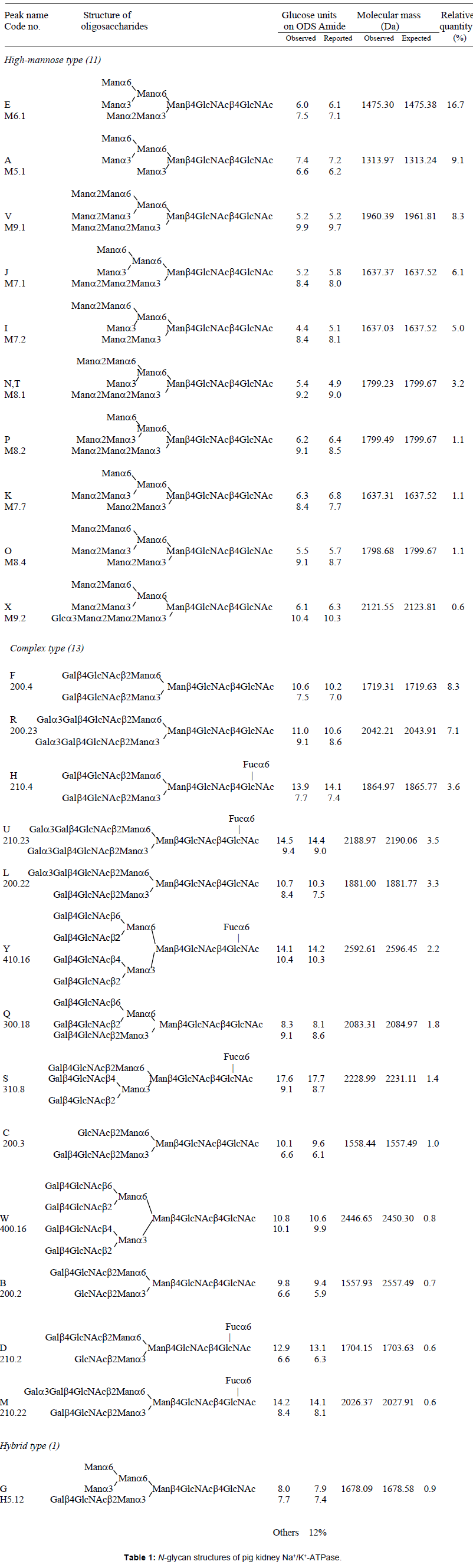 |
Table 1: N-glycan structures of pig kidney Na+/K+-ATPase.
Identification of complex and hybrid type oligosaccharides
PA-oligosaccharides B, C, D, F, H, L, M, Q, R, U, W and Y were identified as complex-type and G as a hybrid-type oligosaccharide. GU coordinates of N-glycans (B, C, D, F, G, H, L, M, Q, R, U, W and Y) coincided again within 15% error with those of known oligosaccharides on the 2D map. The observed molecular mass of the glycans are identical to the expected molecular masses within 2 Da. Complex-type PA-oligosaccharides B, C, D, F, H, L, M, Q, R, U, W and Y were assigned to code numbers 200.2, 200.3, 210.2, 200.4, 210.4, 200.22, 210.22, 300.18, 200.23, 400.16 and 410.16, respectively (Table 1). Most of the complex-type glycans were composed of biantennary oligosaccharides; tri/tetraantenary glycans were detected to a lesser extent. PA-oligosaccharides R, U, L and M contain one or two terminal α-1,3-galactose residues and the existence of α-1,3-galactose residues was confirmed by the changes in GUs upon α-galactosidase digestion. After α-galactosidase digestion, the elution positions of oligosaccharides R and L shifted to a position identical to that of oligosaccharide F. In a similar way, α-galactosidase digestion shifted the elution positions of fucosylated oligosaccharides U and M to a position identical to that of oligosaccharide H.
Mass spectrometric analysis of trypsin-digested pig kidney Na+/ K+-ATPase showed that the major subtype is α1ß1, consistent with the findings of Ovchinnikov et al. [54]. We found partial modification of Lys456 of α subunit with 4-hydroxynonenal (HNE). HNE is known to be one of the major end products of lipid peroxidation in cells and recent studies suggest that HNE is a key signaling molecule to regulate stress-mediated signaling [55]. Previous reports suggest that HNEreacts withcysteine sulfhydryl groups of Na+/K+-ATPase and inhibit the enzyme activity [56-58]. It was also found that HNE reacts with other amino acids such as lysine to form adducts that also interfere with the protein function [56]. Lys456 is close to the ATPbinding site in the cytoplasmic domain [2]. The HNE modification at Lys456 thus conceivably inhibits enzymatic activity, but this should be investigated further in detail. Deglycosylation with PNGase F resulted in the time dependent appearance of three intermediate bands (corresponding to the β subunit with 2, 1 and 0 glycans), indicating that all the three N-glycosylation sites of the pig kidney β subunit are occupied with glycans. Full glycosylation has also been observed in other species [28,37]. Glycosylation analysis showed that pig kidney Na+/K+-ATPase has essentially no sialic acid. N-glycans attached to pig kidney Na+/K+-ATPase were composed of high-mannose type (60%), complex type (30%) and unidentified (10%). Oligosaccharides from lamb and dog kidney ß subunit have been reported to be complex type [28], and those from rat kidney Na+/K+-ATPase are dominated by tetra-antennary complex-type glycans [40]. These differences suggest that the glycans expressed on Na+/K+-ATPase ß subunit may be species specific. Three N-glycosylation sites are conserved among sheep, dog, rat, and pig and the sequence homology of the ß-subunit is high (Supplementary Figure 1). The observed differences in N-glycan structure are thus possibly attributed to the different expression levels of glycosyl transferases/glycosidases among species. Characteristics of complex-type oligosaccharides include capping of the non-reducing ends with a lactosamine unit and some with α-(1-3)-linked galactose. Lactosamine was also found on N-glycans derived from Na+/K+- ATPase-enriched rat kidney membrane fraction [40]. Capping with α1-3 linked galactose is not found in human N-glycans. Kitamura et al. [25] have reported that Na+/K+-ATPase ß subunit from mouse brain binds to GlcNAc, and trans and cis β-β interactions were predicted. In our analysis, most glycans were capped with α-(1-3) or ß-(1-4) linked galactose; GlcNAc-terminating oligosaccharides were hardly detected. Therefore, pig kidney Na+/K+-ATPase seems not to show significant β-β interactions. The oligomeric state of Na+/K+-ATPase may regulate cell-cell interactions. Although the α and β-subunits are non-covalently linked in a minimal αβ-protomer structural unit [59], solubilized enzyme solutions contain other oligomers, such as (αβ)2, (αβ)3 and (αβ)4 as shown by chemical crosslinking [60]. HPLC analysis of solubilized Na+/K+-ATPase reveals that K+ induces the conversion of protomer into diprotomer and/or higher oligomers while Na+ has the opposite effect [61,62]. Cation and hence, conformation-dependent alteration in the oligomer equilibrium may occur through changes in exposure of the transmembrane domain of the ß-subunit as mutational analysis has shown that this portion of the protein mediates β-β homooligomerization, as well as α/β assembly [63]. Oligomerization of Na+/ K+-ATPase through β-β contact would concentrate or induce the close assembly of N-glycans which may facilitate cell-cell adhesion. Experiments are underway in our laboratory to investigate the role of glycans in the oligomerization of Na+/K+-ATPase during cell-cell adhesion.
We thank Drs. Naoyuki Taniguchi, Eiji Miyoshi, and Takashi Saito for kindly providing the GnT-V construct and Ms. Akemi Ikeda for purification of GnT-V. We also thank Dr. Yoshikazu Tahara for his contribution to the purification of Na+/K+-ATPase. One of the authors (YH) thanks Dr. Kazuya Taniguchi for his encouragement during the course of this study. This work was supported in part by Grants-in-Aid from the Ministry of Education, Culture, Sports, Science and Technology (MEXT).