GET THE APP

Raphides in Food - An Unsafe Menu
Journal of Plant Biochemistry & Physiology

Journal of Plant Biochemistry & Physiology
Open Access

ISSN: 2329-9029

+44 1478 350008

Review Article - (2015) Volume 3, Issue 2

Raphides in Food - An Unsafe Menu

Naveen Tripathi1, Chandra Bose1, Srijoni Basu1, Nabajit Das1, Susmit Maitra2, Arindam Sikdar1 and Sukant Khurana1*
1Indian Institute of Science Education and Research Kolkata (IISER-K), Mohanpur-741246, India
2Kalinga Institute of Industrial Technology, Odisha-751024, India
*Corresponding Author: Sukant Khurana, Kalinga Institute of Industrial Technology, Odisha-751024, India, Tel: +91-33-2587-3017, Fax: +91-33-2587- 3028 Email: ,

Abstract

Calcium oxalate in plant bodies lead to stones in kidney, upon consumption. Calcium oxalate is frequently found in plants in the form of tiny needle like raphides. Out of the 5 types of calcium oxalate crystals, raphides are the predominant ones. Calcium oxalate gets incorporated in our body through plant derived food that contains them; a little amount of them is also synthesized in humans endogenously. Both the sources contribute to kidney problems. Occurrence of calcium oxalate is not limited to higher plants only but also extends to algae, fungi and lichens. Out of all the 3 forms of calcium oxalate, monohydrate form is the one widely reported to cause kidney problems. In this study, we review raphides and explore their possible remediation in order to utilize plants of food and medicinal importance the better way. We also review traditional knowledge of raphide neutralization and point to the methods of removal of calcium oxalate and raphides.

Introduction

Plants are the key sources of food and medicine. Calcium oxalate, a potential causative agent of human kidney stones, can range from 3 to 80% of the dry weight of various plants [1-3]. Calcium oxalate exists in varying crystal shapes and sizes in plants, with raphides being the predominant crystal form [2,4-8]. Deleterious influence so raphides, in addition to promoting kidney stone formation, include irritation to throat, mouth and skin [7,9-18]. Excess presence of raphides, in conjugation with cytotoxic compounds [5,19], can render the food poisonous and is responsible for mentionable fatalities every year [20-22]. Calcium oxalate can contribute up to 70% or 75% of the composition of kidney stones [ 23,24]. It i s present in mono, di and trihydrate forms [25-28]. Monohydrate form is the least soluble and the main constituent of nephroliths [23,24,29]. Monohydrate form readily attaches to the cell surface of the renal tubules [24,30]. In addition to major absorption of calcium oxalate from plant food sources, smaller amount of calcium oxalate can be synthesized endogenously from free oxalic acid or directly from several other biosynthetic precursors [23,31-33]. Liver is the main site of endogenous synthesis of oxalic acid [34]. Inter-conversion rates between monohydrate and dihydrate state governs the attachment of calcium oxalate to renal membrane [24]. Oxalate formation can deplete the human body of divalents, including calcium [35,36]. This depletion of calcium due to calcium oxalate formation aids in osteoporosis [37-39].

In addition to medicines [40,41], dietary control is frequently recommended in the treatment of kidney stones[33,37]. Some proteins that can regulate kidney stone formation [24,42,43] have been identified, which raises a hope for novel drug development. Preclinical studies indicate that few plant extracts can inhibit the growth of calcium oxalate crystals [44], as well as block their adhesion to renal epithelial cells [45,46]. Given that the removal of calcium oxalate from diet can have such a large impact on human health, we review crystals of calcium oxalate in plants, calcium oxalate biosynthesis, and possible ways of calcium oxalate neutralization.

Crystalline Calcium Oxalate; Importance of Raphides

Calcium oxalate is found in several crystal shapes in plants. In some plants presence or absence of a certain kind of crystals can help in taxonomic identification [9] and evolutionary studies [47]. The major kinds of crystals include: needle shaped “raphides” [5-7,48,49], cuboidal or pencil shaped “steroids” [6,7], block shaped aggregates called “crystal sand” [2,11], prism shaped structures [8], and mace macehead or rosette shaped aggregates called “druses” [4]. Raphides are found to have subdivisions in their shapes. According to their shapes, they are classified from type 1 to 4 [9] and a recent study has reported 2 more additional kinds [50]. The calcium oxalate crystals are formed in specialized vacuoles of idioblast cells. The shape of idioblasts, which is under genetic influence, governs the shape of the crystal [51] and is species specific [52]. There can be several kinds of crystals present in the same plant [4,9]. Figure 1 shows the common kinds of Calcium Oxalate crystals [4,17,22,53-59].

plant-biochemistry-physiology-calcium-oxalate

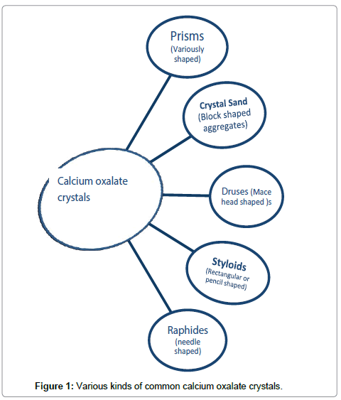
Figure 1: Various kinds of common calcium oxalate crystals.

Raphides provide plant defense against herbivore [60,61]. Herbivore has been demonstrated to increase the amount of raphides in plants [60]. Frequently raphides are co-present with cysteine proteases and other chemical defense [5]. The needle shaped raphides bruise the lining of the throat, gut, and intestine, while cysteine proteases add to the irritation [15]. Studies using larvae and caterpillars have shown additive effect of the irritants such as proteases and raphides [62]. Raphides can act as needles or syringes to deposit cysteine proteases in herbivore cells [4]. More acridity measurement studies are needed to evaluate the synergistic action of raphides and cysteine proteases on human taste perception [5].

Raphide crystals also play a role in reducing metal toxicity. This suggestion has largely been based on the observation that such crystals can have many other divalents [63-65]. Raphides have also been implicated in light scattering and increasing efficiency of photosynthesis [12,18] but work is required on other crystal types to see if it is exclusively a phenomenon limited to raphide containing plants. Some studies have also suggested a clear role of raphides in structural support to the plant [7,66]. So far, other crystal shapes have not been explored much in defense. Several crystal shapes have been implicated in calcium [67-70] and play an important role in ion regulation.

Biosynthesis of Calcium Oxalate

Many recent studies have established that several precursors can be used to synthesize calcium oxalate. The major precursors are ascorbate, glycolate, glyoxylate, oxaloacetate and isocitrate [53,71-75]. Different precursor use by the different parts of a plant remains to be explored. Biosynthetic pathways have been very well reviewed at length in several recent works [53,71-76] so we are merely summarizing the key findings and focusing on amelioration in the next section. The synthesis of ascorbate, a precursor of calcium oxalate takes place in the vacuoles of idioblast cells itself. Concentration of oxalate is higher in idioblasts and calcium is incorporated to make calcium oxalate [18,25]. This higher oxalate in idioblasts results in predominantly the monohydrate form and not the di or trihydrate form [2]. The process of calcium oxalate crystal formation takes place in a matter of hours [77], rising hopes of its possible quick removal. Figure 2 summarizes the common biosynthetic pathways and possible amelioration approaches.

plant-biochemistry-physiology-biosynthetic-pathways

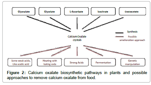
Figure 2: Calcium oxalate biosynthetic pathways in plants and possible approaches to remove calcium oxalate from food.

Traditional Remediation of Raphides

Not much modern analysis has been conducted of the traditional approaches, creating a problem of lack of peer-reviewed scientific material on the particular topic of traditional methods. We are bringing forward such information with the hope that modern analysis of these approaches follows soon. Traditional approaches work largely by neutralizing the cysteine proteases. Milk and coconut milk have been used in India to ameliorate the acridity of raphides and they likely work by providing protein substrates to interact with the active poisons present along with raphids. Tamarind [78], lime [79] and various other acidic treatments have also been used because acid can neutralize toxins. Heating, boiling, frying [80], baking [81], battering, mashing, fermentation and sun drying [82] have also been used to the same effect of neutralization of cysteine proteases and release of raphides from idioblasts. Changes in crystal structures of calcium oxalate itself due to these traditional approaches should be explored systematically but this topic has not received attention yet.

Removal of Calcium Oxalate

While several methods neutralize raphides, very few have addressed the neutralization of the long-term damage of kidney stones that increases by eating foods rich in calcium oxalate. There are few traditional approaches that offer hope and few established methods that are yet to gain popularity. Peeling of foods where plant pericarp has high calcium oxalate [83] seems to be an effective and economical way of reducing calcium oxalate. In addition, there are emerging pieces of evidence that raise the hope that several plants with huge drug and food potential can be better utilized by reduction or removal of calcium oxalate. Few approaches, such as treatment with strong acid, sodium bicarbonate and tetracycline treatment have been shown to work but it is not clear as of now that any of these approaches can be applied at mass scale due to unacceptable alteration of food and the associated costs. Some fungi have been shown to be able to degrade calcium oxalate [84] and they offer a hope for future biotech applications to remove calcium oxalate. The Flamnulina velutipes study shows formation of formic acid but formic acid is extremely unpleasant and additional enzymatic steps might be required to use the enzymes from this fungus [85]. Transgenic tomatoes have been made using OXDC gene from Flamnulinavelutipes Like fungi, are port from bacteria also suggests break down of calcium oxalate [86-91]. CoD genes, as observed in Medicago trancatula, regulate the formation of calcium oxalate crystals and this gene might also have good biotechnology potential [14]. Heat at the levels used in traditional cooking removes only a small fraction of calcium oxalate, if any, [92] but we suspect that its potential might lie in combination with other approaches. Fast synthesis of calcium oxalate in idioblasts [77] raises hope that an equally fast removal of calcium oxalate might be a possibility. We would expect next few years of research to provide us with signaling and extrinsic factors. To summarize, we have briefly evaluated the kidney stone formation due to calcium oxalate, explored the various crystalline forms of the compound and looked at its biosynthesis, with the end goal of evaluating novel possible calcium oxalate amelioration methods for better utilization of plants by humans.

Conclusion

To summarize, we have briefly evaluated the kidney stone formation due to calcium oxalate, explored the various crystalline forms of the compound, with a focus on raphides and looked at its biosynthesis, with the end goal of evaluating novel possible calcium oxalate amelioration methods for better utilization of plants by humans. Modern evaluation of traditional methods has been largely lacking but from knowledge of known ingredients and limited published work, one can infer that milk, heating, boiling, frying, baking, battering, mashing, fermentation and sun drying, likely work by neutralization of cysteine proteases or through release of raphides from idioblasts or both. Neutralization of calcium oxalate is a bigger health question than the neutralization of specific crystal form of raphides. A traditional approach of peeling plant pericarp rich in calcium oxalate has beneficial effects. Discovery of fungi and bacteria that can break down calcium oxalate and plant genes that regulate calcium oxalate formation have all offered hope and some initial promising results in genetic engineering to counteract calcium oxalate toxicity. We expect the next few years to witness more chemical, physical and genetic engineering approaches to make food safer for the health of kidneys.

References

  1. Zindler E (1976) Oxalate biosynthesis in relation to photosynthetic pathway and plant productivity A survey. ZeitschriftfürPflanzenphysiologie 80:1-3.
  2. Webb MA (1999) Cell-mediated crystallization of calcium oxalate in plants. Plant Cell 11: 751-761.
  3. Ilarslan H, Palmer R, Imsande J, Horner H (1997) Quantitative determination of calcium oxalate and oxalate in developing seeds of soybean (Leguminosae).Am J Bot 84: 1042.
  4. Coté GG (2009) Diversity and distribution of idioblasts producing calcium oxalate crystals in Dieffenbachia seguine (Araceae).Am J Bot 96: 1245-1254.
  5. Konno K, Inoue TA, Nakamura M (2014) Synergistic defensive function of raphides and protease through the needle effect.PLoS One 9: e91341.
  6. Crowther A (2009) Morphometric analysis of calcium oxalate raphides and assessment of their taxonomic value for archaeological microfossil studies. In: Archaeological science under a microscope: studies
  7. Prychid CJ, Rudall PJ (1999) Calcium Oxalate Crystals in Monocotyledons: A Review of their Structure and Systematics. Annals of botany 84: 725-739
  8. Chairiyah N, Harijati N, Mastuti R (2013) Variation of Calcium Oxalate (CaOx) Crystals in Porang (AmorphophallusmuelleriBlume). AJPS 4:1765-1773.
  9. Saadi S,Mondal AK (2011) Studies on the calcium oxalate crystals of some seleted aroids (Araceae) in Eastern India. Advances in Bioresearch 2:134-143.
  10. Salinas M L, Ogura T,Soffchi L (2001) Irritant contact dermatitis caused by needle-like calcium oxalate crystals, raphides, in Agave tequilana among workers in tequila distilleries and agave plantations. Contact dermatitis 44: 94-96.
  11. Franceschi VR, Nakata PA (2005) Calcium oxalate in plants: formation and function.Annu Rev Plant Biol 56: 41-71.
  12. Franceschi VR, Harry THSJS (1980) Calcium Oxalate Crystals in Plants. Botanical Review 46: 361-427.
  13. Holloway WD, Argall MEJealous WT (1989) Organic acids and calcium oxalate in tropical root crops. J Agric Food Chem 37: 337-341.
  14. Korth KL, Doege SJ, ParkSH , Goggin FL (2006) Medicagotruncatula mutants demonstrate the role of plant calcium oxalate crystals as an effective defense against chewing insects. Plant Physiology 141:188-195
  15. PereraCO,Hallett I (1990) Calcium oxalate crystals: the irritant factor in kiwifruit. Journal of Food Science 55: 1066-1069.
  16. Coelho EG, Amaral A, Ferreira J, Santos DAG, et al. (2007) Calcium oxalate crystals and methyl salicylate as toxic principles of the fresh leaves from Palicourealongiflora, an endemic species in the Amazonas state. Toxicon 49:407-409.
  17. Sunell LA, Healey PL (1979) Distribution of calcium oxalate crystal idioblasts in corms of taro (Colocasiaesculenta). AmerJBot 66: 1029-1032.
  18. Nakata PA (2003) Advances in our understanding of calcium oxalate crystal formation and function in plants. Plant Science 164: 901-909.
  19. Yu HL, Zhu FG, Wu H (2011) Study of toxic proteins on raphides from Pinelliaternata and Pinelliapedatisecta Schott. China J Tradit ChinMed Pharm.
  20. Chen CL, Fang HC, Chou KJ, Wang JS, Chung HM (2001) Acute oxalate nephropathy after ingestion of star fruit.Am J Kidney Dis 37: 418-422.
  21. Bhandari A, Koul S, Sekhon A, Pramanik SK, Chaturvedi LS, et al. (2002) Effects of oxalate on HK-2 cells, a line of proximal tubular epithelial cells from normal human kidney.J Urol 168: 253-259.
  22. Kausch AP, Horner HT(1983) The development of mucilaginous raphide crystal idioblasts in young leaves of Typhaangustifolia L.(Typhaceae). Amer JBot 70: 691-705.
  23. Williams HE, Wandzilak TR (1989) Oxalate synthesis, transport and the hyperoxaluric syndromes.J Urol 141: 742-749.
  24. Wesson JA, Worcester EM, Wiessner JH, Mandel NS, Kleinman JG (1998) Control of calcium oxalate crystal structure and cell adherence by urinary macromolecules.Kidney Int 53: 952-957.
  25. Frey A (1981) Crystallography of the two hydrates of crystalline calcium oxalate in plants. Amer J Bot 68: 130-141.
  26. ArnottHJ, PautardF,Steinfink H (1965) Structure of calcium oxalate monohydrate. Nature 208: 1197-1198.
  27. Gardner GL (1975) Nucleation and crystal growth of calcium oxalate trihydrate. Journal of Crystal Growth 30: 158-168.
  28. GrasesF, Millan A, ConteA (1990) Production of calcium oxalate monohydrate, dihydrate or trihydrate. Urological research 18: 17-20.
  29. Evan A, Lingeman J, Coe FL, Worcester E (2006) Randall's plaque: pathogenesis and role in calcium oxalate nephrolithiasis.Kidney Int 69: 1313-1318.
  30. Mandel N1 (1994) Crystal-membrane interaction in kidney stone disease.J Am SocNephrol 5: S37-45.
  31. Chai W, Liebman M, Kynast-Gales S, Massey L (2004) Oxalate absorption and endogenous oxalate synthesis from ascorbate in calcium oxalate stone formers and non-stone formers.Am J Kidney Dis 44: 1060-1069.
  32. Holmes RP, Assimos DG (1998) Glyoxylate synthesis, and its modulation and influence on oxalate synthesis.J Urol 160: 1617-1624.
  33. Massey LK, Roman-Smith H, Sutton RA (1993) Effect of dietary oxalate and calcium on urinary oxalate and risk of formation of calcium oxalate kidney stones.J Am Diet Assoc 93: 901-906.
  34. Holmes RP, Knight J,Assimos DG (2007) Origin of urinary oxalate. AIP ConfProc, Indianapolis, Indiana USA.
  35. Weaver CM, HeaneyRP, Nickel KP (1997) Calcium bioavailability from high oxalate vegetables: Chinese vegetables, sweet potatoes and rhubarb. Journal of food Science 62: 524-525.
  36. Heaney RP, Weaver CM, Recker RR (1988) Calcium absorbability from spinach.Am J ClinNutr 47: 707-709.
  37. Curhan GC, Willett WC, Rimm EB, Stampfer MJ (1993) A prospective study of dietary calcium and other nutrients and the risk of symptomatic kidney stones.N Engl J Med 328: 833-838.
  38. DomrongkitchaipornS,Ongphiphadhanakul B (2002) Risk of calcium oxalate nephrolithiasis in postmenopausal women supplemented with calcium or combined calcium and estrogen. Maturitas41:149-156.
  39. Harvey JA, Zobitz MM, Pak CY (1985) Calcium citrate: reduced propensity for the crystallization of calcium oxalate in urine resulting from induced hypercalciuria of calcium supplementation.J ClinEndocrinolMetab 61: 1223-1225.
  40. Finkielstein VA, Goldfarb DS (2006) Strategies for preventing calcium oxalate stones.CMAJ 174: 1407-1409.
  41. Ettinger B, Citron JT, Livermore B, Dolman LI (1988) Chlorthalidone reduces calcium oxalate calculous recurrence but magnesium hydroxide does not.J Urol 139: 679-684.
  42. Wesson JA, Johnson RJ, Mazzali M, Beshensky AM, Stietz S, et al. (2003) Osteopontin is a critical inhibitor of calcium oxalate crystal formation and retention in renal tubules.J Am SocNephrol 14: 139-147.
  43. Hoyer JR, Asplin JR, Otvos L (2001) Phosphorylated osteopontin peptides suppress crystallization by inhibiting the growth of calcium oxalate crystals.Kidney Int 60: 77-82.
  44. Joshi VS, Parekh BB, Joshi MJ,Vaidya AB (2005) Herbal extracts of Tribulusterrestris and Bergenialigulata inhibit growth of calcium oxalate monohydrate crystals in vitro. Journal of Crystal Growth 275: e1403-e1408
  45. Atmani F, Khan SR (2000) Effects of an extract from Herniariahirsuta on calcium oxalate crystallization in vitro.BJU Int 85: 621-625.
  46. Atmani F, Farell G, Lieske JC (2004) Extract from Herniariahirsuta coats calcium oxalate monohydrate crystals and blocks their adhesion to renal epithelial cells.J Urol 172: 1510-1514.
  47. Arroyo SC, Cutler DF (1984) Evolutionary and taxonomic aspects of the internal morphology in Amaryllidaceae from South America and Southern Africa. Kew Bulletin 39:467-498.
  48. Sakai WS, Hanson M, Jones RC (1972) Raphides with barbs and grooves in Xanthosomasagittifolium (Araceae).Science 178: 314-315.
  49. Prychid CJ,Rudall PJ (2000) Distribution of calcium oxalate crystals in monocotyledons. In: Monocots: systematics and evolution, pp. 159-162.
  50. Raman V, Horner HT, Khan IA (2014) New and unusual forms of calcium oxalate raphide crystals in the plant kingdom.J Plant Res 127: 721-730.
  51. McConn MM, Nakata PA (2002) Calcium oxalate crystal morphology mutants from Medicagotruncatula.Planta 215: 380-386.
  52. BouropoulosN, Weiner S (2001) Calcium oxalate crystals in tomato and tobacco plants: morphology and in vitro interactions of crystal-associated macromolecules. Chemistry-A European Journal 7:1881-1888.
  53. Kostman TA, Tarlyn NM, Loewus FA, Franceschi VR (2001) Biosynthesis of L-Ascorbic Acid and Conversion of Carbons 1 and 2 of L-Ascorbic Acid to Oxalic Acid Occurs within Individual Calcium Oxalate Crystal Idioblasts. Plant physiology125: 634-640.
  54. MazenAMA, Zhang D, FranceschVR (2004) Calcium Oxalate Formation in Lemna minor: Physiological and Ultrastructural Aspects of High Capacity Calcium Sequestration. New Phytologist161: 435-448.
  55. Tilton VR (1980) A new type of specialized cell in the gynoecium of Ornithogalumcaudatum (Liliaceae) with notes on specialized cells in carpels of other taxa. Annals of botany46: 527-532.
  56. Sunell LA, Healey PL (1985) Distribution of calcium oxalate crystal idioblasts in leaves of taro (Colocasiaesculenta). AmerJ Bot 72: 1854-860.
  57. Zindler E (1975) On the formation of the pattern of crystal idioblasts in Canavaliaensiformis DC. VII. Calcium and oxalate content of the leaves in dependence of calcium nutrition.ZeitschriftfürPflanzenphysiologie 77: 80-85.
  58. Ciler M, Dane F (2004) Calcium oxalate crystals in floral organs of Helianthus annuus L. and H. tuberosus L.(Asteraceae). ActaBiologicaSzegediensis 48:19-23.
  59. Franceschi VR, Horner HT(1979) Use of Psychotriapunctata callus in study of calcium oxalate crystal idioblast formation. ZeitschriftfürPflanzenphysiologie 92: 61-75.
  60. MolanoB (2001) Herbivory and calcium concentrations affect calcium oxalate crystal formation in leaves ofSida (Malvaceae). Ann Bot 88: 387-391.
  61. Ward D, Spiegel M,Saltz D (1997) Gazelle herbivory and interpopulation differences in calcium oxalate content of leaves of a desert lily. Journal of Chemical Ecology 23: 333-346.
  62. Lucas J, Lewis SA (2003) Kiwi fruit allergy: a review. Pediatric allergy and Immunology 14: 420-428.
  63. FranceschiVR,SchuerenAM (1986) Incorporation of strontium into plant calcium oxalate crystals. Protoplasma 130: 199-205.
  64. Yang YY, Jung JY, Song WY, Suh HS, Lee Y (2000) Identification of rice varieties with high tolerance or sensitivity to lead and characterization of the mechanism of tolerance.Plant Physiol 124: 1019-1026.
  65. Ma JF, Ryan PR, Delhaize E (2001) Aluminium tolerance in plants and the complexing role of organic acids.Trends Plant Sci 6: 273-278.
  66. Lane BG1 (1994) Oxalate, germin, and the extracellular matrix of higher plants.FASEB J 8: 294-301.
  67. FranceschiVR, Loewus FA (1995) Oxalate biosynthesis and function in plants and fungi. In: Calcium oxalate in biological, pp. 113-130.
  68. Borchert R (1986) Calcium acetate induces calcium uptake and formation of calcium-oxalate crystals in isolated leaflets of Gleditsiatriacanthos L.Planta 168: 571-578.
  69. Borchert R (1985) Calcium-induced patterns of calcium-oxalate crystals in isolated leaflets of Gleditsiatriacanthos L. and AlbiziajulibrissinDurazz.Planta 165: 301-310.
  70. PennisiSV, McConnell DB (2001) Inducible calcium sinks and preferential calcium allocation in leaf primordia of Dracaena sanderiana Hort. Sander ex MT Mast.(Dracaenaceae). Hortscience 36: 1187-1191.
  71. Nuss RF, Loewus FA (1978) Further Studies on Oxalic Acid Biosynthesis in Oxalate-accumulating Plants.Plant Physiol 61: 590-592.
  72. Yu L, Jiang J, Zhang C, Jiang L, Ye N, et al. (2010) Glyoxylate rather than ascorbate is an efficient precursor for oxalate biosynthesis in rice.J Exp Bot 61: 1625-1634.
  73. Debolt S, Melino V, Ford CM (2007) Ascorbate as a biosynthetic precursor in plants.Ann Bot 99: 3-8.
  74. Keates SE, Tarlyn NM, Loewus FA, Franceschi VR (2000) L-Ascorbic acid and L-galactose are sources for oxalic acid and calcium oxalate in Pistiastratiotes.Phytochemistry 53: 433-440.
  75. FujiiN, Watanabe M, Watanabe Y (1993) Kate of oxalate biosynthesis from glycolate and ascorbic acid in spinach leaves. Soil Science and Plant Nutrition 39: 627-634.
  76. Nakata PA (2012) Plant calcium oxalate crystal formation, function, and its impact on human health. Frontiers in biology 7:254-266.
  77. Franceschi VR (1989) Calcium oxalate formation is a rapid and reversible process in Lemna minorL. Protoplasma148: 130-137.
  78. Hallson PC, Rose GA (1985) The additive effects of magnesium and tartrate upon inhibition of calcium oxalate crystal formation in whole urine. Urolithiasis and related clinical research 847-850.
  79. Penniston KL, Nakada SY, Holmes RP, Assimos DG (2008) Quantitative assessment of citric acid in lemon juice, lime juice, and commercially-available fruit juice products.J Endourol 22: 567-570.
  80. Iwuoha CI, Kalu FA (1995) Calcium oxalate and physico-chemical properties of cocoyam (Colocasiaesculenta and Xanthosomasagittifolium) tuber flours as affected by processing. Food chemistry 54 : 61-66.
  81. Oscarsson KV, Savage GP (2007) Composition and availability of soluble and insoluble oxalates in raw and cooked taro (Colocasiaesculentavar. Schott) leaves. Food chemistry 101: 559-562.
  82. Agwunobi LN, Angwukam PO, Cora OO, Isika MA (2002) Studies on the use of Colocasiaesculenta (taro cocoyam) in the diets of weaned pigs.Trop Anim Health Prod 34: 241-247.
  83. Rassam M, Laing W (2005) Variation in ascorbic acid and oxalate levels in the fruit of Actinidiachinensis tissues and genotypes.J Agric Food Chem 53: 2322-2326.
  84. Mattos J, Santos TB, Alemanno L (2007) Involvement of calcium oxalate degradation during programmed cell death in Theobroma cacao tissues triggered by the hemibiotrophic fungus Moniliophthoraperniciosa. Plant Science 173:106-117.
  85. Chakraborty N, Ghosh R, Ghosh S, Narula K (2013) Reduction of oxalate levels in tomato fruit and consequent metabolic remodeling following overexpression of a fungal oxalate decarboxylase. Plant Physiology 162:364-378.
  86. Allison MJ, Daniel SL,Cornick NA (1995) Oxalate-degrading bacteria. Calcium oxalate in biological.
  87. Allison MJ, Cook HM, Milne DB, Gallagher S, Clayman RV (1986) Oxalate degradation by gastrointestinal bacteria from humans.J Nutr 116: 455-460.
  88. Allison MJ, Cook HM (1981) Oxalate degradation by microbes of the large bowel of herbivores: the effect of dietary oxalate.Science 212: 675-676.
  89. Sidhu H, Allison MJ (2013) Compositions and methods for treating or preventing oxalate-related disease, US Patent Office.
  90. Kaur J, Rajkhowa R, Afrin T, Tsuzuki T, Wang X (2014) Facts and myths of antibacterial properties of silk.Biopolymers 101: 237-245.
  91. Hoppe B, Beck B, Gatter N, von Unruh G, Tischer A, et al. (2006) Oxalobacterformigenes: a potential tool for the treatment of primary hyperoxaluria type 1.Kidney Int 70: 1305-1311.
  92. Savage GP, Vanhanen L, Mason SM (2000) Effect of cooking on the soluble and insoluble oxalate content of some New Zealand foods. Journal of Food Composition and Analysis 13: 201-206.
Citation: Tripathi N, Bose C, Basu S, Das N, Maitra S, et al. (2015) Raphides in Food - An Unsafe Menu. J Plant Biochem Physiol 2:143.

Copyright: © 2015 Tripathi N, et al. This is an open-access article distributed under the terms of the Creative Commons Attribution License, which permits unrestricted use, distribution, and reproduction in any medium, provided the original author and source are credited.
Top