Journal of Hematology & Thromboembolic Diseases
Open Access
ISSN: 2329-8790
ISSN: 2329-8790
Research Article - (2014) Volume 2, Issue 3
The aim of this study was to investigate the therapeutic effects of photothermal targeting of hollow gold nanospheres on lymphoma. Hollow gold nanospheres were modified by PEG (polyethylene glycol) and conjugated to anti-CD138 antibody. The conjugates were then injected into the A20 mouse lymphoma model followed by nearinfrared laser radiation (NIR). By examining the level of immunoglobulin G2a secreted by A20 cells and measuring the weight of specimen biopsy, tumorgenesis was determined. Finally, the possibility of lymphoma-targeted photothermal therapy was evaluated. Anti-CD138 antibody-conjugated hollow gold nanospheres (HAuNs-CD138) were prepared and the A20 mouse lymphoma model was established. After NIR treatment, A20 lymphoma growth was significantly suppressed. HAuNs-CD138 treatment represents a potential therapeutic strategy for CD138+ lymphoma.
Keywords: Lymphoma; A20 lymphoma cells; Gold nanosphere; Photothermal therapy; Cancer-targeting therapy
Traditionally, treatments for lymphoma include chemotherapy, radiotherapy, and immunotherapy, among which chemotherapy is the principal treatment for blood tumors represented by lymphoma, but the emergence of drug resistance to chemotherapy makes it an urgent task to seek new treatments. Recently, photothermal therapy based on nanoparticles has become an attractive novel cancer treatment strategy [1-7]. One of the most striking innovations is the combination of noble metal nanoparticles and photics. Noble metal nanoparticles show strong extinction and strong scattering effects by near-infrared laser radiation (NIR, near-infrared region, wavelength 700–850 nm), due to surface plasmon resonance (SPR) [8-11]. It is remarkable that the wavelength range shows minimum absorbance for biological tissue and good penetration. These properties make gold nanoparticles a vital option for cancer treatment.
In recent years, second-generation hollow gold nanospheres (HAuNS) were developed [12]. This nanostructure combines small size (outer diameter is only 30 to 60 nm), spherical shape, hollow interior, and strong and tunable absorption band (520–950 nm), constituting a highly unique nanostructure [12,13]. When this type of nanoparticle is coated by polyethylene glycol (PEG), HAuNS under 100 nm in diameter show significantly prolonged half-life in the circulatory system [14,15]. By enhancing permeability and retention [16], HAuNS with the ability of long cycle have a greater probability of penetrating tumor vascular structures, reaching tumor tissue eventually. Because passive diffusion of nanoparticles to tumor tissue is mainly affected by the perforation diameter of the tumor vessel wall, obviously, smaller HAuNS have an advantage over the bigger nanostructure formed by a silica core in terms of penetrating the tumor vessel wall. Indeed, it has been confirmed that the perforation diameter of tumor vessel wall is only about 7 to 100 nm in gliomas and ovarian cancer [17,18].
Here, we describe a new type of active targeting photothermal therapeutic agent, HAuNS-CD138, applied in lymphoma mouse model treatment. CD138 (also known as syndecan-1), a 220 kDheparan sulfate proteoglycan, acts as an adhesion molecule and is widely expressed on the cell surface of multiple myeloma, leukemia, and lymphoma [19], which makes CD138 a potential therapeutic target for lymphoma. The A20 lymphoma mouse model established here was intraperitoneally injected with HAuNs-CD138, and after NIR treatment, A20 lymphoma growth was significantly suppressed. In conclusion, we have successfully prepared an active targeting photothermal therapeutic agent of HAuNs-CD138 nanoparticles and results in an animal model have suggested that HAuNs-CD138 nanoparticles are a potential therapeutic approach in CD138+ malignancies.
Main materials
The A20 mouse lymphoma cell line was provided by the Department of Myeloma and Lymphoma, MD Anderson Cancer Center (USA). A20 cells were maintained at 37°C in a humidified atmosphere containing 5% CO2 in RPMI (Roswell Park Memorial Institute) 1640 medium and 10% fetal bovine serum (Life Technologies, Inc., Grand Island, NY, USA). Female 6- to 8-week-old BALB/c mice were purchased from the Animal Facility, Soochow University (China), and anti-CD138 antibody was purchased from BD Biosciences (USA). Trifluoroacetic acid, triethylsilane and piperidine were purchased from Chem-Impex International (USA). Methoxy-PEG-SH (MW 5000) was purchased from Nektar (USA). NH2-PEG-COOH.HCl (MW 5000) was purchased from JenKem Technology (USA). Cobalt chloride hexahydrate, sodium borohydride, chloroauric acid trihydrate, hydroxylamine, and dialysis bag were purchased from Fisher Scientific (USA). PD-10 Spin Column was purchased from Amersham-Pharmacia Biotech (China).
Synthesis of HAuNS-CD138
PEG-modified HAuNS were provided by Pro. Chun Li (MD Anderson Cancer Center, USA). The antibodies were first conjugated to one end of the PEG chain to prepare CD138-PEG-SH [13], and then HAuNS (8.5×10¹² particles/mL) were added to an argon-purged aqueous solution containing CD138-PEG-SH (50 μg/mL) and PEG-SH (500 μg/mL). The reaction was allowed to proceed overnight at room temperature. For purification, the reaction mixture was centrifuged at 7000 rpm for 15 min, and the resulting pellet was resuspended with deionized water. The process was repeated twice to remove unreacted PEG molecules.
Establishment of mouse model
Each mouse was injected intraperitoneally with A20 lymphoma cells (1×10⁶ cells/mL) and mice in the experimental group were injected intravenously with HAuNS-CD138 gold nanoparticles. All mice received NIR laser radiation through the skin surface at 24 h post injection. A group without injection of nanoparticles, a group injected with nanoparticles without NIR laser treatment, and a group injected with nanoparticles without anti-CD138 antibodies were prepared and used as controls.
Detection of Immunoglobulin G2a (IgG2a) by ELISA
Heavy chain-specific, polyclonal rat anti-mouse IgG2a (ABD Serotec, Kidlington, UK) at 5 μg/mL in phosphate buffered saline (PBS) pH 7.2 was coated overnight at 4°C and blocked for 1 h at 37°C using 2% bovine serum albumin. Plates were washed four times with PBS/0.05% Tween 20 between all steps. Serial dilutions of purified mouse IgG2a (R&D System, Minneapolis, USA) were used to generate a standard curve for detection. After addition of primary antibody or standard, the plates were incubated for 1 h at 37°C. Then o-phenylenediamine (Sigma-Aldrich, St. Louis, USA) was added, followed by four washes and blocking for 1 h. After 20 min, the ELISA was terminated by the addition of 50 μl 2M H2SO4and quantified at 490 nm using a Molecular Devices Thermo Max Microplate Plate Reader (USA). Finally, the concentration of IgG2a was determined according to the standard curve.
The amount of IgG2a antibody produced and secreted by A20 tumor cells is an important indicator of A20 lymphoma growth. Therefore, the growth condition of A20 lymphoma in mice can be evaluated by detecting IgG2a concentrations in sera. The mice were first divided into three categories and each category contained seven groups (each group had 10 female BALB/c mice). These three categories were: before NIR; 1 day after NIR treatment; and 4 days after NIR treatment. The seven experimental groups were as follows: Q1, no nanoparticles and no NIR treatment; Q2, targeted nanoparticles and no NIR treatment; Q3, no nanoparticles but NIR treatment; Q4, non-targeted nanoparticles and NIR treatment, sampling after 6 h; Q5, targeted nanoparticles and NIR treatment, sampling after 6 h; Q6, non-targeted nanoparticles and NIR treatment, sampling after 24 h; and Q7, targeted nanoparticles and NIR treatment, sampling after 24 h (Table 1). Subsequent to treatments, A20 cells in the logarithmic phase were washed twice and suspended in PBS, and then injected into BALB/c mice tail vein at 105 cells/mouse. The mice were checked for deaths and abnormal behavior at 24 h post injection. On the basis of previous work, A20 lymphoma would evidently be touched at 6 weeks post injection. At 27 days after inoculation, various nanoparticles were injected into anesthetized mice by the caudal vein. After 72 h, PEG-modified gold nanoparticles in the blood vessels were removed, and at the same time (on the 30th day), mice began to receive radiation therapy.
| Before NIR | 1 day after NIR | 4 days after NIR | |
| Q1 | -nanoparticles | -nanoparticles | -nanoparticles |
| -NIR | -NIR | -NIR | |
| Q2 | +targeted nanoparticles | +targeted nanoparticles | +targeted nanoparticles |
| -NIR | -NIR | -NIR | |
| Q3 | +nanoparticles | +nanoparticles | +nanoparticles |
| -NIR | -NIR | -NIR | |
| Q4 | -targeted nanoparticles | -targeted nanoparticles | -targeted nanoparticles |
| +NIR | +NIR | +NIR | |
| sera collected after 6 hours | sera collected after 6 hours | sera collected after 6 hours | |
| Q5 | +targeted nanoparticles | +targeted nanoparticles | +targeted nanoparticles |
| +NIR | +NIR | +NIR | |
| sera collected after 6 hours | sera collected after 6 hours | sera collected after 6 hours | |
| Q6 | -targeted nanoparticles | -targeted nanoparticles | -targeted nanoparticles |
| +NIR | +NIR | +NIR | |
| sera collected after 24 hours | sera collected after 24 hours | sera collected after 24 hours | |
| Q7 | +targeted nanoparticles | +targeted nanoparticles | +targeted nanoparticles |
| +NIR | +NIR | +NIR | |
| sera collected after 24 hours | sera collected after 24 hours | sera collected after 24 hours |
Table 1: Design scheme of experimental groups
Simultaneously, serum levels of IgG2a in each group were determined and the time of determination was set as the 7th, 14th, 21st, 28th, 30th, 31st, and 35th days after inoculation with A20 cells (set as day 0). The experimental results showed that the basal levels of IgG2a averaged between 0.05 mg/mL and 0.08 mg/mL on day 0. In the first 30 days, all A20 lymphomas of experimental mice without NIR treatment showed a similar growth rate, with no significant difference (Figure 1), although IgG2a levels rose to 0.3 to 0.38 mg/mL. On the first day of NIR treatment, that is, 31 days post inoculation of A20 lymphoma cells, growth delay of the tumor in the Q5 and Q7 experimental groups began to emerge. Compared with the day before, IgG2a levels in the Q5 group was slightly decreased, from 0.31 mg/mL to 0.28 mg/mL, while IgG2a levels in the Q7 group remained at 0.35 mg/ml. Meanwhile, IgG2a levels of the rest of the experimental mice obviously kept increasing. At 4 days after NIR treatment, namely, 35 days after inoculation with A20 cells, lymphomas in Q5 and Q7 showed significant growth delay, and the concentration of IgG2a decreased to 0.24 mg/ml and 0.26 mg/ml, respectively. At the same time, the other groups, including those injected with non-targeted nanoparticles (i.e., HAuNS) and treated by NIR, and those injected with targeted nanoparticles (i.e., HAuNS - CD138) but were not treated by NIR, did not show any delay in tumor growth (Figures 1 and 2). In addition, IgG2a levels reached 0.4–0.5 mg/mL, demonstrating that HAuNS-CD138 nanoparticles have great potential in lymphoma targeted therapy.
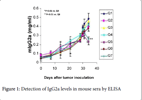
Figure 1: Detection of IgG2a levels in mouse sera by ELISA
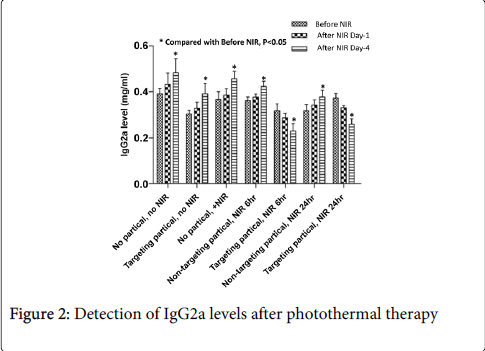
Figure 2: Detection of IgG2a levels after photothermal therapy
In order to investigate the growth of lymphoma further, A20 lymphomas were collected and comparison of their weight showed that it was consistent with IgG2a levels, except that the tumor weight of the Q5 and Q7 groups was significantly decreased (0.25 ± 0.06 g), while the tumor weight of the other experimental groups reached 0.45 ± 0.09 g (Figure 3).
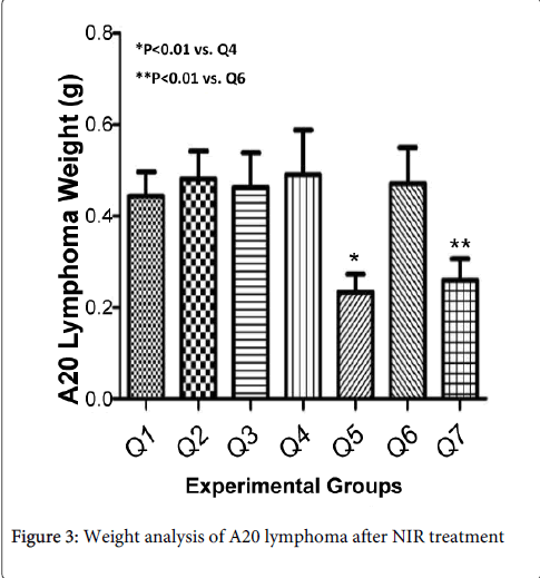
Figure 3: Weight analysis of A20 lymphoma after NIR treatment
Cancer is one of the main diseases that affect human health. There are 2.2 million new cases of cancer each year, and deaths caused by cancer are 1.6 million per year, making cancer the top cause of death in China. However, traditional treatments for cancer cannot achieve the goal of tumor eradication because of their side effects and limitations. In recent years, with the rapid development of nanotechnology, it provides a bright prospect for cancer treatment [1,20-25]. In particular, unique optical tunable gold nanomaterials show extensive and important potential in the field of biomedical application. They can be used as a carrier for drugs and biological molecules, as well as an active reagent for biological imaging and photothermal treatment in early diagnosis and treatment [2,26-30].
Most successful applications of nanoparticles for diagnosis and treatment depend on their aggregation at tumor sites [31,32]. The aggregation mainly depends on several molecular mechanisms. Conventionally, nanoparticles can passively aggregate at tumor sites by abnormal leakage of blood vessels around tumors. New angiogenesis is a major feature of tumors and the newly generated blood vessels around the tumor have an irregular shape and lumen diameter (up to 2 μM), which can allow macromolecules and nanoparticles in the blood vessels to be absorbed by the tumor [17,18]. However, nanoparticles without proper coating would be quickly metabolized in blood circulation. Therefore, chemical modification of nanoparticles can effectively improve their retention period in blood circulation.Recent research has shown that PEG-modified nanoparticles have a greatly improved retention period in blood circulation, enhancing their absorption by the tumor. Besides being passively absorbed by cancer cells, nanoparticles are able to recognize proteins or ligands expressed on the surface of tumor cells, facilitating their active and selective targeting of tumor cells [14-16]. For example, many solid tumors always abnormally overexpress HER2(human epidermalgrowth factor receptor-2), 5α-integrin receptors, or interleukin receptors. Conjugating an antibody to these proteins or ligands to nanoparticles will actively facilitate nanoparticle targeting of these tumor cells. Nanoparticles used for cancer killing can absorb near-infrared light so as to generate heat that will ablate tumor cells under laser radiation. In this study, anti-CD138 antibody was covalently conjugated to gold nanospheres and the conjugates would specifically target A20 lymphoma. With the application of photothermal therapy, gold nanoparticles treated by NIR laser radiation generate heat that is able to destroy tumors [33], achieving the purpose of treatment of the tumor.
In terms of the choice of target molecules, previous experiments showed that A20 lymphoma cells had an high affinity for anti-CD19, anti-CD20, and anti-CD138 antibodies, which reached 92.1%, 88.5%, and 95.2%, respectively (Figure 4). Anti-CD20 monoclonal antibody (rituximab) has been widely used in the treatment of B lymphoma and is currently one of the most successful monoclonal antibody drugs. However, it was found in our study that anti-CD138 antibody had a higher affinity for A20 cells than CD20, so anti-CD138 antibodies were chosen for preparation of antibody-conjugated gold nanospheres. Anti-CD138 antibody recognizes CD138 antigen, which is also called syndecan-1. CD138 is a transmembrane glycoprotein molecule that interacts with extracellular matrix proteins, cell surface molecules, and other soluble proteins [19]. It is also a plasma cell marker that can be used for recognition of B lymphoma and myeloma. The results of this study have confirmed that anti-CD138 antibody is a good targeting molecule for A20 lymphoma, and anti-CD138 antibody-conjugated hollow gold nanoparticles show great potential in clinical therapy.
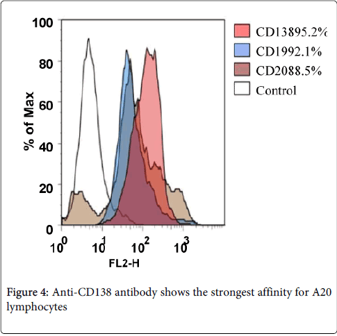
Figure 4: Weight analysis of A20 lymphoma after NIR treatment
Our studies raise a number of interesting questions that should be addressed in the future. For instance, it is important to confirm whether the suppression of A20 lymphomas will be sustained over time and whether sustained suppression will require repeated treatment, so long-term studies to evaluate the antitumor activity of targeted HAuNS in combination with near IR-region laser irradiation will need to be carried out to further confirm the short-term results obtained in the current studies. Using a small-molecular-weight(CD138) peptide as atarget and attaching it at the end of PEG chains, we found that receptor-mediated active targeting of lymphoma in vivo. Although our current study has shown promising results inselective photothermal ablation of lymphoma using targeted HAuNS, much work remains to be done to advance thistechnology further into the clinic. For example, more detailedpreclinical studies with regard to clearance, safety, and efficacy of targeted HAuNS need be documented.
We thank Dr. Chun Li for helpful discussions and Dr. Wei Lu for assistance with nanoshell preparation. This work was supported by the Priority Academic Program Development of Jiangsu Higher Education Institutions, the National Natural Science Foundation of China (81272476), and Science and Technology Support Program Project of Jiangsu Province (BE2010649, BE2011682).