Angiology: Open Access
Open Access
ISSN: 2329-9495
ISSN: 2329-9495
Clinical image - (2021)Volume 9, Issue 2
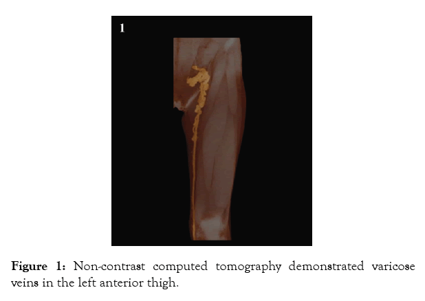
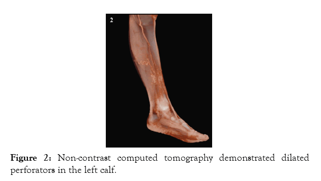
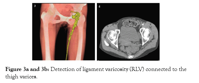
A 70-year-old woman presented with pain, cramps, and hyperpigmentation on her left leg. Dilated and tortuous veins were observed in the left lower extremity. Non-contrast Computed Tomography (CT) demonstrated varicose veins in the left anterior thigh (Figure 1) and dilated perforators in the left calf (Figure 2). In addition, Round Ligament Varicosity (RLV) connected to the thigh varices was also detected (Figure 3a and b). The precise locations of major perforators were intraoperatively marked using Duplex Ultrasound (DUS). The patient underwent saphenous vein stripping and ligation of two dilated perforators and RLV. Surgery remains the standard care for varicose veins, and an accurate evaluation is very important to determine the most appropriate procedure. DUS is considered the gold standard for evaluation of the varicose vein and is widely used to assess the anatomical site of reflux, as well as to quantify the amount of reflux. However, DUS has several limitations such as operator-dependent variable results and difficulty in evaluating pelvic vessels. CT venography is another powerful tool for evaluating varicose veins and can reveal unusual anatomic variations, including those in tributary veins and pelvic vessels. Our strategy for varicose vein surgery involve the use of non-contrast CT to create a three-dimensional (3D) vein map and to detect major perforators. In the present case, RLV was also revealed, and an adequate procedural planning was executed preoperatively. RLV is mostly found in young women, especially during pregnancy or the postpartum period but is rare in elderly women.Non-contrast CT is useful for the appropriate evaluation of varicose veins, as well as and for designing operations while providing clear 3D images and screening for possible co-existing abnormalities. The patient consented to the publication of this report.

Figure 1: Non-contrast computed tomography demonstrated varicose veins in the left anterior thigh.

Figure 2: Non-contrast computed tomography demonstrated dilated perforators in the left calf.

Figure 3a and 3b: Detection of ligament varicosity (RLV) connected to the thigh varices.
Citation: Tamagawa Y, Kawamura M, Tsuchida T, Monta O, Tsutsumi Y (2020) Non-contrast Computed Tomography Volume Rendered Imaging in an Elderly Woman with Round Ligament Varicosity and Lower Extremity Varicose Veins. Angiol Open Access. 9:239.
Received: 06-Nov-2020 Accepted: 16-Feb-2021 Published: 23-Feb-2021 , DOI: 10.35248/2329-9495.21.9.239
Copyright: © 2021 Tamagawa Y, et al. This is an open-access article distributed under the terms of the Creative Commons Attribution License, which permits unrestricted use, distribution, and reproduction in any medium, provided the original author and source are credited.