Journal of Hematology & Thromboembolic Diseases
Open Access
ISSN: 2329-8790
ISSN: 2329-8790
Review Article - (2014) Volume 2, Issue 5
Immunity is typically taken to be the defense that one organism projects against injury from another biological organism, such as infection or parasitization. However, the mechanisms of immunity against biological organisms are fundamentally the same as those that protect an organism from stress and injury of all sorts, including biological, physical and chemical. Any organism’s first line of defense against infection, or other insult, is a physical barrier that delineates self from non-self. Evolution has produced many systems to augment and repair barrier immunity when, inevitably, it is breached. Among these systems are molecules that exist pre-formed in the fluid phase and are instantly available to make temporary patches and initiate long-term healing. Serine protease cascades have arisen as a common mechanism for such systems. Beginning from a primordial molecular with mixed functionality including elements of coagulation, and coagulative targeting of foreign bodies including biological infection, distinct pathways have formed. The coagulation cascade, which helps to maintain the integrity of numerous tissues, including the vascular system, is among the more essential of these systems. Other phylogenetically derivative pathways have arisen, including innate immune systems, that play allied roles in the maintenance of organismal integrity, and which therefore interact extensively with the coagulation systems. In this sense the coagulation system and related, derivative pathways are molecular extensions of barrier immunity that are essential for the maintenance of homeostasis.
Keywords: Immunity; Coagulation; Homeostasis
A defining feature of living organisms is the capacity to defend themselves from their environment and to maintain the integrity of their own structures. Consequently, systems have evolved to perform these duties, which feature considerable overlap between host defense and maintenance of homeostasis. The more complex the organism, and the greater the range of environmental threats that organism faces, the more sophisticated the defense systems must be. For example, simple unicellular organisms have relatively simple systems based on small molecules, macromolecules and subcellular organelles. These systems serve primarily to maintain the cell from non-biological physiochemical environmental influences such as heat, cold, dehydration and mechanical stresses. More complex multi-cellular organisms, particularly those with central nervous systems to sense and respond to the environment, cardiovascular systems to circulate nutrients, and other assorted organ systems to support these functions, must have defense systems with much greater versatility. These defense systems must maintain the physiologic integrity of the much more complex organ systems, taking into account the requirements of specialized environments such as central nervous systems with selective blood/brain barriers and enteral tracts that harbor rich microbiomes essential to the health of the organism. Additionally, more complex organisms face more numerous biological threats ranging from viruses, unicellular prokaryotes and eukaryotes, and multicellular organisms ranging from parasites to predators.
A fundamental function of host defense systems is the establishment of a barrier to maintain the integrity and separation of self from non-self. These barriers apply across the scale of biological structures. Cell membranes define and protect subcellular organelles and cells. Cell walls protect cell membranes and give rise to structures that may define the organism, as in woody plants. In more complex organisms, epithelial cells form skin and its appendages such as scales, feathers and fur to protect interior spaces from the exterior environment. Within the body, specialized barriers consisting of endothelial layers define and protect organs systems such as the enteric tract, hematic and lymphatic vascular systems, and the central nervous system.
Barrier defense, or equivalently, barrier immunity, includes elements that function as fixed barriers, such as cell walls on the subcellular level and large organs such as skin formed by epithelium and supporting tissues and vascular systems formed by endothelial cells and supporting tissues. Barrier immunity in more complex organisms also includes mobile elements such as lymphocytes and macrophages, which may be both resident in tissues as well as more mobile within fluid phases such as blood and lymph.
Because fixed barriers are susceptible to injury and breach, an additional and critical element of barrier immunity in many organisms consists of soluble, fluid phase macromolecules that circulate, both within vascular and extravascular spaces. The hemostasis system, which in the fluid phase consists of a series of serine protease cascades, is the best characterized of these systems. In addition to the macromolecular fluid phase components, the hemostasis system also includes solid phase elements, such as vascular endothelium and acellular tissue matrices, and other larger cellular and subcellular fluid phase components, including platelets. These pathways have numerous control points regulated by inputs from a wide range of physiologic processes.
Barrier immunity is the first line of host defense against the environment, and for a given organism it is adapted to respond to the physical, chemical and biologic stresses that are typical for that organism. When elements of fixed barrier immunity, such as skin and cell walls, are damaged, repair must begin immediately. The response to this injury involves a variety of different systems that can be broadly considered elements of immunity, including hemostasis pathways, humoral immunity (that is, acquired immunity such as immunoglobulins) and innate immunity such as the complement pathways. Together the fixed and fluid immune elements act to restore the barrier defenses and repair damaged tissues. While these different elements of the immune system are frequently regarded as independently functioning systems, they have evolved in concert and have highly interdependent and coordinately regulated functions (Figure 1).
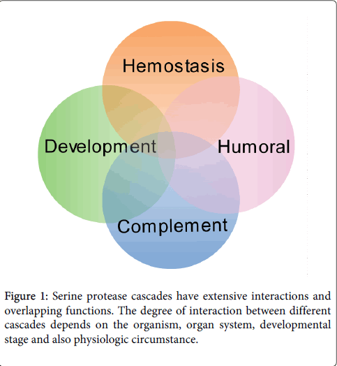
Figure 1: Serine protease cascades have extensive interactions and overlapping functions. The degree of interaction between different cascades depends on the organism, organ system, developmental stage and also physiologic circumstance.
Fluid immunity plays a critical role in sealing and repairing damaged elements of barrier immunity. Additionally, and perhaps equally essential, elements of fluid immunity are important regulators of both fluid and cellular elements of the immune systems. In performance of these functions, molecules of the fluid immune systems have three fundamental purposes: 1) Recognize and bind molecules and cells of the host system in order to maintain physical integrity and physiologic homeostasis; 2) Identify host structures, both cellular and molecular, as self; and 3) Recognize and remove non-self elements from the organism. There is necessarily extensive coordination between these functions, as well as overlap in activity within the relevant biological pathways. Fundamentally, these functions are also the same as those required for the developmental processes of organogenesis and ontogeny. These processes require the specification of patterning of tissues to create organs, and the patterning of organs to make organisms. This requires the organization of cells with appropriate polarity and organization, typically achieved by a combination of cell migration as well as programmed cell death. These processes require systems for establishing pathways for cell migration and for removing apoptotic cell debris, respectively. Consequently, deficiency in particular elements of fluid immunity that participate in these processes may result in severe developmental disorders, including embryonic lethality.
As evolution is fundamentally a process of derivation and variation, many of the sub-systems that perform immune functions share a common heritage, and in particular the serine protease cascades of hemostasis, complement and ontogeny are phylogenetically related [1]. A schematic depiction of the evolutionary development of these systems is shown in Figure 2. In light of this shared evolutionary heritage, it may be expected that there is considerable coordination of the functionality of these systems. In particular, these systems each contain elements that are capable of distinguishing self from non-self structures, with the latter including both damaged and transformed cells arising from injury and disease, as well as foreign materials, such as pathogens. There are multiple, inter-related regulatory mechanisms in these systems, and there are also many areas of interaction and coordinate regulation with cellular elements of these systems, including chemokines and cytokines. Although it is beyond the scope of this current discussion, it is worth noting that serine protease cascades have been adapted to many other homeostatic processes. Among the more prominent of these are the kallikrein pathways, which are involved in a wide range of homeostatic functions, for instance the maintenance of blood pressure by the kinin-kallikrein system [2].
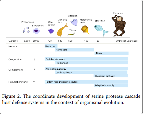
Figure 2: The coordinate development of serine protease cascade host defense systems in the context of organismal evolution.
The ability to distinguish self from non-self can be classified into two categories: Innate immunity and acquired, or adaptive, immunity. The innate immune system consists the complement pathways and a number of molecules that recognize specific molecular patterns. These may be carbohydrates, lipids, genetic material such as DNA and RNA, and some proteins motifs, and hence these are regarded as pattern recognition molecules. These pattern recognition molecules are pre-formed and are not altered in their sequence by the organism’s history of antigenic exposure. The acquired immune system recognizes specific epitopes, most commonly of proteins. In contrast to the relatively small number of pattern recognition molecules with a broad spectrum of the innate immune system, acquired immunity is capable of generating high affinity recognition molecules, including immunoglobulin and major histocompatibility complex proteins, in response to a vast number of epitopes. This ability to respond to antigenic exposure requires a period of time for the immune system to adapt, commonly on the order of weeks.
Adaptive immunity generates molecules that recognize specific antigens with very high affinity. The mechanisms whereby this specific recognition is translated to immune function, such as the elimination of foreign bodies or damaged self structures, are in many instances the same as the innate immune effector systems. These include cells, such as macrophages, and soluble factors that can be mobilized to destroy foreign cells, such as the membrane attack complex. These effector mechanisms provide the functional and regulatory links between acquired immunity and innate immunity. The concerted action of these systems demonstrates the lack of meaningful biological distinction between immune systems targeted to invading organisms and homeostatic systems that serve to patch and heal self.
Considering that fluid phase immunity can be broadly considered to include elements of the hemostasis system including coagulation cascades, the innate immune system, and the acquired immune system, some consistent features are: 1) Phylogenetic conservation; 2) Present in states of normal health; and 3) Available for immediate neutralization of injury or physiologic derangement (given prior antigenic exposure for development of acquired immunity). In particular, proper control of hemostasis requires that numerous factors are present in circulation and on cell surfaces. These factors can be instantly mobilized in case of injury, and may induce formation of a fibrin/platelet clot as well as resolve such clots following adequate repair of the underlying tissue injury.
For many elements of fluid phase immunity, an additional feature is notable: They are required for proper embryological patterning and development. In particular, coagulation enzymes and pattern recognition molecules of the innate immune system demonstrate this property. Certain deficiencies in these systems result in developmental abnormality, including lethality [1].
Hemolymph is a primordial precursor of the hematic and lymphatic circulatory systems that are found in a wide variety of more complex organisms. For example, the horseshoe crab, a living fossil of sorts, has a hemolymph system that dates back approximately 450 millions years ago. Horseshoe crab hemolymph contains serine protease cascades with coagulation activity. In addition, this hemolymph also contains pattern recognition molecules that bind to α-glucans found in many fungi and lipopolysaccharides found in many bacteria [3]. When bound to specific ligands, these pattern recognition molecules also activate the coagulation cascades, providing a host defense against invading and infecting organisms. While the effector mechanisms are different, there are homologous proteins in Drosophila and mammals, as discussed by Krem and colleagues [1,4]. For example, Drosophila has a serine protease cascade that functions in dorsal-ventral patterning and another serine protease cascade that functions in immunity and coagulation, with significant homologies between these cascades and to coagulation and complement cascades in higher order organisms, including mammals. Similarly, there are specific pattern recognition molecules in horseshoe crab hemolymph, such as tachylectins, ficolin homologs that are found in more primitive organisms such as Ascidians (sea squirts) as well as other invertebrate marine mammals [5]. Ficolins are found in mammals including primates, in which they appear to function primarily as elements of the innate immune system, although they also have demonstrated some effect on coagulation [5,6]. Consequently, although complement and coagulation are regarded as distinct systems, it might be predicted based on the shared evolutionary heritage and on the overlapping function that there may be substantial interaction and coordinate regulation.
Several studies, including our own, have shown that mannose-binding lection associated serine protease (MASP) activates complement cascades through the lectin pathway [7-9]. Additionally, we have shown that MASP also has a thrombin-like activity, resulting in fibrin deposition following blood vessel injury and exposure to certain pathogens [7]. There are three enzymatically functional MASPs, which form complexes with ficolins in circulation [6]. Deficiency in one of these MASPs, MASP1, results in small body size in mice and other developmental abnormalities in humans and zebrafish [10].
The functional and evolutionary versatility of the combination of pattern recognition molecules and serine proteases is further revealed by additional examples. For example, zebrafish have a pattern recognition molecule, Collectin Kidney 1 (CLK1, also known as COLEC11) that forms a complex with MASP. This CLK1/MASP complex subsequently plays a role in the determination of polarity in development and in neurons [11].
In addition to a common evolutionary heritage of multiple serine protease cascades, there is also biological evidence for a coordinate regulation and a degree of overlapping activity for some of these systems. For example, knockout mice that are null for certain coagulation factors, and in particular thrombin activators, have a high degree of embryonic lethality or significantly impaired development for those animals that survive birth [12]. In contrast, the genetic absence of complement components and their regulatory factors generally results in a less severe phenotype, in some cases including no evident abnormality under normal physiological conditions [13-16]. However, when challenged with a specific physiologic stress, such as ischemia reperfusion injury or infection, complement knockout animals may develop a more severe phenotype, including derangements of hemostasis in addition to defects in the affected complement pathways [13-18]. These observations support the idea that the more primitive serine protease cascades of the coagulation system may be the primary fluid phase barrier immunity system, while the complement pathways may both modify the hemostasis systems by means of related functions and coordinately regulated activities. Phylogenetic analysis of serine proteases in these pathways has revealed common ancestry and related functional domains, providing some insight into the coordinated function and regulation of these pathways [1,4,19].
Barrier immunity as a homeostatic mechanism
Barrier immunity is essential to maintaining the integrity of an organism in the face of physical, physiologic and biological threats. The fixed, structural elements of barrier immunity are supported by fluid phase elements, which include the coagulation system, complement pathways and a subset of the acquired immune system. These fluid phase elements are pre-formed and instantly ready to respond to acute injury. The coagulation and complement pathways comprise cascades of serine proteases that share a common evolutionary heritage. Consequently there is both considerable overlap in their function and regulation, which interact extensively with other cellular and fluid phase elements of the immune system. Of these systems, the hemostasis pathways may serve a primary role, including hemostasis, repair of barrier immunity and first response to invasive pathogens. The complement pathways, in addition to their role in the identification of non-self structures, supplement and modify the coagulation system, which in turn supplements and modifies the function of the complement pathways. Taken together, these immune systems function to maintain homeostasis. This is achieved through a combination of balancing opposing processes, such as thrombosis and thrombolysis as well as inflammation and resolution, and by directly regulating other homeostatic processes, such as tissue and organ patterning, apoptosis and clearance of necrotic debris, and maintenance of blood pressure.
While these systems have been extensively investigated, they have traditionally been regarded primarily as independent processes. A more complete understanding of the mechanisms underlying immunity and maintenance of homeostasis will be achieved through the investigation of the coordinated function and regulation of these systems.