
Journal of Clinical and Experimental Ophthalmology
Open Access
ISSN: 2155-9570
ISSN: 2155-9570
Case Report - (2025)Volume 17, Issue 4
Strabismus is often associated with amblyopia. The prevalence of amblyopia is variable in different studies ranging from 2.0% to 2.5% in the general population. Amblyopia refers to decreased visual acuity in one or sometimes both eyes due to abnormal binocular interaction during visual maturation without any pathological changes. Amblyopia causes ten times greater monocular visual impairment as compared with other causes of visual impairment in children like ocular pathologies and trauma. Amblyopia has a greater impact on psychological and socio-economics including poor academic performance as amblyopia causes a reduced perception which impairs learning. Constant strabismus causes a greater degree of suppression compared with alternating strabismus or anisometropic (different refractive error between the eyes) amblyopia.
Anisometropic; Amblyopia; Strabismic; Proparacaine; Binocular
The non-dominant eye is suppressed to abolish diplopia and binocular rivalry followed by amblyopia. Strabismic amblyopia does not limit itself to the ocular level, the orientation and response of the cortical fibers change according to the response of the dominance between the eyes at the V1 level [1]. Diagnosis of strabismic amblyopia can be done by test-retest of visual acuity, detection of strabismus in cover test, detailed observation to rule out the presence of ocular pathologies, and cycloplegic refraction for each patient. After diagnosis our primary professional and ethical duty is to provide management but for strabismic amblyopia no preferred guideline is available for a child older than 7 years of age [2]. Strabismic amblyopia is associated with vision loss of the non-dominant eye and the loss of stereoacuity. Present available standard management strategies to improve visual acuity for strabismic amblyopia is occlusion of the dominant eye with standard refractive correction. However, it fails to gain the stereoacuity as binocular participation is necessary to stimulate or reactivate the cells responsible for binocular vision mainly present in the lateral geniculate nucleus. Dichoptic therapy has shown its effectiveness with partial dominant eye patching therapy. Binocular participation through different levels of contrast uses the active involvement of the neural cells of the brain which can accelerate the prognosis for a child outside the sensitive period for visual development [3].
This unmasked prospective case report evaluates the effect of an alternative approach to manage strabismic amblyopia based on monocular fixation in binocular field, dichoptic training and accommodation and vergence training. The effect of our therapy is measured as improvement in visual acuity and stereoacuity for distance and near.
A ten-year-old boy presented to our hospital complaining of blurred vision in his right eye noticed two days ago when he suddenly closed his left eye while playing. Parents gave a history of inward deviation of the same eye since early childhood. He was born by vaginal delivery after a normal pregnancy with normal prenatal, perinatal and postnatal medical history. No one in their family, including their second child, has manifest deviation. On examination, his unaided Snellen’s visual acuity for distance was measured as 20/60 (0.50) in the right eye and 20/25 (0.10) in the left eye. Objective refraction and un-dilated retinoscopy revealed findings of 0.00/-1.50 × 180° in the right eye and 0.00/-0.50 × 180° in the left eye. The Hirschberg test indicated a 30° esotropia and there was -1 abduction deficit in both eyes. Worth four-dot testing was positive for suppression of the right eye at both distance and near. Randot stereo acuity was <400 seconds of arc. Modified Krimsky was performed (as the child was not able to maintain fixation for the cover test), which revealed a 70-prism diopter esotropia at both distance and near. The pupillary examination revealed equal, round, and reactive pupils with no afferent pupillary defect noted. Slit lamp examination of the anterior segment revealed a normal eyelid for both eyes. Upon everting the upper lid, the conjunctiva was normal for both eyes. Normal eyelashes were noted in both eyes, with all puncta patent and appearing normal. The bulbar conjunctiva appeared clear and quiet, and the sclera appeared white in both eyes. The cornea was intact and clear in all layers, with flat and dark brown irises. The anterior chamber was deep without cells or flare, and open angles were estimated at 4+ using the Von Herrick method for both eyes. One drop of Proparacaine 0.5% and a sterile fluorescein strip soaked with one drop of saline water were applied to the inferior conjunctiva in both eyes before intraocular pressure measurements. Goldmann application tonometry at 9:15 A.M. measured 10 mmHg OD and 09 mmHg OS.
Refractive error was confirmed following the administration of 1% cyclopentolate administered twice at 15-minute intervals. After 40 minutes, retinoscopy was conducted using a Heine Beta 200 retinoscope. Subsequently, post-cycloplegic autorefractometry was performed with the Potec PRK-7000 autorefractor. The retinoscopy values, after deducting tonus allowance (+0.75 D) and working distance (+1.50 D), for the right and left eyes were respectively +0.50/-1.50 × 180 and +0.25/-0.50 × 180. The autorefractor showed a refractive error of +0.50/-1.50 × 180 and +0.25/-0.75 × 180 for the right and left eyes, respectively. Upon full dilatation, an examination of the posterior segment was conducted using a slit lamp with a 90D lens and a binocular indirect ophthalmoscope with a 20D lens. The crystalline lens and vitreous were optically clear in both eyes. The optic nerves exhibited distinct margins and pink, healthy rim tissue, with cupping graded at 0.2 horizontally and vertically for both the eyes. The macula appeared flat and even with a healthy appearance to the posterior pole in both eyes. Blood vessels showed normal caliber and contour, and the peripheral retina was observed to be flat and intact 360 degrees in both eyes.
Based on the clinical presentation the diagnosis was made of strabismic amblyopia in the presence of large angle esodeviation. The diagnosis of anisometropic amblyopia was excluded as the amount of anisometropia was less than -2.50 diopter, meridional amblyopia was excluded in the absence of mild astigmatism for the right eye, ocular media were visible so stimulus derivational amblyopia was ruled out and there was no discernible hypermetropia at the cyclo-refraction so accommodative esotropia was also ruled out. At this visit, we advised his parent to do Magnetic Resonance Imaging (MRI) to rule out any IntraCranial-Space-Occupying-Lesion (ICSOL) and for detection of any anomalies in the 6th or abducens cranial nerve pathway.
The patient returned one week later with the neuro-imaging report. It revealed no evidence of any ICSOL and bilateral cisternal segments of the sixth nerves appeared normal. The post-mydriatic test was conducted at this visit, and any necessary adjustments to the eyeglass power were finalized. Eyeglasses were prescribed with a final acceptance of 0.00/-1.50 × 180,20/60 (0.50, N8) OD and 0.00/-0.50 × 180, 20/20 (0.00, N6) OS.
At the visit, we advised his parent of the need for strabismus correction surgery followed by usage of the eyeglasses. On the very next day, bilateral medial rectus recession of 7.00 millimeters along with right eye 11-millimeter lateral rectus resection was performed under general anesthesia [4]. The next day we evaluated only the Hirshberg reflex demonstrated in Figure 1 which revealed a centered corneal reflex. We asked his parents to return for in one week for reassessment. At the reassessment, vision with his eyeglasses remained the same as pre-surgery, OD 0.00/-1.50 × 180, 20/60 (0.50, N8) and OS 0.00/-0.50 × 180, 20/20 (0.00, N6). Hirshberg test OD revealed a central corneal reflex with mild abduction limitation during extraocular motility testing. Cover test measurement was 6 prism diopters exophoria. Worth four dot test for distance and near showed right eye suppression and Randot stereo acuity was <400 secs.
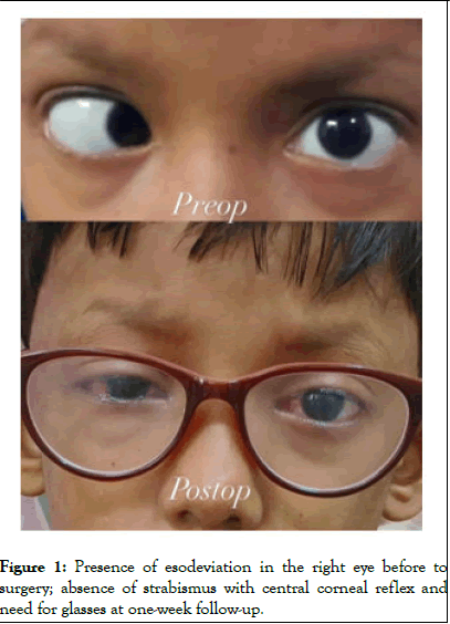
Figure 1: Presence of esodeviation in the right eye before to surgery; absence of strabismus with central corneal reflex and need for glasses at one-week follow-up.
We advised the patient to patch the non-amblyopic eye daily for three hours while performing near activities like drawing at home. Follow up was scheduled in 8weeks. After 8 weeks the child came to us with no improvement in visual acuity. It remained the same at OD 20/60 (0.50, N8) and OS 20/20 (0.00, N6). We repeated the objective refraction and it remained OD 0.00/-1.50 × 180 and OS 0.00/-0.50 × 180. Worth four-dot testing and Randot stereo acuity showed no improvement with right eye suppression and <400 secs of arc stereopsis. When we asked the parents, they reported the child was not wearing his eyeglasses and there was non-compliance with the patching therapy. We then advised daily office-based vision therapy for two hourly sessions with one being from 9 am to 10 am and another from 4 pm to 5 pm.
Our treatment strategy focuses on improving monocular visual acuity followed by recovering binocularity in the non-amblyopic state. The treatment comprises a framework to treat the amblyopic eye and additional brain activation activities to reactivate visual processing. Our methodology takes this first step towards enhancing monocular visual acuity. We can practice it as tracing, tracking, searching, and segregating. Such activities can be used to work on the basics before moving to more sophisticated binocular activities like focusing and object processing. The Sanet Vision Integrator (SVI) is a useful therapy device as you can adjust contrast differently in each eye to help stimulate the amblyopic eye [5,6]. With increased contrast before the amblyopic eye and weaker contrast before the nonamblyopic eye, the neural pathways of the weaker eye are more activated, contributing immensely to improving visual acuity of the amblyopic eye. A child needs to actively participate in this key component of therapy for it to work. This style of practice engages the child, ensuring their loyalty to the therapy and the positive outcomes of treatment.
Daily therapy sessions are essential for improving visual acuity and for the use of perceptual therapy to train the brain to better use the non-amblyopic eye and hasten the brain’s acceptance of using the amblyopic eye more actively. Perceptual skill predicts whether an activity will build a stronger neural structure in brain regions related to vision. We introduce training in accommodation and convergence by simple means such as the accommodative flipper (± 2.00) and Brock string. These techniques are designed to improve the focus power of the eye and make harmony with both eyes [7]. Binocularity is promoted by the dichoptic principle, in which various images are shown to each eye through the red-green or blue screens. The technique is reported in several studies to train the eyes to cooperate and circumvent the suppression of the amblyopic eye. A total of 30 hours of therapy was given and pre- and post-therapy values are demonstrated in the below Table 1.
|
|
Pre-therapy |
Post-therapy; After 30 hours over 15 days |
|
Best-corrected-visual-acuity |
Amblyopic eye (OD): Distance acuity-0.50; Near acuity-0.14 |
Amblyopic eye (OD): Distance acuity-0.2; Near acuity-0.11 |
|
Fellow eye (OS): Distance acuity-0; Near acuity-0.11 |
Fellow eye (OS): Distance acuity-0; Near acuity-0.11 |
|
|
Refraction |
OD: 0.00/-1.50 × 180 |
OD: 0.00/-1.50 × 180 |
|
OS: 0.00/-0.50 × 180 |
OS: 0.00/-0.50 × 180 |
|
|
Worth-four-dot test |
Distance and near OD suppression |
Distance and near fusion present |
|
Stereopsis |
<400 secs-of-arc |
120 secs-of-arc |
Table 1: At baseline and after 30 hours (15 days) of active vision therapy, the best-corrected visual acuity at both distance and near, refraction, worth four-dot testing for both distance and near, and stereopsis.
At the end of fifteen days of office-based therapy, (30, 1-hour sessions) we moved to a home-based therapy program including the use of a Hart chart, Marden ball, anti-suppression red-green glasses, and a bar reader for 1-hour daily activities. We asked the parents to have him return in 4 weeks. After four weeks no further improvement and even some deterioration was noted. We decided to stop home-based therapy by tapering it from daily the first week to one less day each week (6-5-4-3-2-1 day each week) until it was stopped after seven weeks. We followed up with the patient after 12 weeks at which time the stereopsis and best corrected visual acuity remained the same. Proper monitoring of the home-based therapy was done with twice weekly video calls to the patient’s parents and having the family maintain a daily dairy of the therapy performed.
The result of our study suggests that supervised office-based vision therapy using monocular fixation in the binocular field, dichoptic therapy, vergence and accommodative training improves visual and stereo acuity. Standardized treatment for amblyopia focuses on the improvement of monocular acuity but our approach involves both eyes and focuses on the establishment of binocularity and enhancement of stereopsis. Post-strabismic surgery it is rare to have improved stereoacuity as motor fusion is established but sensory fusion can only be reestablished by the equal engagement of both eyes. Binocular dysfunction is the primary challenge and monocular function is the secondary consequence of strabismic amblyopia. The depth of suppression is deeper for strabismic amblyopia compared with another form of amblyopia due to false projection of the retinal image [8]. After gaining binocularity, it is rare for the improved visual acuity to deteriorate or regress in the amblyopic eye. As conventional patching therapy, in our case, failed due to poor compliance, we applied active vision therapy which facilitated the visual recovery and reduced the suppression for the amblyopic right eye. Active office-based therapy involves engaging children in interactive and stimulating exercises oractivities to achieve therapeutic goals while minimizing resistance. We followed the case after one month of no intervention, and there was no deterioration of best corrected visual acuity as well as in binocular vision parameters including stereopsis, demonstrating the effectiveness of our combined treatment strategy for this case. Table 2 describes therapy protocol and Table 3 describes several vision therapy activities with goal and description.
| Therapy days 1 to 5 |
| 1. Amblyopia iNet according to his vision –Find the target (MFBF) |
| 2. SVI eye-hand–proactive, rotator 1 and 2 (under 100% contrast) |
| 3. Marsden ball, drawing with sound eye patching |
| 4. Tracing on the whiteboard – pursuit therapy |
| 5. Synoptophore with Haidinger’s brush for OS 15 min |
| 6. Beading activities |
| Therapy days 6 to 10 |
| 1. SVI Rotator 3, eye-hand (under 60% contrast) |
| 2. Amblyopia iNet-letter jump, laser ball, follow the letter |
| 3. Pegboard for eye-hand coordination with balance board |
| 4. Saccades therapy with the saccadic fixator |
| 5. Hart chart |
| 6. Synoptophore with autoflash on for OS |
| Therapy Days 11 to 15 |
| 1. Pursuit with tracing, saccades with SVI |
| 2. Amblyopia iNet increased difficulty, focused on traffic jams and capturing target |
| 3. Cheiroscope – cheiroscopic tracing with picture before the dominant eye |
| 4. Anti-suppression therapy - bar reader with red and green filter with 20/40 print |
| 5. Dart throwing with dominant eye patched |
| 6. Brock sting with accommodative flipper +/- 1.50 |
Table 2: Therapy protocol for 15 days.
| Name of the task | Goal | Description |
| Amblyopia iNet | Saccades, pursuit, accommodative training, finding target of different stimulus through monocular fixation in binocular field | Finding a target using saccades, and pursuits. Whole brain is involved as both the dorsal and ventral streams are activated. |
| Sanet Vision Integrator | Anti-suppression therapy under variable contrast | Different contrast is presented to each eye, follows the principle of dichoptic therapy which influences the reactivation of binocular neural pathway for regaining binocularity. |
| Cheiroscope | Tracing geometrical figure or images under binocular condition | Enhance concentration and eye hand coordination. |
| Rotating Pegboard | Dynamic visual acuity enhancer | Dynamic training of the amblyopic eye, the level of difficulty for a visual task can be increased, representative of real-world experience. |
| Brock string, Accommodative flipper, Hart chart | Awareness of physiological diplopia, Changes of focusing ability | Binocularity training must include both accommodation and vergence, Hart chart facility at distance and near viewing, Accommodative flipper to increase the amplitude of accommodation and enhance accommodative facility, Brock string to maintain binocularity by the awareness of physiological diplopia. |
| Bar reader with red and green filter | Anti-suppression therapy | Binocular participation is mandatory to complete the task. |
Table 3: Vision therapy activities with goal and description.
Amblyopia is a brain-origin disorder, and to address this, our therapy approach involves not only the visual cortex (colour, contrast, visual field, visual acuity) but also the frontal lobe (executive function, problem-solving, organizing, decisionmaking) and temporal and parietal lobes respectively by tasks involving laterality, and number and letter recognition in variable sizes in different loci. Mezad Kourash, Kelly, Li, et al. showed the effectiveness of active vision therapy compared with patching therapy and they have suggested that active vision therapy accelerates the visual recovery and psychological acceptance of vision therapy due to active participation of the child. Functional MRI results suggest that striate area V1 and extrastriate area V2 are anomalous in amblyopia [9]. The strongest features of strabismic amblyopia are positional uncertainty and distorted spatial perception which can be managed by strabismic correction through surgical management but reactivation of V1 and extra striate areas can only be done by the active involvement of the amblyopic eye under binocular conditions. Improvements in Pattern Visual Evoked Potential (PVEP) amplitude and P100 delay show how dichoptic training affects the cortical networks that process visual information and enables two-eye interaction through varied contrast.
This case emphasizes that patients with strabismic amblyopia can benefit from active vision therapy to maximize monocular visual acuity, sensory fusion and binocularity. Modifying the traditional approach to amblyopia therapy with the addition of active vision therapy can allow for better management of binocular vision disorders with recovery that includes restoration of a high level of binocular vision and stereopsis. Vision recovery is both better and faster with active vision therapy. Further research is required to study strabismic amblyopia with a greater sample size to determine predictable visual prognoses in different types of amblyopia. Our case showed restoration of binocularity with 30 hours of active vision therapy over 15 days combined with spectacle correction.
Nil.
All authors have declared that they have no financial relationships at present or within the previous three years with any organizations that might have an interest in the submitted work.
[Crossref] [Google Scholar] [PubMed]
[Crossref] [Google Scholar] [PubMed]
[Crossref] [Google Scholar] [PubMed]
[Google Scholar] [PubMed]
[Crossref] [Google Scholar] [PubMed]
[Crossref] [Google Scholar] [PubMed]
[Crossref] [Google Scholar] [PubMed]
Citation: Chakraborty S, Parida S, Hossain S, Hajira R (2026) An Alternate Method of Managing Strabismic Amblyopia-A Case Report. J Clin Exp Ophthalmol. 17:1052.
Received: 24-Nov-2024, Manuscript No. JCEO-24-35398; Editor assigned: 27-Nov-2024, Pre QC No. JCEO-24-35398 (PQ); Reviewed: 11-Dec-2024, QC No. JCEO-24-35398; Revised: 15-Jul-2026, Manuscript No. JCEO-24-35398 (R); Published: 22-Jul-2026 , DOI: 10.35248/2155-9570.26.17.1052
Copyright: © 2026 Chakraborty S, et al. This is an open-access article distributed under the terms of the Creative Commons Attribution License, which permits unrestricted use, distribution, and reproduction in any medium, provided the original author and source are credited.