Journal of Clinical and Experimental Ophthalmology
Open Access
ISSN: 2155-9570
ISSN: 2155-9570
Short Communication - (2021)Volume 12, Issue 5
Aid to Fixation
At the start of a surgical case under topical anesthesia, patients are usually asked to look at a microscope light prior to the initial incision. Most microscopes have a few illumination bulbs making it difficult for patients to find a single fixation point. The use of a single red fixation light makes it easier for patients to find a fixation point and decreases the chance of a wandering eye especially for the initial incision.
Centration of Capsulorehexis
If the patient fixes on the fixation light the light reflex can be used to guide the surgeon in centering the capsulorehexis. This enhances the chance of the surgeon performing a 360-degree symmetric peripheral overlap of the implant with the anterior capsule and centration of the implant on the visual axis. If the white rings are also projected on to the eye this can aid the surgeon with the sizing of the capsulorehexis [1].
Centration of an Intraocular Implant
After insertion of an intraocular lens implant and with the patient instructed to gaze on the fixation light, the lens implant position can be adjusted so it is centered on the corneal light image. This allows the implant to be positioned over the visual axis. There is evidence that this can enhance the quality of vision especially in aspheric, toric, and multifocal implants [2-5]. The use of one-piece acrylic implants allows the surgeon some flexibility in centering the optic of the lens. This is in contrast to 3-piece implants, both silicone and acrylic materials, or plate lenses in which the position of the optic cannot be modified.
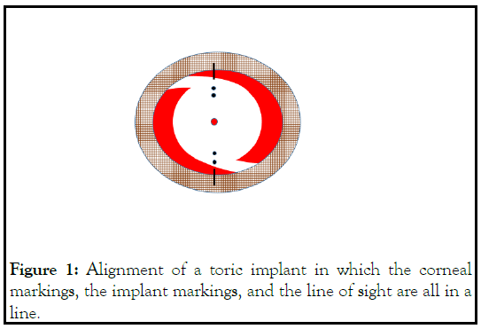
Surgeons typically mark the cornea prior to insertion of a toric implant. After the toric implant is inserted and aligned to the corneal markings the surgeon can determine if the implant is centered on the visual axis by using the reflected corneal image from the fixation light. The corneal markings, the toric implant marks, and the corneal fixation light reflex should all line up (Figure 1). If the corneal markings are off and not positioned over the visual axis, the surgeon can usually adjust the position of the implant so it is centered on this line. This results in the implant markings being equal distance from the corneal markings (Figure 2). The degree of angle kappa will often determine the success rate in adjusting the optic of the implant to the visual axis. It may be difficult to perfectly align the optic with the visual axis in eyes with a high angle kappa.

Figure 1: Alignment of a toric implant in which the corneal markings, the implant markings, and the line of sight are all in a line.
Figure 2: Implant markings being equal distance from the corneal markings.
• Initial misalignment of a toric implant from the line of sight in that the corneal markings and implants marks are not aligned with the visual axis.
• Adjustment of the position of the implant so the implant marks and visual axis are in alignment. The markings of the cornea and the implant are equal distance apart.
The use of a patient fixation light on a surgical microscope can allow for improved patient fixation and enhances the ability of a surgeon to perform a capsulorehexis and align the center of an implant with the visual axis. Alignment of intraocular lens implants especially multifocal lenses on the visual axis may reduce glare and halos but is not fully supported in the literature. Studies confirm that patients with high angle kappas and multifocal implants have a tendency to be less satisfied with their outcome [4]. Adjusting the position of an implant to be on or closer to the visual axis may improve patient satisfaction levels. There are however a number of factors that can influence the final resting position of an implant other then intentional decentration towards the visual axis. These factors include capsular contraction, memory of the haptics and IOL rotation.
LASIK studies have found that decentration of an excimer ablation over the corneal light reflex in patients with high angle kappas can provide excellent outcomes in terms of safety, accuracy, induced astigmatism, contrast sensitivity, or night vision disturbances [6-8]. Further research is required to guide surgeons’ approach to angle kappa and the results of intentional decentration of an implant on the visual axis. Nevertheless, there is some evidence that centering of an implant on the visual axis can improve the quality of vision by decreasing the induction of higher-order aberrations. This relatively inexpensive device can be a valuable tool in performing surgery under topical anesthesia.
Citation: Stein R (2021) Advantages of a Microscope Fixation Light for Topical Intraocular Surgery. J Clin Exp Ophthalmol. 12:p371.
Received: 22-Apr-2021 Accepted: 27-Aug-2021 Published: 06-Sep-2021 , DOI: 10.35248/2155-9570.21.12.888
Copyright: © 2021 Stein R. This is an open-access article distributed under the terms of the Creative Commons Attribution License, which permits unrestricted use, distribution, and reproduction in any medium, provided the original author and source are credited.