Journal of Clinical and Experimental Ophthalmology
Open Access
ISSN: 2155-9570
ISSN: 2155-9570
Case Report - (2020)Volume 11, Issue 3
Rhabdomyosarcoma is a malignant tumour of mesenchymal origin with an aggressive pattern of growth. Its occurrence is quite rare in children and adolescents, and even rarer in adults. We discuss the case of a young adult female diagnosed with embryonal rhabdomyosarcoma.
Rhabdomyosarcoma; Proptosis; Adult
Rhabdomyosarcoma is a rare soft tissue malignant neoplasm which arises from the undifferentiated mesenchyme and histologically resembles the normal fetal skeletal muscle before innervation [1]. Four histological varities of rhabdomyosarcoma are: - embryonic, alveolar, botryoidal and pleomorphic. Of these, 60% are embryonal type, which have predilection for young children [2]. Rhabdomyosarcoma is a rarely occurring neoplasm of the childhood and therefore its occurrence in an adult makes it further rare tumour accounting for 3% of soft tissue sarcomas, which themselves have a prevalence of less than 1% [3].
According to the literature reports, Rhabdomyosarcoma has an estimated incidence of 4.3 million cases per million occurring in the head and neck region and of these 10% occur in the orbit [4,5].The prevalence is more in males as compared to females [2].
The main Head and Neck sites include-orbit, parameningeal sites (nasopharynx, nasal fossa, para- nasal sinuses, infratemporal fossa, pterygoid fossa, middle ear and mastoid) and non-parameningeal sites (scalp, face, parotid, oral cavity, oropharynx, larynx and neck). Tumours which are arising from the parameningeal sites tend to invade the cranial cavity via basal skull foramina and are therefore having the worst prognosis [2].
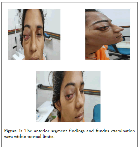
A 26 years old female presented to our ophthalmic OPD with sudden onset, paraxial proptosis of the right eye (RE) with lid swelling since 5 days associated with ipsilateral periorbital pain of 15 days duration. Patient denied history of ocular trauma, surgery or any other systemic illness. On further probing, patient mentioned about the symptoms of intermittent episodes of nasal blockage along with occasional nasal bleed for the past 4-5 months aggravated since last 2 weeks for which no previous consultation was taken. Ocular examination of the affected eye (RE) revealed best corrected visual acuity of finger counting at 4 metres with total ophthalmoplegia. Anterior segment findings revealed conjunctival congestion with chemosis along with sluggish pupillary reaction. Fundus examination revealed disc oedema with tortuous vessels. In the Left eye (LE), the unaided visual acuity was 6/6. The anterior segment findings and fundus examination were within normal limits (Figure 1).

Figure 1: The anterior segment findings and fundus examination were within normal limits.
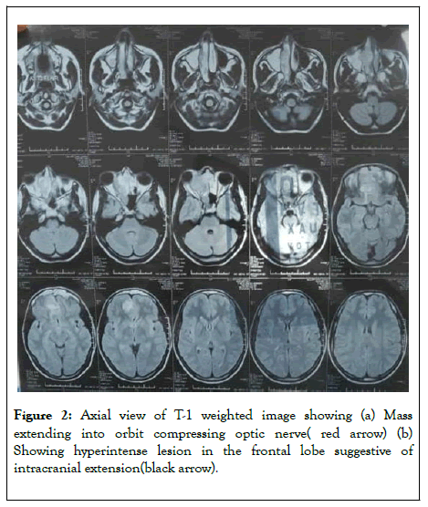
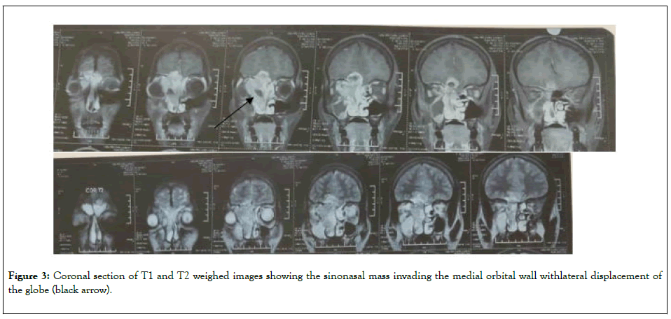
MRI revealed a multi-lobulated sino-nasal mass which was isointense on T1and hyperintense on T2 weighted images. It was invading the surrounding structures like the frontal sinus, ethmoidal sinus and the olfactory recess. Intracranial extension was seen through base of the skull (Figures 2 and 3).

Figure 2: Axial view of T-1 weighted image showing (a) Mass extending into orbit compressing optic nerve (red arrow) (b) Showing hyperintense lesion in the frontal lobe suggestive of intracranial extension(black arrow).

Figure 3: Coronal section of T1 and T2 weighed images showing the sinonasal mass invading the medial orbital wall withlateral displacement of the globe (black arrow).
Soft tissue punch biopsy specimen taken from the right nostril showed round hyperchromatic cells with oedematous stroma which favoured the diagnosis of rhabdomyosarcoma-embryonal type. Diagnosis was confirmed with immunohistochemistry. Patient was referred to the oncology department of atertiary care center for further management. Unfortunately, the patient could not be saved.
RMS is an uncommon malignancy of childhood comprising about 4% of total pediatric malignancies [1]. Its occurrence in adults is extremely rare. It is a soft tissue sarcoma with a prevalence of 3% and among these incidence of RMS is around 1% [1]. Adult RMS differs from childhood RMS in terms of natural history, response to treatment and prognosis. Due to differences in the morphological structures in adults, the response and prognosis of RMS to therapy in adults is not good.
Around 40-50% RMS involve head and neck region. In the head and neck region, it can either arise from parameningial sites (nasal fossa, nasopharynx, paranasal sinuses, middle ear) ornon paramengial site (oral cavity, oropharynx, larynx and neck). RMS arising from parameningial sites have a propensity to spread intracranially and have the worst prognosis [2,3]. There are four subtypes:- alveolar, embryonal (most common), pleomorphicand botryoid. In adults, the most common type seen is alveolar, which has the worst prognosis.
RMS invading orbit can present in the form of proptosis, lid mass or an orbital mass mimicking orbital cellulitis, lymphangioma, hemangioma, metastasis to orbit, lymphoma, dermoid cyst, or chalazion [6,7]. In adults, RMS shows increased tendency to invade cranial cavity even after treatment with radiotherapy and chemotherapy [6] and therefore the unfavourable prognosis. A multi-displinary approach is mandatory in such cases.
Staging of rhabdomyosarcoma according to intergroup rhabdomyosarcoma study (IGRS) GROUP:
• Localized disease, tumor resected completely, regional lymph nodes not involved.
• Localized disease with microscopic residual disease or regional disease with or without microscopic residual disease.
• Incomplete resection with gross residual disease.
• Metastatic disease.
These guidelines for staging help in deciding the management protocol which could be in the form of combination therapy (radiotherapy, chemotherapy and sometimes surgical debulking). Metastasis occurs by hematogenous or lymphatic route. The most common sites are lungs, bones and brain [8].A thorough systemic evaluation for metastasis should be performed. The prognosis is influenced by the anatomic location at the time of presentation, patient's age, completeness of surgical resection of the mass, extent of metastasic disease and tumor histology [9,10].
In this case, even though the patient seeked immediate medical advice for her proptosis along with timely referral to a tertiary care center, due to the poor survival rates of RMS in adults, she succumbed.
Citation: Sharada S (2020) Adult Rhabdomyosarcoma: A Rare Case Report and the Associated Challenges. J Clin Exp Ophthalmol. 11:838. DOI: 10.35248/2155-9570.20.11.838
Received: 27-Apr-2020 Accepted: 11-May-2020 Published: 18-May-2020 , DOI: 10.35248/2155-9570.20.11.838
Copyright: © 2020 Sharada S. This is an open-access article distributed under the terms of the Creative Commons Attribution License, which permits unrestricted use, distribution, and reproduction in any medium, provided the original author and source are credited.