
Journal of Clinical Trials
Open Access
ISSN: 2167-0870

ISSN: 2167-0870
Case Report - (2022)Volume 12, Issue 6
Background: Large Cell Neuroendocrine Carcinoma (LCNEC) is a highly aggressive but rare cancer, especially when it first presents as shoulder pain. However, shoulder pain as a possible initial manifestation of lung cancer has been frequently reported.
Case Presentation: An 84 year-old male, an ex-smoker, presented with left shoulder pain for two weeks. On examination, his clinical findings showed minor rotator cuff pathology and cervical spine degenerative changes. However, 6 months later, he was diagnosed of primary LCNEC lung cancer.
Conclusion: The potential aetiologies of shoulder pain in a lung cancer case include: 1) Pain due to tumor invasion or supraclavicular lymph nodes compression of the brachial plexus; 2) Somatic referred pain due to stimulation of phrenic nerve or cervical structure; 3) Pain due to shoulder metastatic disease; 4) Radicular pain due to lower cervical nerve root impingement. Smoking and past cancer history should draw attention and lead to early investigations and regular follow-up reviews. Cautious interpretations of clinical examination and imaging findings are essential to increase diagnostic accuracy.
Large cell neuroendocrine carcinoma; Aging; Shoulder pain; Lung cancer; Tumor
Shoulder pain is a common musculoskeletal problem, with prevalence between 6.6 to 25 cases per 1000 patients annually [1-4]. It has been challenging for physicians to evaluate shoulder pain due to the wide range of aetiologies and the lack of consensus on diagnostic criteria [1,5-7]. The common causes of shoulder pain include rotator cuff disorders, glenohumeral disorders, acromioclavicular disease, and referred pain from the neck, mediastinal pathology, or heart disease. Occasionally, shoulder pain may be caused by a sinister condition such as cancer or osteomyelitis [7,8]. Lung cancer cases manifesting with symptoms resembling simple musculoskeletal pain, although uncommon, are the usual pitfalls causing delays in making a diagnosis, and compromising treatment outcomes and overall prognosis [5].
LCNEC is a rare but highly malignant cancer, first reported by Travis et al. in 1991 [9-11]. This cancer accounts for 1 % of lung cancer cases, and has a close association with cigarette smoking [11]. LCENC has a significantly poor 5-year survival compared with other types of lung cancer (30.2% vs 71.3%, P=0.013) [12]. In patients with more advanced diseases, the survival is even worse when surgery and adjuvant therapies become unsuitable [13]. This study presents an 84-year-old patient who was diagnosed of left apical LCNEC lung cancer, however manifested initially with only left shoulder pain. The purpose of this study is to raise the awareness of the possibility that shoulder pain may be the initial sole presenting symptom of lung cancer. The patient's family has given consent for this report.
An 84 year-old Chinese male presented with posterior left shoulder pain for two weeks. The pain felt dull, aching, and radiated to the left upper trapezius and lateral lower neck region. The onset of the symptoms was insidious, with no trigger event recalled. The patient denied paraesthesia, weakness, or pain in his left arm or hand. There were no other symptoms, such as night sweats, fever, cough, or haemoptysis. The patient had a history of upper lobectomy 25 years ago for right apical large cell lung cancer. A routine follow-up chest CT six months ago was remarkable. The patient was on Metoprolol for Hypertension and Allopurinol for gout. Apart from a 40-pack-year ex-smoking history, there was no history suggestive of asbestos or silica dust exposure. He had no family history of cancer. He had no known adverse drug reactions.
On palpation, there was mild pain located at the posterior left shoulder and left lateral lower neck. Range of motion test revealed mild restriction in his left shoulder with abduction (150˚ vs 180˚) and flexion (150˚ vs 180˚) compared with the opposite side. Lift off test; Hawkins-Kennedy test and Jobe test were positive. There was no evidence suggesting left shoulder instability, acromioclavicular joint arthropathy, or glenohumeral labral tear. His neck exam showed no restriction of movement in all directions. The Spurling test was negative on both sides. There were no positive findings from the neurological and vascular examination suggestive of cervical radiculopathy or vascular involvement. A shoulder ultrasound confirmed supraspinatus tendinosis and a small interstitial tear in the subscapularis tendon (Figure 1). Plain cervical X-rays reported no destructive osseous lesion. However, there were multilevel degenerative changes involving the mid-cervical spine with facet joint arthrosis on the left, resulting in moderate neural foraminal narrowing on the left side from the C4 to C6 level (Figure 2). The patient was treated with simple analgesia and referred for physiotherapy.
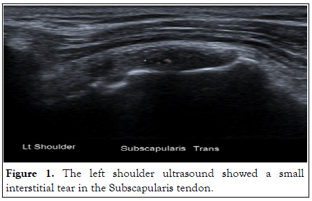
Figure 1: The left shoulder ultrasound showed a small interstitial tear in the Subscapularis tendon.
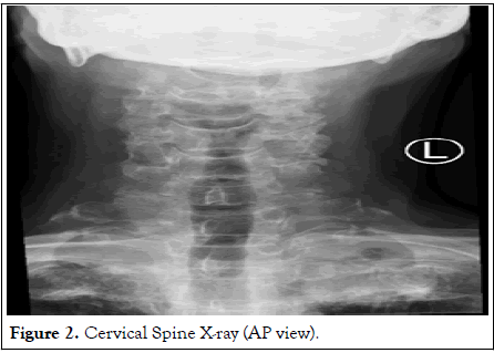
Figure 2: Cervical Spine X-ray (AP view).
Six months later, the patient returned with weight loss, hoarseness, and focal sweats around the left scapular and posterior shoulder region. His left shoulder and neck pain had progressively worsened, and he became frail. Four days before the second review, he had one episode of gross haemoptysis. Chest X-rays and Computed Tomography (CT) showed a welldefined 6 cm mass in the apex of the left lung (Figures 3 and 4).
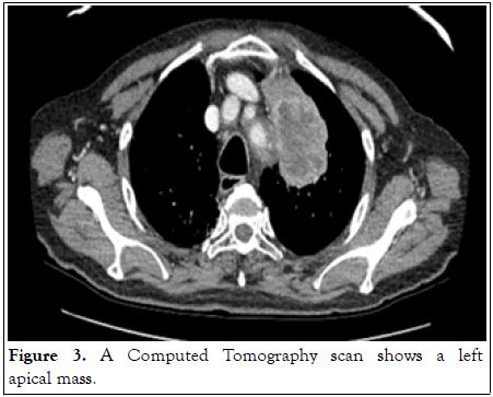
Figure 3: A Computed Tomography scan shows a left apical mass.
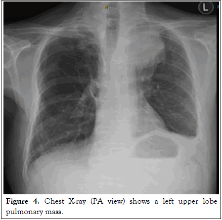
Figure 4: Chest X-ray (PA view) shows a left upper lobe pulmonary mass.
Core biopsy under CT guidance suggested typical features of LCNEC (Figures 5 and 6). Primary lung cancer was considered more likely. Unfortunately, molecular studies showed negative mutations in Epidermal Growth Factor Receptor (EGFR), which excluded his options for immunotherapy. Surgery or radiation therapy options were unsuitable due to the patient’s frailty and poor lung function. The patient received Palliative chemotherapy and passed away within one year of diagnosis.
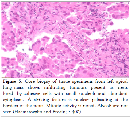
Figure 5: Core biopsy of tissue specimens from left apical lung mass shows infiltrating tumours present as nests lined by cohesive cells with small nucleoli and abundant cytoplasm. A striking feature is nuclear palisading at the borders of the nests. Mitotic activity is noted. Alveoli are not seen (Haematoxylin and Erosin; × 400).

Figure 6: Immunohistochemistry study of the left apical lung mass shows strongly positive for Cytokeratin (CK) 7 (× 20). Napsin A, TTF-1, CK5, p40, synaptophysin and chromogranin A are negative.
Clinical manifestations of lung cancer are usually cough, haemoptysis, chest pain and dyspnoea, however the chief symptoms are closely linked to the cancer location, classification, and stage [14]. Unfortunately, patients with LCNEC during the early stage are poorly symptomatic and may present solely with non-pulmonary symptoms [10]. Bisbinas et al. reported an average delay of 18.5 months in their case study [5]. Therefore, extrapulmonary cues of lung cancer have clinical importance for an early diagnosis.
Possible pathophysiology of lung cancer presenting as shoulder pain
Brachial plexus involvement: An American Roentgenologist, Professor Henry K. Pancoast, first named “the Pancoast tumor,” a tumor found in the superior pulmonary sulcus, causing posterior shoulder and neck pain. Other symptoms that may also occur in Pancoast tumors include ptosis, miosis, and anhidrosis [6]. The symptoms of Pancoast tumors are presumed to be due to the invasion of the tumor to the posterior division of the first and second thoracic common trunks of the brachial plexus, leading to ipsilateral shoulder and neck pain [6].
However, Onuigbo’s necropsy study in 1964 did not identify the primary lung tumor in the ipsilateral superior pulmonary sulcus. Metastatic lymph nodes were exclusively in contact with the brachial plexus in the thoracic inlet of the ipsilateral side [15]. Therefore, he believes the metastatic lymph nodes via the lymphatic spread pathway can also provide compression to the brachial plexus [15].
Somatic referred pain: Recent studies found that phrenic nerve stimulation can cause shoulder pain in cancer patients [16]. This pain is called a “somatic referred pain”, described as the pain felt in the territory innervated by nerves which are not those that innervate the source of the pain. The confusion is related to the convergence of the nociceptive afferents on second-order neurons in the spinal cord that also innervates the region where the pain is felt [17]. Brown described that the phrenic nerve arises from the third, fourth and fifth cervical nerve roots. It provides sensory innervation to the mediastinal, diaphragmatic peritoneum, as well as the skin on the shoulder, neck, and supraclavicular area (16). Therefore, stimulation of the diaphragm or pleura due to lung cancer can feel as though the pain is in the shoulder, neck, or supraclavicular region [16]. The phenomenon was agreed upon by Welch’s study on monkeys in 1994 [18].
Pancoast tumors may also affect other structures, including adjacent vertebrae [5,6]. If cervical zygapophysial joints or intervertebral discs at C4-5, C5-6 or C6-7 levels are involved, a somatic referred pain may feel like the shoulder, scapula or lateral neck [19,20] (Figures 7 and 8). The pain is often dull, aching, and poorly localized [21].
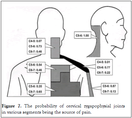
Figure 7: The probability of cervical zygapophysial joints in various segments being the source of pain.
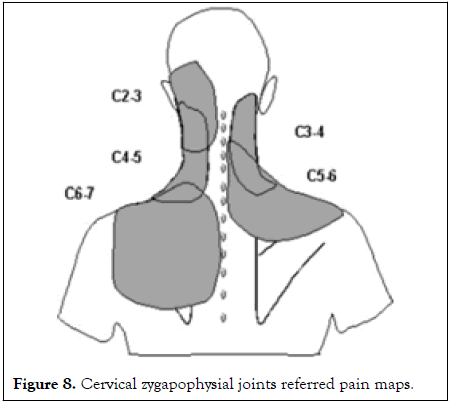
Figure 8: Cervical zygapophysial joints referred pain maps.
Nociceptive pain: Metastatic lung cancer to the humeral head is another commonly reported cause of cancer-related shoulder pain. This shoulder pain is an example of nociceptive pain, which is the pain caused by the noxious stimulation of the structure in the shoulder joint [21]. Argyriou et al. reported a lung LCNEC case metastatic to the shoulder joint, resulting in shoulder pain and suprascapular nerve palsy [22]. Other primary cancers, such as colon adenocarcinoma [23], or breast cancer [24], can also metastasize to the shoulder and presents with shoulder pain as the only initial presentation. X-rays of the affected shoulder are helpful screening tools [25].
Other cause: Radicular pain due to the impingement of the lower cervical nerve root in a Pancoast tumor may also cause ipsilateral shoulder pain. However, radicular pain is more likely to be sharp and lancinating rather than dull and aching [21].
High alert to red flag background information is essential for an early diagnosis
At the time of the first presentation, this patient did not have the typical pulmonary symptoms suggestive of lung cancer. However, the suspicion for underlying lung cancer should be raised based on the history of being a substantial ex-smoker who also had a lung cancer history. A strong association between cigarette smoking and lung cancer was recognized almost a century ago [26]. The overwhelming evidence suggested that about 85% of lung cancer cases were associated with tobacco smoking, including passive smoking [27]. In Travis et al.’s study in 1991, 100% (9 out of 9) of cases diagnosed with highly aggressive lung cancer, including LCNEC or Small Cell Undifferentiated Carcinoma (SCUC), were heavy smokers [11]. The risk for cancer remains for several years, even after smoking has been discontinued [28]. Patients diagnosed with cancer in the past have a higher risk of developing another primary cancer [27].
Clinical examinations and imaging findings are essential to increase diagnostic accuracy
High-quality diagnostic accuracy studies have shown that commonly used shoulder examination tests lack diagnostic accuracy [1,29,30]. For example, the sensitivity and specificity for the Hawkins-Kennedy test in detecting impingement were 79% and 59% respectively [29]. Thus, a positive or negative finding from a shoulder exam does not necessarily rule out the tested condition. Some shoulder examinations serve multiple purposes. For example, Jobe test is a test to detect rotator cuff tears, with the sensitivity and specificity being 84-89% and 50-58%, respectively [1,29,30]. The Jobe test is also a test to check for subacromial impingement. The sensitivity and specificity for subacromial impingement are 52% and 33%, respectively [29]. Therefore, having a positive result does not differentiate between the two clinical conditions. More frustratingly, shoulder pain aetiology can be multifactorial. For example, diagnosis of a rotator cuff tear does not exclude referred somatic shoulder pain due to a malignant cause.
The diagnostic accuracy of an imaging study can be affected by its false negative rate. Hamilton reported that 21% of lung cancer cases were unrecognized in the patients who presented with haemoptysis but had a negative chest X-ray [31]. The diagnostic accuracy of imaging tool may also be affected by the clinical experience of the interpreting practitioner. Initial cervical X-rays of this patient may have shown subtle evidence of increased attenuation in the limited view at the apex of the left lung. However, this was not acknowledged in the formal report. Lack of awareness of the possibility that shoulder pain could be an initial symptom of lung cancer, in the clinically setting of having no typical pulmonary symptoms suggestive of lung cancer, deprived the patient of a potential opportunity for a full chest X-Ray or other investigations.
Preoccupation with minor positive findings from the shoulder ultrasound and neck X-rays may give the clinician a false reassurance, however, the imaging findings may not directly correlate to the corresponding clinical presentation [32-34]. Tempelholf, et al. claimed that up to 23% of patients with rotator cuff tears are asymptomatic. The figures are higher in elderly patients (>80 years) to about 51% [32]. Degenerative cervical spine X-ray changes are also common in asymptomatic people [33-35]. In this case study, the patient had rotator cuff pathology and minor degenerative cervical spine changes, however, those findings do not exclude other coexisting causes of his shoulder pain.
LCENC lung cancer presented with shoulder pain is rare in current literature. However, shoulder pain as a possible initial manifestation of lung cancer has been frequently reported. Awareness of this unique feature of lung cancer may lead to early chest X-rays, regular clinical monitoring, and eventually an early diagnosis.
[Google Scholar] [Pubmed]
[Crossref] [Google Scholar] [Pubmed]
[Crossref] [Google Scholar] [Pubmed]
[Crossref] [Google Scholar] [Pubmed]
[Google Scholar] [Pubmed]
[Google Scholar] [Pubmed]
Citation: Wang T (2022) A Rare but Sinister Cause of Shoulder Pain: A Case Report. J Clin Trials. 12:515.
Received: 07-Sep-2022, Manuscript No. JCTR-22-19487; Editor assigned: 12-Sep-2022, Pre QC No. JCTR-22-19487 (PQ); Reviewed: 28-Sep-2022, QC No. JCTR-22-19487; Revised: 10-Oct-2022, Manuscript No. JCTR-22-19487 (R); Accepted: 12-Oct-2022 Published: 18-Oct-2022 , DOI: 10.35248/2167-0870.22.12.515
Copyright: © 2022 Wang T. This is an open-access article distributed under the terms of the Creative Commons Attribution License, which permits unrestricted use, distribution, and reproduction in any medium, provided the original author and source are credited.