Journal of Osteoporosis and Physical Activity
Open Access
ISSN: 2329-9509
ISSN: 2329-9509
Commentary - (2015) Volume 3, Issue 3
Objective: Evaluate long term whole body vibration training effects on bone mineral density in postmenopausal osteoporosis.
Background: Osteoporosis has been defined as a skeletal system disease characterized by low bone density and deterioration of bone microarchitecture which results in increased risk for fracture occurrence and predisposes the person to injury. Whole body vibration therapy showed positive effects on Bone mineral density.
Materials and methods: Literature and scientific papers review of was conducted through the use of several databases: Science Direct, Web of Science, SCIdirect, PubMed, Taylor and Francis Online, Springer Link, SAE publications, JAMA Pediatr Idea: drexler e-repository and archives, Google Scholar and City Library Marko Marulic Split University Library in Split Online catalog.
Conclusion: Whole body vibration training is a relatively new and promising non-pharmacological method for bone mineral density decline prevention.
<Keywords: Vibration therapy; Training; Low bone mineral density; Menopause
WBV: Whole Body Vibration; BMD: Bone Mineral Density
The World Health Organization has defined osteoporosis as a skeletal system disease characterized by low bone density and deterioration of bone microarchitecture which results in increased risk for fracture occurrence and predisposes the person to injury. In modern times, sedentary lifestyle is the main reason for the increasing incidence of osteoporosis, and this problem will be further increased [1]. The disease itself, and complications associated with it, already have an epidemiological character [2-5], and as a consequence of these events, many different pharmacological and non-pharmacological treatments are being developed.
There are several sub-populations, such as astronauts [6], the elderly [7], postmenopausal women [8], and people with muscle dystrophy [9,10] and neurological disorders, such as spinal-cord injury [11], that have an increased risk for the occurrence of osteoporosis. In addition to above, osteoporosis can also occur in men, young adults and even children [12]. However, it is most commonly occurring in postmenopausal women, primarily, due to hormonal changes and, in addition, reduced physical activity. After menopause, bone density in healthy women decreases up to 3% per year [13].
The bone mineral density is being evaluated by using densitometric methods. The results of bone densitometry are made on the basis of two findings, primarily the T-score that shows bone density compared with the reference group of young healthy people with an average bone density. T-score above -1 is normal, between -1 and -2.5 indicates that there has been a significant reduction in bone density, a condition called osteopenia, and below -2.5 diagnosed with osteoporosis. In addition to T-score, there is a Z-value, which indicates bone density compared with others of similar age [14]. This value is used primarily for treatment progress monitoring.
Physical activity with and without external loads is important for maintaining bone mass. Reduced activity is one of the main reasons for the gradual loss of bone mass in old age. In cases of severe trauma and strict immobilization one can develop a form of rapid and progressive (disuse) osteoporosis, which is the result of a sudden imbalance between bone resorption and new bone formation. Bone loss can be up to 1% per week [15] and can last up to six months before it is being gradually reduced. As a sign of increased bone resorption, high concentration of calcium in the blood and increased calcium excretion in urine is prominent. Diffuse osteoporosis, which occurs during long stays in zero gravity conditions, is similar.
The classic treatment of osteoporosis involves specific lifestyle changes which help patients, and can improve their condition. This includes general steps for the patients’ protection, dietary changes and application of additives in diet, certain drugs and the inclusion of exercise in daily life habits of the patient.
As for the general measures, it is important to remove the factors that are conducive to bone loss. One should be advised to quit smoking, pay attention to the drugs that can cause orthostatic hypotension and drugs that can accelerate bone loss and, it is necessary to pay attention to the environment of patients. It is important to pay attention to patients’ diet. Adequate intake of calcium and vitamin D can greatly slow bone loss [16]. A positive calcium balance can be maintained by daily intake of 800-1000mg, while many people have less than 500 mg of elemental calcium daily. Vitamin D deficiency causes a decrease in bone mass whilst increasing the risk of fractures.
Osteoporosis pharmacological treatment purports hormone replacement therapy (estrogen administration in postmenopausal women), followed by a selective estrogen receptor modulator other than hormones which keep the bone mass and affect the breast and uterine tissue as antiestrogens. Bisphosphonates can stimulate the maturation of osteoblasts (creates a new bone) and to increase their number. Calcitonin is a hormone that prevents bone loss by inhibiting osteoclast (which decompositions the bone) activity. Various other medicamentous treatments like fluroide, tibolone, parathyroid hormone and osteoprotegerin can be used, and represent an important factor in osteoporosis treatment [17,18]. So far, the prevention of BMD loss is mainly treated by using various medicaments, however, the risk involved with the use of the same is not completely investigated [19].
Daily physical activity in older people slows down or, in some cases, stops bone loss and increases muscle coordination and thus reduces the risk of falls. Given that the fractures caused by osteoporosis is associated with bone fragility (low BMD), and that less than a third of people suffering from osteoporosis who experience hip fracture recover functional state to level before the injury [20], most of this article will be directed precisely to the studies which presented the effects of physical activity on bone mineral density (BMD) in subject suffering from osteoporosis.
As bone density decreases with falling levels of hormones, which are just like the bone density decrease with age, and that every form of exercise has a positive effect manifesting through the increasing hormones levels [21], it can be concluded that, generally, physical activity has a positive effect on bone density. Therefore, mechanical loading, which occurs during physical activity, provides an endogenous anabolic stimulus for bone [22,23] or a method that can inhibit bone degradation and, respectively, oppose the osteoclastogenesis [24]. Thus, the mechanical leading is very important for improving and maintaining bone mass [25,26], it is important to note that the mechanism resulting with the best effects are still unknown [27]. In contrast, the reduction of the magnitude and/or frequency of physical activities lead to increased bone resorption and therefore causes the reduction in bone mineral density [28].
Since the mechanical loading leads to bone structure adaptation [29], which is known as Wolff law [30], physical activity is often recommended for people with osteoporosis as a means of preventing bone loss [31-33].
Gutin and Kasper [34] in their review article suggested that both, high intensive aerobic training and strength training results in good osteogenic effects and it is therefore recommended. However, it is questionable whether people diagnosed with osteoporosis should be exposed to high intensity load because, as mentioned Lanyon [22], this may increase the risk of injuries. In fact, just running and jumping can cause ground reaction force 3 to 6 times the body weight [35], and it is this excessive burden, particularly in the elderly, that can increase the risk of injury [36].
Because of these and similar conflicts of opinion, scientists and practitioners are constantly looking for new training modalities for people with diagnosed osteoporosis, especially postmenopausal women.
According to previous researchers [37] mechanical stimulus should be different from that which the person (patient) has to meet in everyday life. Some authors suggest that the magnitude of the stimulus is not important if the frequency and distribution of the load increases, and that the osteogenic effect can be caused in this way also. In regards to this, there are a number of studies that have attempted to evaluate the effects of whole body vibration training on humans [38-41] and even animals [42-45].
Vibration is a mechanical stimulus characterized by oscillatory motion. Biomechanical parameters which determine the intensity of the stimulus itself are amplitude, frequency and magnitude of oscillation. The range is determined by the oscillatory movement representing the total amplitude of vibration displacement in millimeters (mm), the rate of repetition of the oscillation cycle is determined by the frequency of vibration is measured in Hertz (Hz), while the acceleration indicates vibration magnitude [46].
There are two methods of applying vibration to the human body during exercise. In the first method, vibration is applied directly to the muscle that is trained via vibrating unit that is directly attached to the muscle itself. In another method, which is used more often, the vibration is indirectly applied to the working muscles. In this case, vibrations are transmitted from the vibration platform to the targeted muscles through other body parts, e.g., during training the m. quadriceps one can stand on the platform which oscillates vertically and perform various exercises (such as squat). In this case, the vibration is transmitted from the platform on to lower extremities, including m.quadriceps. This method is called “whole body vibration training”.
Whole body vibration involves a person standing on a vibrating platform in such way that the vibrating stimulus is being directly sent to the plantar surface and then transmitted through the body to bones and muscles [47,48].
Since the deterioration of bone tissue does not arise solely because of reduced levels of physical activity, but also because of the monotony of muscle dynamics [49], whole body vibration training is studied primarily because it can maintain, and even, in some cases, of course, if properly used, improve bone structure in people with diagnosed osteoporosis. In addition to being osteogenic, vibration training improves muscle strength, and balance [50-52]. Also, the positive effects of vibration training have been observed in patients with chronic low back pain. Namely, Iwamoto et al. [53] in their study intended to evaluate the effects of vibration training on bone mineral density in postmenopausal women while establishing an eligibility criterion which included the presence of chronic low back pain. All subjects felt relief in leg muscles and back after one year treatment, and, besides that, they reported less pain in the lumbar region of the spine. This study showed that the pain in the lumbar region of the spine had significantly higher reduction rate in the group that, in addition to alendronate, underwent WBV training than the group that had exclusively used pharmacological therapy which included alendronate. One could say that the whole body vibration training combined with medical treatment causes greater reduction in pain in the lumbar region of the spine than medications application exclusively.
It is because of these facts that the whole body vibration training is considered to be a potential means for osteoporosis intervention which is, in fact, appropriate for this population [54,55].
Five factors affect the type and intensity of the response of the body to WBV training. These factors are: 1) the direction of vibration (vertical, oscillating and horizontal), 2) vibration frequency (expressed in Hz) and 3) the amplitude of vibration (usually expressed in mm), which together account for the magnitude of vibration, the acceleration, which is usually expressed in gravities (g, 1 g ~9.81m /s2) 4) the duration of the vibration stimuli and 5) body position.
Depending on the device (commercially available vibrating platforms) there are three types of mechanical vibrations: 1) horizontal, 2) vertical and 3) pivotal (Scheme 1). According to current knowledge, the transmission of vibrations through the body is greater in vertical than pivotal vibrations [56]. It is noteworthy that there are no available studies that applied horizontal vibration.
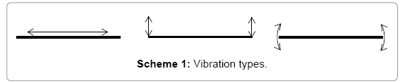
Scheme 1: Vibration types.
In addition to the above said, according to some authors [57,58], the use of frequency lower than 20 Hz is not recommended in training, and therefore in therapeutic purposes also. Other authors [48] stated that the mechanical signals of high frequency and low amplitude are successfully transferred to the hip and spine, and because of this it is necessary to apply frequency greater than 20Hz. They came to this conclusion based on the results of their study in which the subjects were standing on the vibrating platform (vertical or pivotal) with the knee flexed at 20° angle. Also, the same authors stated that mechanotransduction is primarily dependent on the angle at the knee joint.
Safety of the trainees, especially when it comes to this population, must be ensured. With this in mind, the contraindication factors for the use of vibration training, enlisted by the manufacturers themselves [59-61] are: the existence of kidney or bladder stones, epilepsy, cancer, pacemaker, recent implantation or surgery, thrombosis, hernia, rheumatologic arthritis, migraine, some cardiovascular problems and spinal injuries.
Slatkowska et al. [62] stated that there is no risk of using vibration training, and that it represents a new, promising modality for improving the characteristics of the skeletal system in postmenopausal women, respectively, same authors stated that the whole body vibration training increases muscular strength and power and slows down bone mineral density loss. Also, Russo et al. [38] reported that there were no adverse effects of vibration training for women in menopause and post menopause. The only side effect that was observed was increased itching in the lower extremities, more specifically, 6 of 17 participants, who were included in high magnitude vibration training reported these side effects. Moawed and Mohammed [63] have explained in their study that the itching that occurs during the first use of the vibrating stimulus is the result of an increase in skin blood flow. In addition, in that same study, knee pain was reported by two obese subjects diagnosed with osteoarthritis, but it withdrew after a few days allowing subjects to continue with the treatment. Besides that, it is noteworthy that some authors [64] claimed that at frequencies above ~50 Hz severe muscle soreness and even hematoma may emerge in untrained subjects, but this hasn’t been empirically tested due to the ethic issue.
Literature and scientific papers review of was conducted through the use of several databases: Science Direct, Web of Science, SCIdirect, PubMed, Taylor and Francis Online, Springer Link, SAE publications, JAMA Pediatr Idea: drexler e-repository and archives, Google Scholar and City Library Marko Marulic Split University Library in Split Online catalog. Keywords used during the time were: whole body vibration exercise, whole body vibration therapy, vibration intervention, all combined with the terms of osteoporosis, postmenopausal women and bone mineral density (BMD).
The work includes all available research related to the effects of vibration training the whole body in postmenopausal women diagnosed with osteoporosis.
Low bone density and osteoporosis are health problems mainly occurring in elderly population and individuals with some kind of physical or neurological impairment, and movement difficulties. Changes in bone structure primarily manifested as the decline in bone mineral density, predisposes this population to bone fractures. Bone tissue needs mechanical stimulus to stay healthy [29], and the main reason for the change in bone mineral density which occurs in these individuals is reduced physical activity (hypokinesia) followed by reduction of mechanical stimuli/strain.
Gravitational force, muscular force and ground reaction forces are major forces which are encountered daily by the human skeleton, and thus, participate in the bone modeling and remodeling, so, consequently, the increase in bone mineral density is generally associated with longterm physical activity [35]. Some authors [65,66] suggest that highly intensive training has positive effects on bone mineral density even in well-trained athletes, but however, it is questionable whether people with diagnosed osteoporosis should be exposed to high intensity load. (Table 1)
| Author (s) | Pattern | Duration of study | Characteristics of vibration | Characteristics of treatment | Results | ||
| (Mm) | (Hz) | (G) | |||||
| Russo et al. [38] | 29 postmenopausal women; Age 61 ± 7 |
6 months | nn | 12- 8 | 0,1-10 | Standing on the platform; 3x2 minutes; 2 x week |
No statistically significant differences |
| Verschueren et al. [75] | 70 postmenopausal women; Age 58-74 |
6 months | 1,7-2,5 | 35-40 | 2,28–5,09 | Squat and isometric squat; up to 30 minutes, 3 times per week |
Hip BMD +0.93%; +1.5% compared to the control group |
| Rubin et al. [49] | 56 postmenopausal women; Age 47-64 |
12 months | nn | 30 | 0,2 | Standing on the platform; 2x10 minutes, 7 x week |
No statistically significant differences |
| Iwamoto et al. [53] | 50 postmenopausal women; Age 55-88 |
12 months | 0,7-4,2 | 20 | nn | Standing on the platform; 1x4 minute, 1 x per week |
No statistically significant differences |
| Gusi et al. [77] | 28 postmenopausal women; Age 66 ± 5 |
8 months | nn | 12,6 | 0,7–3,3 | Standing on the platform; 6x1 minutes, 3 times per week |
Hip BMD: +4.3% compared to the control group |
| Corrie et al. [83] | 37 postmenopausal women; Age 65-95 |
3 months | nn | nn | nn | nn 6x1 minutes, 3 times per week |
P1NP: Oscillating vibration ± 20.2% The vertical vibration ± 15.2% |
| Ruan et al. [79] | 28 postmenopausal women; Age 61 ± 8 |
6 months | 5 | 30 | 18 | Standing on the platform; 1x10 minutes, 5 times a week |
Hip BMD +3.2% Spine BMD +4.3% |
| Von Stengel et al.[78] | 108 post-menopausal women; Age 60-75 | 12 months | 1,7-12 | 12,5-35 | 8g | Unilateral, bilateral and isotonic squat; 7x1,5 minutes, 3 times a week |
Spine BMD +0.5 - 0.7% Hip BMD +0.3 - 11% |
P1NP - Marker of Bone Formation; BMD – Bone Mineral Density
Table 1: Whole-body vibration (WBV) exposure as an intervention for improving BMD among postmenopausal women.
Apparently, the skeletal system’s answer to whole body vibration training, or the mechanotransduction which it activates, is osteogenesis [67,68], respectively, increased bone density after mechanical stimulus indicates that skeletal system adapts to the type of physical activity [30,69]. Frost [37] stated that the mechanical inputs must result in a heavy load in order to influence the bone morphology. It should be noted that, although the existence of osteogenic effect of vibration training is proven, the mechanisms underlying the same have not been fully explored. It is assumed that the vibrations cause micro trauma to the bone which is later repaired by the osteoblast [70]. Only thing that could be claimed without doubts is that the osteogenic effect, which occurs as a result of adaptation to mechanical stimuli, decreases with age [71], and therefore, it is recommends that the vibration treatment begins as early as possible.
Whole body vibration training also affects bone remodeling indirectly through the endocrine system response. As stated in some previous researches [72], it increases growth hormone and testosterone levels in the body. The positive effect of testosterone on bone mineral density was detected in the forearm, the lumbar spine and hip in healthy men and women [73-75]. It is important to admonish that there are no studies that have showed the same or similar effect on a sample of postmenopausal women. In general, the effects of vibration training on bone mineral density have not been fully explored in this population.
Of all the studies that have dealt with this issue only three groups of authors [38,49,53] did not record statistically significant positive whole body vibration training effects on the bone characteristics in postmenopausal women. It is important to specify that, in this case, the lack of statistically significant results can be seen as a positive effect because, as stated above, after menopause, bone mineral density declines rapidly. In addition, other studies have shown that the whole body vibration training is a potential non-pharmacological method for bone mineral density decline prevention, and even causes an increase in bone mineral density in population suffering osteoporosis. The reasons for this variability are manifold. As stated by some authors [47,48] mechanotransduction varies among the body regions because, firstly, the nonlinearity of the musculoskeletal system, and, secondly, because of the different body positions during vibration application in previous studies. In fact, this could explain the differences between the effects on the hip and spine which could be found in the results of some previous studies. Along with the previously mentioned, a group of authors [62] stated that these differences could occur due to inadequate sample sizes in individual studies.
Slatkowska et al. [62] concluded in their meta-analysis that whole body vibration training causes small but statistically significant improvements in bone mineral density in postmenopausal women and children and adolescents, but not in young adults. They also stated that whole body vibration training, in some cases, does not cause significant bone mineral density changes in shin bone and the spine, and they have attributed this to vibrations transmission variability which occurs throughout the body.
In a study by Verschueren et al. [76], in which the subjects were exposed to vertical vibrations 3 times per week for 24 weeks (35-40 Hz; 1, 7-2, 5 mm; 2, 28-5, 09 g), reported an increase bone mineral density of the femur. One could state that it is precisely this type of training/ treatment that caused changes similar to those recorded by Yamazaki et al. [77] after a 12-month program consisted of exclusively walking at a moderate pace, but the study of Gusi et al. [78], in which the vibration effects (12.6 Hz; 30 mm; knee angle 60°) after 8-month treatment were higher than those recorded in the group which had only walking treatment is indicating that greater positive effects are caused by the use of using vibration treatment. Gusi et al. [78], after 8-month (3x week) treatment recorded 4.3% larger increase in femur bone mineral density in vibration group, compared to the walking group which had a treatment that included one hour walk with five minute stretching thereafter. Furthermore, Verschueren et al. [76] recorded 1.5% increase in bone density in the WBV group compared to the control and fitness group after 6-month treatment. Both experimental groups had 3 sessions per week (knee extensor strength training) with progressively increased load, except that the fitness group trained knee extensors by using dynamic leg press and leg extension exercises (20-8 RM), while the WBV group performed static and dynamic knee-extensor exercises on a vibration platform (20-30 min; 35-40 Hz; 2.28-5, 09 g), which mechanically loaded the bone and evoked reflexive muscle contractions.
In addition to these, the study of the Von Stengel [79], which included 108 postmenopausal women randomly assigned to three groups: 1) pivotal vibration platform (12.5 Hz; 12 mm), 2) vertical vibration platform, which had three 15-minute treatments per week (both groups had a magnitude of vibration of 8 g) and 3) control/fitness group which had two low-intensity workouts per week. They came to the conclusion that bone density increased significantly when it comes to the lumbar spine (PVT + 0.7% ± 2.2% and VVT + 0.5% ± 2%), while, when it comes to the femur, progress which was recorded in both vibration groups (PVT + 0.3% ± 2.7% and VVT + 1.1% ± 3.4%) was not statistically significant. The control group’s bone mineral density was decreased in the lumbar spine (-0.4% ± 2%), whilst the femur bone mineral density was maintained at the same level (-0.0% ± 2.1%). It is important to note that all respondents were taking additional supplements, calcium (up to 1200 mg) and vitamin D (800 international units - IU), which further raises the question of whether the vibration training is, in fact, better than some pharmacological methods used in medical practice in improving and/or maintaining bone mineral density. The authors of this study [79] indicate that these vibration parameters, though, the frequency and amplitude differ, but the acceleration is equal, had osteogenic effect. On the basis of this study, it is evident that the effects of different vibration platforms are different, but they are generally better from all the other forms of exercise.
The study of Ruan et al. [80] showed the highest increase in bone mineral density. In fact, after 6 months of 10-minutes, 5 times per week, vibration training at a frequency of 30 Hz and amplitude of 5 mm (overall magnitude 18 g), on a sample of 116 postmenopausal women, these authors recorded an increase in bone mineral density at the lumbar spine by 6.2% and hip by 4.9%, compared to control (n = 50) group. Although the authors did not report any side effects, application of magnitude this high raises the question whether patients suffering osteoporosis should be involved in this kind of treatment and how safe it is for them.
Osteogenic effect (adaptation) depends on various parameters such as the number of strains (frequency), the magnitude and direction of the load distribution. If the law of bone remodeling (Wolff ’s law) was thoroughly analyzed, it could be concluded that only mechanical stimulus, in this case, vibration, with large magnitude can result in improving bone mineral density, i.e., as stated by Rubin et al. [69], the larger the magnitude, the greater the effect of the treatment are. Some authors state that the large magnitude, as a result of large frequency [39,49,81], induces the best osteogenic effects and, therefore, the increase in bone mineral density is proportional to the magnitude of vibration stimuli [82]. Other [37] in turn indicate that the amplitude is most important in order to improve bone structure. For example, Tanaka et al. [83,84] stated that low frequency and large amplitude vibration stimuli can quadruple the osteogenic response.
The best proof that the correct determination of the frequencies and amplitudes can be very important can be found in a study by Rubin et al. [49]. In their study, after 12-month treatment that included daily, 2x10 minutes, vibration training (30 Hz; 0.2 g; 0, 5 μm), authors [49] showed a decrease in bone mineral density in experimental (-0.69%) and placebo (-0.27%) group at femur (-0.69%; -0.27%), the trochanter (-0.07%; -0.19%) and the lumbar spine (-0.51%; -0.65%). Such small differences after this extensive treatment (12 months x 7 days x 2 x 10 min) can be found in the intensity that was not enough to, not only increase the anabolic effects, but also inhibit the resorptive ones. This can be primarily attributed to very small amplitude, which was less than one millimeter.
Also, noteworthy is that, in that same study, after a post hoc analysis, authors [49] found statistically significant differences in women with low body weight (<65 kg) as opposed to the heavier ones, where difference was not statistically significant. These authors found that women with lower body weight, in this case, below 65kg, recorded the best effects. Namely, compared to the placebo group, they have achieved a relative benefit in bone mineral density values of 2.1% at femoral neck, 1.92% at trochanter and 3.35% in lumbar spine.
This could be attributed to very small vibration magnitude which is probably not sufficient enough to cause osteogenic response in women with higher body mass. Based on these findings, the question is whether vibration magnitude (primarily amplitude) should somehow be normalized with body mass. On the basis of this study, it can be concluded that vibration stimuli of small amplitude can only diminish the fall, but not improve bone mineral density, and that these effects are greatest in women with lower body weight. Increasing the amplitude, which would increase the total magnitude, could result in far greater effects.
There are many uncertainties associated with the application of whole body vibration training in order to increase bone mineral density level. More specifically, the optimum frequency and amplitude, and thus the magnitude of the vibration stimulus have not yet been established. In addition, it is unknown whether the application of pharmacological therapies, together with vibration exercises results in synergistic (additional) effect on bone mineral density than solely pharmacological therapy. Considering that WBV is a relatively new therapeutic modality, no long term (duration over a year) studies were conducted. Therefore additional long term studies on humans are requires in order establishing vibration training parameters that are optimal for bone mineral density preservation and increase in women suffering osteoporosis.
Although there are many ambiguities related to whole body vibration application, there is no doubt that the results are positive. Further studies are still needed to establish the optimal protocol which would incorporate vibration treatment and would be best suited for this population, and that, through three segments. First, establish magnitude limits for this population, secondly, given that there are three types of vibrating platform, it is necessary to determine which type of vibration results in best effects, and third, most important, determine optimal vibrations characteristics that would result in the best effects on bone mineral density in this population.
Generally, in order to create protocols that would be successful in osteoporosis treatment, and considering that there is a small number of studies that have dealt with these issue, large sample, long-term studies are required in order to establishing the optimal vibration parameters that could help patients with osteoporosis and improve their quality of life. One thing is for certain, commercially available platforms and their parameters, amplitude of 2-4 mm and frequency of 30-40 Hz, can be used, and recommended, as a safe and effective type of therapy. It could be used on a daily basis but is best utilized 3 times per week, not longer than 30 minutes.
Based on the foregoing, it can be concluded that whole body vibration training is a relatively new and promising method to oppose osteoporosis. Although the patient cannot be cured by this method, vibration treatment greatly reduces negative effects. It has been shown that whole body vibration training results in positive effect on bone mineral density, improves strength, balance. All these factors combined reduce incidence of falls and therefore reduce the risk of bone fractures.