Journal of Clinical & Experimental Dermatology Research
Open Access
ISSN: 2155-9554
ISSN: 2155-9554
Image Article - (2019)Volume 10, Issue 6
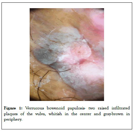
A 54 year-old-female, without medical history, presented with two raised infiltrated plaques of the vulva, whitish in the center and gray-brown in periphery, with papillomatous patches, which had appeared over the last year (Figure 1).
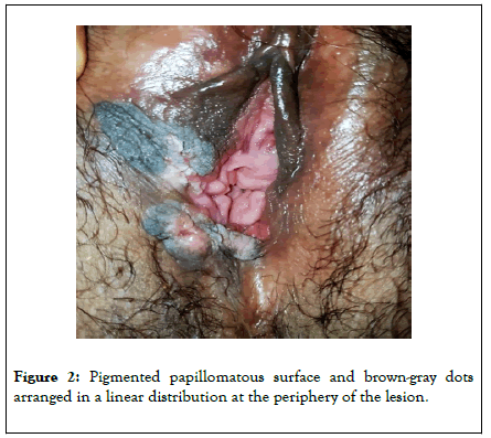
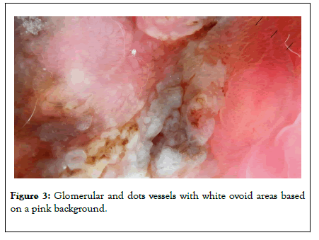
A 54 year-old-female, without medical history, presented with two raised infiltrated plaques of the vulva, whitish in the center and gray-brown in periphery, with papillomatous patches, which had appeared over the last year (Figure 1). Dermoscopic examination revealed a pigmented papillomatous surface and brown-gray dots arranged in a linear distribution at the periphery of the lesion. At the center, we observed a numerous glomerular and dots vessels with white ovoid areas based on a pink background (Figures 2 and 3). Histologic examination of skin biopsy showed an irregular acanthosis, dyskeratosis, and cluster of pseudo-koilocytosis cell with a clear perinuclear halo, cytological atypia and mitosis, melanophages in the papillary dermis, consisting with the diagnosis of verrucous bowenoid papulosis.

Figure 1. Verrucous bowenoid papulosis- two raised infiltrated plaques of the vulva, whitish in the center and gray-brown in periphery.

Figure 2. Pigmented papillomatous surface and brown-gray dots arranged in a linear distribution at the periphery of the lesion.

Figure 3. Glomerular and dots vessels with white ovoid areas based on a pink background.
Bowenoid papulosis can be confused with other diagnostics such as lichen planus, genital warts or seborrheic keratoses. In doubtful cases, dermoscopic examination may show particular signs; the appearance of the grey-brown spots with a linear arrangement at the periphery can be explained by the presence of melanophages in the papillary dermis in histology. This feature was described in a man by Marcucci et al. in 2014 [1] and was also seen in our patient. Other signs have been reported such as whitish-red exophytic papillary structures [2] and pigmented papillomatous surface, which corresponding to irregular acanthosis in histology. Vessels abnormalities were also described by Dong et al. [3] and consist on dots or glomerular vessels. These dermoscopic characteristics remind us the signs of pigmented Bowen's disease, which can only be distinguished from bowenoid papulosis by the histopathological analysis.
To conclude, we report the dermoscopic appearance of an unusual case of verrucous bowenoid papulosis in women. Dermatologist must recognize this features to be able to eliminate others differentials diagnostics.
None.
Citation: Safae M, Myriam M, Laila B, Karima S (2019) Vulvar Verrucous Bowenoid Papulosis in Dermoscopy. J Clin Exp Dermatol Res. 10:514. Doi: 10.35248/2155-9554.19.10.514
Received: 27-Nov-2019 Accepted: 12-Dec-2019 Published: 18-Dec-2019
Copyright: © 2019 Safae M, et al. This is an open-access article distributed under the terms of the Creative Commons Attribution License, which permits unrestricted use, distribution, and reproduction in any medium, provided the original author and source are credited.