Journal of Oceanography and Marine Research
Open Access
ISSN: 2572-3103
ISSN: 2572-3103
Research Article - (2015) Volume 3, Issue 1
A total of 150 fish samples belonging to three families were collected from Mediterranean sea, Egypt and examined for Trypanorhynch Cestode larvae infection from September 2011 till February 2012. Sixty three (42%) of the investigated fish were parasitized with different larval species belonging to Lacistorhynchidae and Tentaculariidae. All Trypanorhynch species larvae were isolated from muscles and abdominal cavities. Results revealed that, 46%; 44% and 36% of the examined fish species Epinephelus gigas; Sciaena umbra and Scomber spp. were infected respectively and were considered as new hosts for the detected parasites. Callitetrarhynchus gracilis larvae (Pintner, 1931) showed the highest prevalence (30%) and maximum intensity (37). The collected C. gracilis larvae and its host muscles (Epinephelus gigas) were analyzed for some heavy metals ( Cu, Pb, and Cd) using atomic absorption spectrophotometer, the results indicated a significance higher concentration of the heavy metals in the parasite tissues compared with its fish host muscles. The present study has concluded for the 1st time that; the Trypanorhynch C. gracilis larvae could be used as bio-indicator of heavy metals pollution and add further evidence to the usefulness of parasites in monitoring environmental impacts.
<Keywords: Trypanorhynch; Egypt; Callitetrarhynchus gracilis; First record; Mediterranean sea; Bio-indicator; Heavy metals
Fish is one of the most important sources among the food products of animal origin because of their content of high quality biological animal protein, lipids, vitamins, essential fatty acids and several kinds of minerals [1,2].
Fish parasites recorded to have a negative impact on the commercial fisheries industry [3],among them Trypanorhynchid cestodes (Order Trypanorhyncha) are considered as one of the main groups which follow three or four host life cycles, before reaching the final host [4]. They are typically found in the stomach and intestine of sharks and rays, whilst their larval forms infect a wide variety of marine invertebrates and mainly use fishes as intermediate or paratenic hosts [4] and their detection among the infected fish may represent marketing problems [5]. Although only few cases of accidental human infections by trypanorhynchs have been reported [4], these worms may cause allergic reactions [6]. Despite their worldwide distribution and importance for commercial fisheries, trypanorhynchids are still a relatively poorly studied group. In aquatic ecosystem, heavy metals are considered as the most important pollutants, they are non biodegradable potent toxins and their bioaccumulation in tissues lead to intoxication, tissue damage and dysfunction of a variety of organs [7]. The most non-essential heavy metals of particular concern to fish and surface water are Cadmium (Cd) Copper (Cu) and Lead (Pb) which have the way to fish flesh mainly via gills [8]. Fish may absorb heavy metals from surrounding water and food, which may accumulate in various tissues in significant amounts leading to toxicological effects. Contaminants accumulation in various fish and other marine species provides an estimate of integrated metal exposure. Muscle of fish is the tissue commonly chosen because of the implications it carries.
For human consumption and health risk. Such studies are imperative since rapid industrialization and urbanization during the last two decades have affected the quality of the Egyptian coastal environment along the Mediterranean Sea [9]. In the recent years, there has been increasing interest in the interrelationship between parasitism and pollution, especially in aquatic habitat and also to the role of parasites as bio-indicator of heavy metals pollution [10-12].
El-Max pay is part of Alexandria coast of Mediterranean sea considered as a main fishing source in the city [13] and subjected to contamination by the drainage of sewage effluents, industrial and agricultural untreated discharges.
In the present study, three common species of marine water fishes from the coastal region of El-Max Bay at Alexandria city were investigated for identifying the infecting trypanorhynchid parasites and determining their prevalence, intensity and abundance. Also, the study aims to detect the concentrations of some heavy metals in the tissues of the trypanorhynchid species that showed the highest prevalence as well as in the muscles of their fish host, so as to evaluate the efficiency of using trypanorhynchids as a bio-indicator for heavy metal pollution.
Collection of fish samples
A total of 150 fish specimens, belonging to 3 Mediterranean Sea fish species (Epinephelus gigas; Sciaena umbra and Scomber sp.) were collected along the coastal region of El-Max Bay at Alexandria city, during the period from September 2011 till February 2012.
Parasitological examination
Fishes were examined for trypanorhynchid larvae in muscles, different organs, mesenteries and body cavity.
The specimens were placed in bags with ice and transported to the laboratory of the Department of Parasitology, Faculty of Veterinary Medicine, Cairo University. In the laboratory, they were filleted and the skin removed and macroscopically examined to detect any gross parasites. Muscle tissues, body cavity and viscera were examined with the help of a stereoscopic dissecting microscope, and the capsulated larvae were removed from the infected tissues. Parasite blastocystic walls were opened to detect the scolices. The collected parasites were washed with saline solution then fixed in 10% buffered formalin .For permanent mount; specimens were stained in acetic carmine, dehydrated in alcoholic grades and then mounted in Canada balsam [14,15].
Prevalence, abundance and mean intensity of infection were calculated according to Margolis et al.[16].
Determination of heavy metals
Larvae of Callitetrarhynchus gracilis and the infected muscle tissues from their host were analyzed to determine the concentration of heavy metals (Cu, Cd and Pb) according to the methods of Sures et al. [17] and by using a Perkin-Elmer Analyst 100 Atomic Absorption Spectrophotometer.
Statistical analysis
Data were presented as Mean ± standard deviation for numerical variables. Pearson correlation coefficient was used to assess the degree of correlation (0-0.3: weak correlation, >0.3-0.6: moderate correlation, >0.6-1: strong correlation). To compare mean, student’s test was used. The significance level was set as (P ≤ 0.05: Significant). Statistical analysis was performed using SPSS © version 16.
Parasitological examination
Out of the 150 investigated specimens, 63 (42%) were found to be infected with trypanorhynchid species larvae. The highest rate of infection was recorded among Epinephelus gigas (46%). (Table 1)
| Fish spp. | Length (cm) |
Weight (g) |
Examined number |
Infected number |
% of infection |
|---|---|---|---|---|---|
| Family Psciaenidae: Epinephelusgigas |
25-38 | 230-525 | 50 | 23 | 46 |
| Family Serranidae: Sciaena umbra |
18-25 | 145-160 | 50 | 22 | 44 |
| Family Scombridae: Scomber sp. |
20-28 | 150-178 | 50 | 18 | 36 |
| Total | 150 | 63 | 42 |
Table 1: Prevalence of trypanorhynch spp. Infection among the investigated fish species.
Four trypanorhynch species larvae were detected from the 3 investigated fish spp. (Figure 1). Larvae of Callitetrarhynchus gracilis showed the highest prevalence (30%) followed by Nybelinia bisulcata (17.33%), Pseudogrilotia sp.(10%) and Floriceps minacanthus (6%) among the total examined fishes (Table 2 and 2.1).
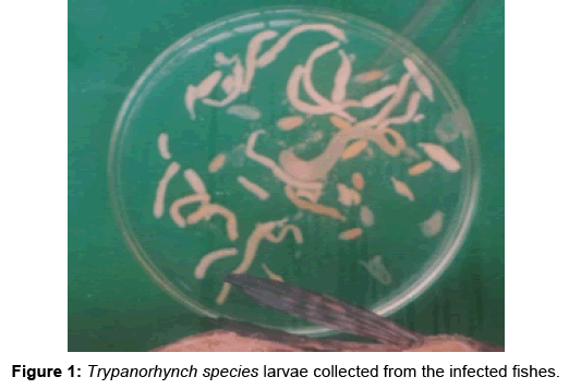
Figure 1: Trypanorhynch species larvae collected from the infected fishes.
| Fishspp | Examined Number |
Detected parasites |
Infected number |
Prevalence | Intensity | Abundance |
|---|---|---|---|---|---|---|
| E. gigas | 50 | C. gracilis | 23 | 46 | 11 | 5.06 |
| Pseudogrilotia | 4 | 8 | 12 | 0.96 | ||
| N. bisulcata | 8 | 16 | 6.25 | 1.00 | ||
| S. umbra | 50 | C. gracilis | 20 | 40 | 6.5 | 2.6 |
| F.minacanthus | 2 | 4 | 11 | 0.44 | ||
| Pseudogrilotia | 5 | 10 | 7.6 | 0.76 | ||
| N. bisulcata | 8 | 16 | 8 | 1.28 | ||
| Scomber sp. | 50 | C. gracilis | 2 | 4 | 9 | 0.36 |
| F.minacanthus | 7 | 14 | 6.4 | 0.9 | ||
| Pseudogrilotia | 6 | 12 | 4.6 | 0.56 | ||
| N. bisulcata | 10 | 20 | 3.5 | 0.70 |
Table 2: Prevalence, intensity and abundance of the detected trypanorhynch spp. Larvae in their host fish spp.
| Larval sp. | Total prevalence % |
|---|---|
| C. gracilis | 30 |
| F.minacanthus | 6 |
| Pseudogrilotia sp. | 10 |
| N. bisulcata | 17.33 |
Table 2.1: Total prevalence of the detected Trypanorhynchid larvae spp.
Morphological description of the detected species
Order Trypanorhyncha
A -Superfamily Lacistorhynchoidea
Family Lacistorhynchidae
1- Callitetrarhynchus gracilisRudolphi, 1819 (Figure 2):
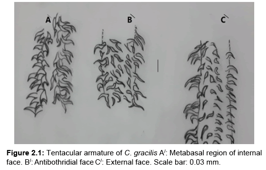
Figure 2a: C. gracilis . Left: Scolex . Right: Cyst Scale Bar=0.5 mm. A. Parsbothridialis B. Parsvaginalis C. Parsbulbosa . D. Pars postbulbosa
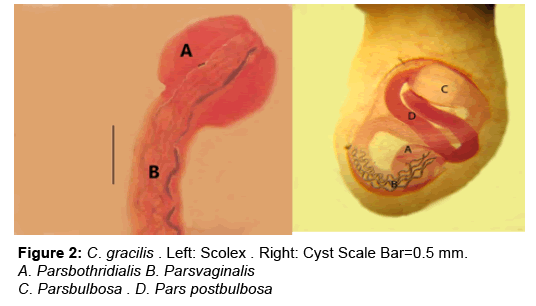
Figure 2: Tentacular armature of C. gracilis A/: Metabasal region of internal face. B/: Antibothridial face C/: External face. Scale bar: 0.03 mm.
Description: The cyst is bladder-like too elongated and of white colour.
The larva has an elongated scolex, measures 5.54-9.67 (7.11) mm long, with two heart shaped bothridia. Bothridial length is 1.24-2.53 (1.88) mm and 0.42-0.83 (0.63) mm wide. Pars vaginalis 2.31-5.12 (3.72) mm. Pars bulbosa 0.75-1.19 (0.97) mm. Pars post bulbosa is long reaches 0.31-0.38 (0.35) mm. The tentacle bulbs are 0.28-0.32 (0.30) mm, reaching the posterior part of the scolex. Tentacular sheaths are spirally coiled and tentacular armatures are shown in Figure. 2.1.
2- Floriceps minacanthus Campbell and Beveridge, 1987 (Figure 3):
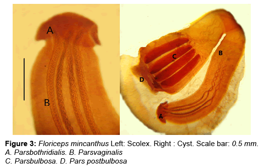
Figure 3: Floriceps mincanthus Left: Scolex. Right : Cyst. Scale bar: 0.5 mm. A. Parsbothridialis. B. Parsvaginalis C. Parsbulbosa. D. Pars postbulbosa
Description: Scolex is 2.93-5.18 (4.05) mm long and 0.69-1.73 (1.21) mm as maximum width at the level of bulbs. Pars bothridialis formed of two bothridia, of 0.52-1.17 (0.85) mm long and 0.45-1.37 (0.91) mm wide. Tentacular sheathes are coiled. Four bulbs, each measures 0.54 -1.13 (0.03) mm long and 0.12-0.27 (0.19) mm wide. Pars post bulbosa measures 0.8-1.23 (1.02) mm long. Pars vaginalis 2.20-3.32 (2.76) mm. Bulbs are elongated and located in the base of peduncululated area of the scolex. The ratio of pars bulbosa to pars vaginalis is 1:4.4 to 1:5.1(1:4.8).
3- Pseudogrillotia sp. (Figure 4):
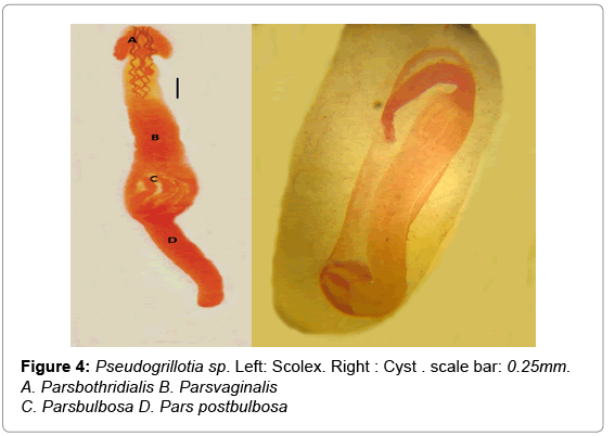
Figure 4: Pseudogrillotia sp. Left: Scolex. Right : Cyst . scale bar: 0.25mm. A. Parsbothridialis B. Parsvaginalis C. Parsbulbosa D. Pars postbulbosa
Description:The scolex is elongated divided into four regions:, Pars bothridialis of 0.61-0.78 (0.69) mm long, and composed of two bothridia notched at its posterior end, Tentacle sheath is spirally coiled. Pars vaginalis is 3.95-7.86 (5.91) mm long. Pars bulbosa measures 1.94- 3.57 (2.75) mm long and consists of four bulbs each of 2.30-3.25 (2.60) mm long and 1.22-1.95 (1.85) mm wide. Pars post bulbosa is long measures 1.3-3.9 (2.6) mm long.
B-Superfamily: Tentacularioidea
Family: Tentaculariidae
4- Nybelinia bisulcataLinton, 1889 (Figure 5):
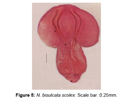
Figure 5: N. bisulcata scolex. Scale bar :0.25mm.
Description: The capsules are small and ovoid in shape. Spine shaped structures are seen on the margins of the bothridia. Tentacles are relatively short. Scolex is subcylindrical, measuring 1.38-2.40 (1.89) mm long. Bothridia are relatively large and of 0.51-1.25 (0.88) mm long. Pars bulbosa is 0.42-0.49 (0.47) mm long and 0.22-0.31 (0.32) mm wide. Pars post bulbosa is 0.28-0.37 (0.33) mm long.
Heavy metals determination
(Table 3) shows the mean concentration of the tested metals in tissues of C. gracilis larvae and their host muscles and revealed that metals accumulated in the investigated muscles were ranked : Cu > Pb> Cd. It was found that the tissues of C. gracilis accumulated considerable higher level of Cu (48.05 times), Pb (147.29 times) and Cd (209.73 times) than that accumulated in fish muscles.
| Heavy metal concentration |
Infected E. gigasmuscles | C.gracilis larvae | Ratio |
|---|---|---|---|
| Cu | 7.56±1.98 | 90.82 ±1.89 | 1 : 48.05 |
| Pb | 1.10 ± 0.35 | 250.40 ±15.21 | 1 : 227.63 |
| Cd | 0.660 ± 0.22 | 180.37 ± 17.5 | 1 : 274.28 |
Table 3: Mean ± SD of Cu, Cd and Pb concentrations in C. gracilis larvae and in their host muscles (μg/g wet weight).
Results of statistical analysis revealed that there is a moderate significant (0.557 P value ≤ 0.05) correlation between the copper content in muscles and the parasite, the lead content in the muscles and the parasite (0.532 P value ≤ 0.05) and the cadmium content in fish muscles and parasites (0.660 P value ≤ 0.05)(Tables 4-6).
| Muscles | Parasite | |
|---|---|---|
| Tissue | 1 | 0.557* |
| Parasite | 0.557* | 1 |
Table 4: Correlation between copper content in infected muscle tissue and their parasite.
| Muscles | Parasite | |
| Tissue | 1 | 0.532* |
| Parasite | 0.532* | 1 |
Table 5: Correlation between lead content in tissue, parasite.
| Muscles | Parasite | |
|---|---|---|
| Tissue | 1 | 0.660* |
| Parasite | 0.660* | 1 |
Table 6: Correlation between cadmium content in tissue, parasite.
There are relatively few studies on Trypanorhynch cestodes at the coast of the Mediterranean sea of Egypt. Previous studies focused mainly on surveying, classifying and identifying larvae species in the Red sea [18-21]. No previous data was available concerning Trypanorhync –pollution relationship. Therefore, the present study was the first to consider them in Egypt.
In the present study, the prevalence of Trypanorhynch cestode larvae was 42% among the examined three fish species.This finding was lower than that reported by Mahmoud et al. [22] (59.06%) among other Mediterranean sea fishes in Egypt while it was higher than that recorded from the Red sea by Abdou and Palm, 2008 [21] (20%) in Egypt, Al-Zubaidy and Mhaisen, 2011 [15] ( 24.3 % in mesenteries and 1.26 % of the flesh of the examined fishes) in Yemen and by Ibrahim, 2000 [23] from the Arabian Gulf (34.5% in Lethrinus sp. and 24.2% In Epinephelus sp) in Saudi Arabia. Variation in the prevalence of Trypanorhynch worldwide are expected and may be attributed to various factors including locality difference, degree of water pollution, the feeding habits of fishes as well as the selected fish species [24].
Concerning the detected Trypanorhynch sp. Larvae; Callitetrarhynchus gracilis in the present study, was morphologically agree with Carvajal and Rego [25]. It recorded the highest prevalence among the examined fish species particularly E.gigas (46%). Its prevalence is higher than those reported by Haseli et al. 2011 [26] in seven teleosts from the Arabian Gulf (21.13%), Carvalho and Luque, 2011 [27] in Trichiurus lepturus (12.5%) from Rio de Janeiro and in seven fish species from the Red Sea in Yemen by Al-Zubaidy and Mhaisen, 2011 [15] (12.5-34.6%). The record of C. gracilis from the three examined fish hosts in the present work supports its record in a wide range of different intermediate hosts in the Red Sea by Abdou and Palm [21], the low host specificity [21] and cosmopolitan distribution [4] of this species.
The distribution pattern of C. gracilic is promoted by the omnipresence of a variety of Carcharhinid elasmobranchs as definitive hosts in coastal tropical regions, combined with restricted host specificity in both second intermediate and definitive hosts [28].
Larvae of Floriceps minacanthus was isolated from Sciaena umbra and Scomber scomber fishes and was identical to that recorded by Campbell and Beveridge,[29] from viscera of few fish species from the coastal waters of Australia. From the Red Sea fishes, Abdou [30] identified F.minacanthus from Egypt with scanning electron microscopy. Floriceps sp. was also isolated from several fish species from the Arabian Gulf by El-Naffar et al. and Al-Ghais and Kardousha [31,32].
The isolated Pseudogrillotia sp. was recorded and identified according to Al-Zubaidy [33] who detected this species from Lethrinus lentjan in Yemeni coast of the Red Sea. Trypanorhynchid cestodes of the this group have been reported from four species of carangid fishes (Carangoides bajad, C. fulvoguttatus, Caranx sexfasciatus and C. melampygus) in Jeddah, Saudi Arabian coast of the Red Sea [34] and in the Australian waters [35].
Larvae of N. bisulcata is widely spread among teleost fishes of the world oceans [36]. It was previously recorded by Mahmoud et.al, 1995 [22] from Epinephelus gigas and Siganus canaleculatus from Mediterranean Sea coast in Egypt with an incidence of 7.09%. It was also reported from the Arab Gulf by Bannai, 2008 [37]. The adult of N. bisulcata occurs in sharks with a wide geographical distribution and its post larvae have low host specificity in combination with a wide oceanic distribution [38]. In the present investigation, N. bisulcata was isolated from the three examined fish species with highest prevalence among Scomer spp.(20%) and found morphologically identical to that described by Al-Zubaidy and Mhaisen, 2011 [15].In this study, The investigated Epinephelus gigas; Sciaena umbra and Scomber sp. were considered as new hosts for all the described trypanorhynchids and also all these trypanorhynchiid species are recorded for the first time from the Egyptian South-Eastern coast of Mediterranean sea as a new geographical record except for N. bisulcata.
As C. gracilis was recorded in the highest prevalence among the examined Epinephelus gigas fish, its role as bio-indicator of heavy metal pollution was studied. In present investigation, the accumulation of Cu, Pb and Cd in both C.gracilis larvae and the muscles of their host E. gigas revealed moderate significant (0.557 P value ≤ 0.05) correlation between the copper content in fish muscles and the parasite, the lead content in the muscles and the parasite (0.532 P value ≤ 0.05) and the cadmium content in muscles and parasites (0.660 P value ≤ 0.05). The mean levels of Pb and Cd concentration (1.10 and 0.66 μg/g respectively) in E.gigas muscles were found higher than all the permissible limits for human consumption while Cu level was found below the recommended levels (Table 7) . Different concentrations of heavy metals in muscle of many fish species from El-Mex Bay was recorded by Abdallah, [13] and reported that the concentrations of several metals in fishes were significantly different among the species.
| References | Cu | Pb | Cd |
|---|---|---|---|
| FAO/WHO l(1992) | 30 | 0.5 | 0.5 |
| E.O.S.(2005) | 30 | 1.0 | 0.10 |
Table 7: Maximum Permissible Limit of heavy metals in fish muscles (μg/g wet wt.) according to international standards.
Industrial and agricultural discharges are the primary sources of Pb pollution in Egypt [39]. Neurological defects, rinal disfunction and anemia are the most characterize of Pb poisoning [40]. Cadmium has effects on fish and their consumer. It acts as fish stressor, leading to metabolic alteration [41]. On human, Cd ingestion can be associated with salivation, choking attacks, abdominal pain, vertigo and loss of consciousness [42]. Metal residues problem in fish flesh are serious as reflect by high metal concentration recorded in the water and sediments [7]. The high lead and cadmium level in fish muscles in the present study could reflect the high degree of water pollution in El-Max area where fish samples were collected; this degree might be attributed to the near location of the examined area to the untreated industrial discharges and sewage effluents. The mean Cu, Pb and Cd concentration in C. gracilis larvae were ~ 48,227 and 273 times respectively higher than that of the E.gigas muscles. The data which supports the previous studies that recorded higher concentrations of heavy metals in some other intestinal fish parasites compared to those found in the tissues of their final hosts. For example, Galli et al. [43] examined the contents of Pb and Cr in one host-parasite system (Leuciscus cephalus-Acanthocephalus anguillae) and demonstrated higher concentrations of these metals in acanthocephalans than in its host. Gabrashanska and Nedeva. [44] examined the contents of Cu, Cr and Zn in two host parasite system (Vimba vimba melanops-Caryophillaeus brachycollis and Alburnus alburnus-Ligula intestinalis) and indicated higher concentrations of these metals in cestodes than their hosts. Additionally, Tenora et al. [45] recorded mean concentrations of Pb, Cr and Cd in Philometra ovata from hosts (Abramis brama, Rutilus rutilus, blicca bjoerkna) to be 160, 43 and 119 times higher than in the muscle of the hosts. It is worthy to mention that this is the first usage of C.gracilis as bio-indicator of heavy metal pollution in aquatic ecosystem [46-48]. It can be concluded that results of the present study add further evidence to the possibility of using parasites particularly of trypanorhynchiid species as indicator of metal pollution and supporting that parasites may serve as useful indicators for aquatic ecosystem pollutants where other methods of water or sediment analysis can not accurately applied.