Biochemistry & Pharmacology: Open Access
Open Access
ISSN: 2167-0501
ISSN: 2167-0501
Research Article - (2012) Volume 1, Issue 4
Bladder toxicity is one of the most troublesome side effects associated with chemotherapeutic treatments of cancer involving the use of cyclophosphamide (CP) and buthionine-SR-sulfoximine (BSO). The present study was undertaken to investigate the effects of pre-treatment with an Aloe vera plant extract (AVE) on the urotoxicity induced by acute doses of CP and BSO using a Swiss albino mice model. The modulation of toxicity was evaluated by measuring lipid peroxidation (LPO), peroxide hydrogen production (H2O2) and antioxidants in the urinary bladder of the animals. The findings revealed that Aloe vera induced remarkable protective effects in terms of both LPO and enzymatic antioxidant activities. The CP-treated mice were noted to undergo significant decreases in the activities of glutathione S-transferase (GST), glutathione reductase (GR), glutathione peroxidase (GP), and catalase (CAT) when compared to the controls. The levels of reduced glutathione (GSH) also decreased with an increase in LPO in the CP-treated animals and BSO treatment exerted an additive toxic effect in the CP-treated animals. Pre-treatment with the Aloe vera herbal extract restored all enzymatic activities back to normal, thus confirming its overall protective effect against the toxic effects of CP and BSO. The potential effect of AVE to prevent CP and BSO induced urotoxicity was also reflected by the histological analysis, indicative of its uroprotective effects. The restoration of GSH through treatment with Aloe vera may play an important role in the reversal of CP-induced apoptosis and free radical mediated LPO in urinary bladder. Due to its widespread availability in nature and relative lack of toxicity, the Aloe vera plant extract presented in the current study could be considered a potential strong candidate for future applications as an adjutant to cancer chemotherapies.
<Bladder cancer is one of the most frequently occurring cancers in the world. According to the current estimates by the American Cancer Society (ACS), this disease is the fourth most common cancer in men and eighth leading cause of death in women in the United States. Although several treatments and therapies, including surgery, radiation, and chemotherapy, have been used to reduce and prevent the rising death rates caused by this deadly disease, a number of serious concerns have often been voiced with respect to the troublesome side effects associated with each type of treatment.
Cyclophosphamide (CP) and buthionine-SR-sulfoximine (BSO) are two of the leading medications often prescribed for the treatment of cancer. CP is an effective alkylating agent widely used in the antineoplastic therapy of a variety of cancers, including lymphoma, myeloma, and chronic lymphocytic leukemia [1]. CP intake has, however, frequently been associated with a variety of troublesome side effects, including alopecia, cardiac damage, gonadotropy, hemorrhagic cystitis, hematopoetic depression, nausea, vomiting, and carcinogenicity [2] as well as lung toxicity [3]. It has also been reported to involve a number of urological side effects, such as transient irritating voiding symptoms, including dypsia, hemorrhagic cystitis, bladder fibrosis, necrosis, contracture, and vesicometral flux [4,5].
The accumulation of CP reactive metabolites in the urinary bladder has also been reported to induce reactive oxygen species (ROS) production, which cause peroxidative damage to the urinary bladder as well as to other vital organs [3]. Additionally, CP treatment has been described to bring about a decrease in the levels of reduced glutathione (GSH), which represents one of the most important organic molecules within the cell that determine its ability to suppress toxic substances, such as chemotherapeutic reactive metabolites [2,6,7].
In fact, GSH depletion is considered as an attractive strategy for the sensitization of tumour cells to certain chemotherapeutic agents. A number of reports suggested that BSO can be used effectively to reduce the level of GSH and may help to restore the sensitivity of resistant tumours to alkylating agents. BSO is a potent inhibitor of r-glutamylcysteine synthetase, has been reported to enhance the in vitro antitumor efficacy of a number of drugs including CP, melphalan, adriamycin, daunomycin and mitomycin C [8]. By inhibiting this essential enzyme, BSO has the capacity to drastically reduce glutathione content and has been shown in model systems to enhance the cytotoxic effects of specific chemotherapeutic agents and radiation therapy [9]. However, CP-induced immunosuppression may incite various types of infectious agents that have serious GSH depleting effects [10]. In other words, CP treatment might decrease the GSH content itself but the secondary infections associated with it might bring additional decreases in the levels of GSH. Accordingly, a patient undergoing CP chemotherapy would need an excessive supply of GSH-restoring antioxidants or compounds that induce GSH production.
In this context, a number of GSH-inducing compounds have been reported to be effective CP and BSO toxicity reducers in animals [8]. Several extracts from plant origin have been described to exert valuable protective and/or restorative effects on BSO and CP-induced decreases of GSH [9]. Of particular interest to the aims of the present study, Aloe plants have long been used to treat a wide array of ailments and diseases. Aloe barbadensis Miller, commonly known as Aloe vera, is one of the most popular varieties of Aloe plants whose healing properties are immense [11]. It belongs to the Liliaceal family, of which about 360 species exist. It is a cactus-like plant that grows readily in hot, dry climates and, due to its great curative properties, has often been cultivated in large quantities. Two distinct preparations of Aloe plants are most used medicinally. The leaf exudate (Aloe latex), which is a yellow, bitter liquid derived from the skin of the Aloe leaf, has strong stimulatory and laxative properties and has, therefore, been commonly used for the treatment of diabetes, coughs, wounds, ulcers, cancer, headaches, arthritis, among various other conditions [12]. The mucilaginous gel (Aloe gel), which is a clear colourless semi-solid gel extracted from the leaf parenchyma, has attractive immunomodulatory properties, and has, therefore, been commonly used as a remedy against a variety of skin disorders [13].
Public interest in Aloe vera has grown quickly, and considerable research has been conducted over the past few decades to explore the various components and properties of this herbal medicine and to find out clues as to its potential cosmetic, pharmaceutical, and therapeutic applications. In fact, although scientific evidence for the cosmetic and therapeutic effectiveness of Aloe vera is very limited, various cosmetic and medicinal formulations have been made from Aloe latex and Aloe gel. Aloe vera is rich in antioxidants and various nutrients. It contains lignin, saponins, and mono and polysaccharides (including Acemannan) [14]. The consumption of Aloe vera leaf formulations has been shown to display anti-arthritic, anti-rheumatoid, and anti-cancer properties [15] as well as anti-diabetic benefits [16]. It has been commonly used for the topical treatment of a variety of ailments and disorders, including chronic wounds, thermal injuries, skin infections, inflammations, oral ulcers, and psoriasis [17] as well as for the prevention of ultraviolet (UV)-induced immunosuppressions [18].
Considering its widespread availability in nature, relative richness in antioxidants, and distinguished healing properties, the present study was undertaken to investigate the effects of the oral administration of an Aloe vera plant extract in terms of antioxidant activities, lipid peroxidation (LPO) and peroxide hydrogen (H2O2) production in the urinary bladder of CP and BSO treated mice that were pre-disposed or concomitantly exposed to a GSH reducing agent in the form of either an infection or antibiotic use.
Aloe vera plant extract (AVE)
The present study used a total aqueous semisolid extract of Aloe vera that was purchased from the Plant Extract Division of the local Central Pharmacy, Tunisia. The plant extract had moisture and ash contents of 12% and 8%, respectively. The pH of 10% aqueous solution of extract was 4.6. The authenticity of the extract was certified by the manufacturer’s expert taxonomist.
Chemicals
Cyclophosphamide monohydrate (2-(bis-(2-chloroethyl) amino) tetrahydro- 2H-1 ,3,2-oxazaphosphorine 2-oxide monohydrate); CAS 6055-1 9-2; BSO (L-buthionine-SR-sulfoximine); CAS 5072-26-4 were purchased from Sigma–Aldrich Co., St. Louis, MO, USA.
Animals
The experimental essays of the present study were conducted on male Swiss albino mice (25 ± 2 g) provided by the animal service of the Pasteur Institute of Tunis, Tunisia. All care and animal handling procedures were performed in accordance with the guidelines of the Institutional Animal Ethics Committee (IAEC). The animals were bred and maintained under standard laboratory conditions (temperature 25 ± 2°C; photoperiod of 12 hrs). Commercial pellet diet and water were given ad libitum.
Dosage and experimental groups
BSO, CP and plant extract of Aloe vera (AVE) were suspended in normal saline. The animals were divided into seven groups, Groups I–VII, of six mice each. Group I (Control) referred to control mice that were administered normal saline p.o. for 10 days and a single i.p. injection on the 10th day of treatment. Group II (BSO) referred to the mice that received BSO (500 mg/kg body wt) and to which a single i.p. injection was administered on the 10th day. Group III (CP) designated the mice that received CP (50 mg/kg body wt.) and a single i.p. dose on the 10th day. Group IV (BSO + CP) referred to animals that were administered BSO i.p. 5 hrs before CP administration. Group V (CP + AVE) referred to animals that were administered plant extract for 10 days and a single i.p. injection of CP on the 10th day. Group VI (BSO + AVE) designated animals that were given plant extract treatment (100 mg/kg body wt.) p.o. for 10 days and a single i.p. injection of BSO on the 10th day. Group VII (BSO + CP + AVE) referred to the animals that were administered plant extract for 10 days and CP and BSO on the 10th day. Dosing was performed in such a way that all of the animals could be sacrificed on the same day, i.e., day 11. The selection of BSO and CP doses was based on pilot experiments that involved the assay of a wide range of doses and on data provided from previously published reports [9,19].
Biochemical investigations
Upon the completion of the treatment, the animals were sacrificed under mild anesthesia and their bladders were removed. The bladder tissue was homogenized in chilled phosphate buffer (0.1 M, pH 7.4) using a Potter homogenizer. The homogenate was centrifuged at 10,500g for 30 min at 4°C to obtain the post-mitochondrial supernatant (PMS), which was used for the biochemical measurements as described below.
Cell culture and peroxide hydrogen production:
Cell culture: The urothelial cell line T24 was employed as an in vitro model of the human bladder because of its ability to react to adequate stimuli, obtained from Pateur Institute of Tunis, Tunisia, was routinely grown in 75 cm2 flasks (Nunc, Denmark) and maintained in minimum essential medium (MEM) (Invitrogen, Glasgow, UK) supplemented with 10% foetal bovine serum (FBS, Hyclone, Logan, UT), 100 units/ ml penicillin and 100 lg/ml streptomycin. Cultures were maintained in a humidified atmosphere with 5% CO2 at 37°C. Cell dissociation was achieved with 0.05% trypsin-0.02% EDTA. Briefly, cells were seeded on 24-well culture plates in medium at an approximate density of 105 cells/cm2 and, after 24 hrs stabilization, bladder urothelial cell line T24 were co-cultured with medium containing various concentrations of CP and BSO (200 and 800 μM) and Aloe vera plant extract (10, 50 and 100μM) for 24 hrs. The concentration of CP was selected based on previously reported cytotoxic levels in cultured cells [20]. For stock solution, CP and BSO were dissolved in MilliQ Plus sterilized water at the concentration of 800 mM and Aloe vera plant extract was dissolved in dimethyl sulfoxide (DMSO) to obtain a 100mM. The experimental concentrations were freshly prepared in the basal medium with a final DMSO concentration of 0.1%.
Measurement of H2O2: Measurement of H2O2 was carried out by the ferrous ion oxidation xylenol orange (FOX1) method [21]. The FOX1 reagent consisted of 25mM sulphuric acid, 250 lM ferrous ammonium sulfate, 100μM xylenol orange and 0.1 M sorbitol. Briefly, 100μl of culture medium were added to 900μl of FOX1 reagent vortexed and incubated during 30min at room temperature. Solutions were then centrifuged at 12000g for 10 min; the amount of H2O2 in the supernatants was determined using spectrophotometer at 560nm.
Lipid peroxidation: LPO was measured using the procedure of Uchiyama and Mihara [22]. The assay mixture consisted of 0.67% thiobarbituric acid; TBA (Sigma-Aldrich), 10 mM; butylated hydroxy toluene BHT (Sigma-Aldrich), 1%; ortho-phosphoric acid (Sigma- Aldrich); and tissue homogenate in a total volume of 3ml. The rate of LPO was expressed as nmol of TBA reactive substances (TBARS) formed/h/g of tissue using molecular extinction coefficient epsilon (?) of 1.56 x 10⁵ M-1 cm-1.
Measurement of GSH: GSH content was measured in the PMS of urinary bladder by the method of Haque et al. [9]. PMS (1ml) was precipitated with 1ml of 4% sulfosalicylic acid (Sigma-Aldrich). The samples were incubated at 4°C for 1 hr and then centrifuged at 1200g for 15 min at 4°C. The assay mixture consisted of 0.2 ml of filtered aliquot, 2.6 ml of sodium phosphate buffer (0.1 M, pH 7.4), and 0.2 ml 100 mM DTNB (dithio-bis-2-nitrobenzoic acid, Sigma-Aldrich) in a total volume of 3 ml. The absorbance of the reaction product was measured at 412 nm, and the results were expressed as nmol GSH/g of tissue.
Antioxidant enzyme measurements: Glutathione-S-Transferase (GST) activity was assayed using the method of Haque et al. [9]. The reaction mixture consisted of 1.675 ml sodium phosphate buffer, 0.2 ml of 1 mM GSH (Sigma-Aldrich), 0.025 ml of 1 mM CDNB (1-chloro- 2,4-dinitrobenzene, Sigma-Aldrich), and 0.1 ml of PMS in a total volume of 2 ml. The change in absorbance was recorded at 340 nm and the enzyme activity calculated as nmol CDNB conjugates formed/ min/mg protein using epsilon (?) of 9.6x10³ M-1 cm-1. GR (glutathione reductase) activity was assayed by the method of Sharma et al. [19]. The assay mixture consisted of 1.6 ml sodium phosphate buffer, 0.1 ml of 1 mM ethylenediamine tetra acetic acid disodium salt (EDTA, Sigma-Aldrich), 0.1 ml NADPH (nicotinamide adenine dinucleotide phosphate reduced, Sigma-Aldrich), and 0.1 ml oxidized glutathione (Sigma-Aldrich) and PMS (0.1 ml) in total volume of 2 ml.
Enzyme activity measured at 340 nm was calculated as nmol NADPH oxidized/min/mg of protein, using epsilon (ε) of 6.22 x10³ M-1 cm-1. Glutathione peroxidase (GP) activity was assayed using the method of Sharma et al. [19]. The assay mixture consisted of 1.49 ml sodium phosphate buffer, 0.1 ml EDTA (1 mM), 0.1 ml sodium azide (1 mM, Central Pharmacy of Tunis, Tunisia), 0.1 ml of 1 mM GSH (Sigma– Aldrich), 0.1 ml NADPH (0.02 mM), 0.01 ml of 0.25 mM hydrogen peroxide (H2O2, CDH Chemicals), and 0.1 ml PMS in a total volume of 2 ml. Oxidation of NADPH was recorded spectrophotometrically at 340 nm. The enzyme activity was calculated as nmol NADPH oxidized/min/ mg of protein using epsilon (ε) of 6.22 x10³ M-1 cm-1. CAT (catalase) activity was assayed using the method of Haque et al. [9]. The assay mixture consisted of 1.95 ml phosphate buffer, 1 ml H2O2 (0.09 M), and 0.05 ml of PMS at a final volume of 3 ml. Change in absorbance was recorded kinetically at 240 nm. CAT activity was calculated in terms of nmol H2O2 consumed/min/mg of protein.
Protein measurement: Protein was measured by the method of Lowry et al. (1951).
Histological studies
Bladder urothelium tissue, extracted from the control and treated mice was fixed in 10% buffered formalin and was processed for paraffin sectioning. Sections of about 5 μm thickness were stained with hematoxylin and eosin to examine under light microscope.
Statistical analysis
Single factor one-way analysis of variance (ANOVA) was performed to determine significant differences in results of various groups. The statistical significance level was set at P values < 0.05. A Student- Newman–Keuls test was then carried out to analyze and compare the significance of the different treatment groups. The values were expressed as mean ± SE.
No mortalities and significant changes in the body weight of the different groups of animals were recorded during the treatment.
Lipid peroxidation (LPO)
The findings revealed that BSO treatment brought about a significant increase (P < 0.01) in the LPO levels in the bladder over the control values (Figure 1). The administration of CP was also noted to induce LPO in significant way. Likewise, the cumulative effect of BSO + CP resulted in significant increase in the LPO as compared to Group I (control). BSO + CP group (Group IV) also underwent a significant (P < 0.01) increase in the levels of LPO when compared the CP group (Group III) alone (Figure 1). The animals pre-treated with Aloe vera plant extract (AVE) and subsequently exposed to BSO (BSO + AVE), on the other hand, were noted to undergo a significant (P < 0.01) reduction in terms of LPO levels in the bladder. When the values obtained for the CP + AVE group (Group V) were compared to those of the CP group (Group II), AVE treatment was also observed to significantly (P < 0.05) reduce the levels of LPO in the bladder. As illustrated in Figure 1, the comparison between the LPO values obtained for the LPO of BSO + CP group (Group IV) and those of the BSO + CP + AVE group clearly shows a significant decrease (P < 0.01) in the case of the latter (Group VII).
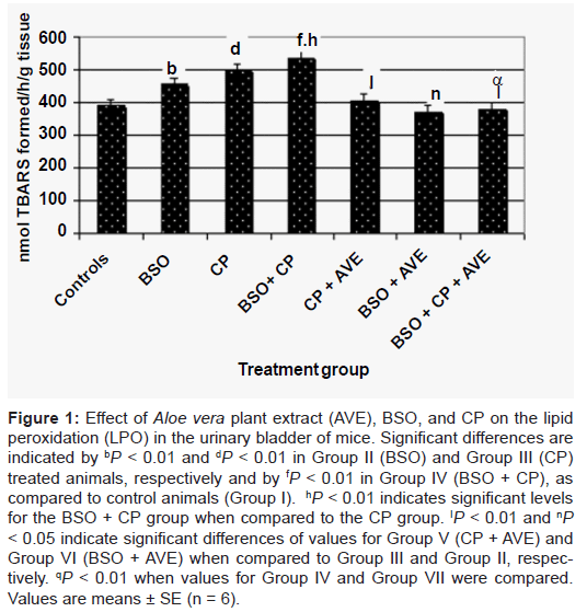
Figure 1: Effect of Aloe vera plant extract (AVE), BSO, and CP on the lipid peroxidation (LPO) in the urinary bladder of mice. Significant differences are indicated by bP < 0.01 and dP < 0.01 in Group II (BSO) and Group III (CP) treated animals, respectively and by fP < 0.01 in Group IV (BSO + CP), as compared to control animals (Group I). hP < 0.01 indicates significant levels for the BSO + CP group when compared to the CP group. IP < 0.01 and nP < 0.05 indicate significant differences of values for Group V (CP + AVE) and Group VI (BSO + AVE) when compared to Group III and Group II, respectively. qP < 0.01 when values for Group IV and Group VII were compared. Values are means ± SE (n = 6).
Reduced glutathione
The findings revealed that, when compared to the control values (5.23 nmol GSH/g tissue), the BSO, CP and BSO + CP-treated groups underwent significant (P < 0.01) decreases of 2.9, 4.1 and 1.9 nmol GSH/g tissue in GSH, respectively (data generated for Cellular GSH of urinary bladder are shown in Figure 2). Likewise and when compared to the CP group (Group III), the BSO + CP group (Group IV) showed a significant (P < 0.01) decrease in terms of GSH levels. As shown in Figure 2, when the BSO and BSO + AVE groups were compared, the GSH content of the latter was noted to undergo a significant increase (P < 0.01). Similarly, the GSH content in the bladder of the CP + AVE group (Group V) showed a significant increase (P < 0.01) when compared to the group given only CP (Group III). When the GSH values obtained for the BSO + CP (Group IV) group were compared to those of the BSO + CP + AVE group (Group VII), a significant (P < 0.01) restoration in GSH was recorded (Figure 2).
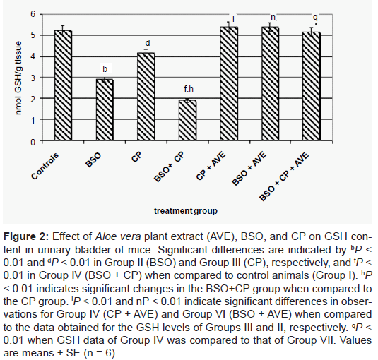
Figure 2: Effect of Aloe vera plant extract (AVE), BSO, and CP on GSH content in urinary bladder of mice. Significant differences are indicated by bP < 0.01 and dP < 0.01 in Group II (BSO) and Group III (CP), respectively, and fP < 0.01 in Group IV (BSO + CP) when compared to control animals (Group I). hP < 0.01 indicates significant changes in the BSO+CP group when compared to the CP group. IP < 0.01 and nP < 0.01 indicate significant differences in observations for Group IV (CP + AVE) and Group VI (BSO + AVE) when compared to the data obtained for the GSH levels of Groups III and II, respectively. qP < 0.01 when GSH data of Group IV was compared to that of Group VII. Values are means ± SE (n = 6).
Antioxidant enzymes
BSO and CP treatments were observed to induce significant (P < 0.01) decreases in terms of GST, GR, GP and CAT activities in the bladder when compared to the control group (Table 1). The BSO + CP group also showed an additive significant (P < 0.01) decrease in the activities of GST, GR, and GP when compared to the CP or control group. However, no significant difference was observed in terms of CAT activity between the animals of group IV (BSO + CP) and group III (CP). The activities of those antioxidant enzymes increased significantly (P < 0.01) in both the BSO + AVE (Group VI) and the CP + AVE (Group V) groups when compared to their respective controls, the BSO (Group II) and CP (Group III) groups (Table 1). The animals treated with AVE and subsequently exposed to BSO + CP (Group VII, BSO + CP + AVE treatment) displayed a significant increase (P < 0.01) in the activities of all the antioxidant enzymes when compared to the BSO + CP group (Group IV).
| Group | Activity of antioxidant enzyme | |||
|---|---|---|---|---|
| GST | GR | GP | CAT | |
| I (Controls) | 144 ± 3 | 143 ± 4 | 155 ± 5 | 104± 3 |
| II (BSO) | 100 ± 2b | 92 ± 4 b | 122 ± 3b | 97± 2b |
| III (CP) | 115 ± 3d | 122 ± 5d | 139 ± 4c | 84± 3d |
| IV (BSO+CP) | 79 ± 3f,h | 86 ± 2 f,h | 118 ± 3f,h | 82± 3f |
| V (CP+AVE) | 153 ± 5I | 152 ± 5I | 162 ± 2I | 107± 5I |
| VI (BSO+AVE) | 146 ± 3n | 149 ± 6n | 156 ± 4n | 113± 7n |
| VII (BSO+CP+AVE) | 145 ± 4q | 148 ± 6q | 154 ± 4q | 104± 5q |
CP: Cyclophosphamide; BSO: Buthionine Sulfoximine; AVE: Aloe vera plant extract Values are means ± SE (n = 6). GST expressed as nmol CDNB conjugates/min/mg protein, GR as nmol NADPH oxidized/min/mg protein, and GP as nmol NADPH oxidized/ min/mg protein. CAT activity is expressed as nmol H2O2 consumed/min/mg protein. Significant differences are indicated by bP < 0.01, dP < 0.01, cP < 0.05, and fP < 0.01 when compared to control animals. (Group I) nP < 0.01 when compared to Group II, hP < 0.01 and IP < 0.01 when compared to CP-treated animals (Group III), and qP < 0.01 when compared to Group IV.
Table 1: Activities of antioxidant enzymes in the urinary bladder of mice in different treatment groups.
Hydrogen peroxide (H2O2) production in vitro
Figure 3 showed the effects of CP and AVE on H2O2 production in urothelial cell line T24. To check if the combination of CP and AVE had any benefits, cells were treated with CP (200 and 800μM) and varying doses of AVE (10, 50 and 100μM) for 24 h. The levels of H2O2 generated in medium of cells were significantly (P < 0.05) increased by 159 and 241% compared to controls after exposure to CP (200 and 800μM) respectively, and were significantly (P < 0.05) deceased by (27%, 41% and 20%) cells co-culture with AVE (10, 50 and 100μM) and CP at dose (200μM) and by (40%, 49% and 25%) at dose (800μM) compared with CP alone (200 and 800μM) respectively.
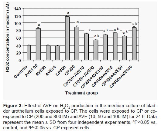
Figure 3: Effect of AVE on H2O2 production in the medium culture of bladder urothelium cells exposed to CP. The cells were exposed to CP or coexposed to CP (200 and 800 lM) and AVE (10, 50 and 100 lM) for 24 h. Data represent the mean ± SD from four independent experiments. aP<0.05 vs. control, and kP<0.05 vs. CP exposed cells.
Histological examination of bladder urethelium
Figure 4 illustrates the histopathological assessments of bladder urothelium tissue in experimental mice. Histopathological examination of the urothelium tissue revealed that CP treatment caused abnormal cellular arrangement with few pyknotic nucleus, vacuolated spaces and hemorrhage. However, co-administration of AVE at 100 mg/kg body weight prevented these changes and maintained normal architecture with less number of pyknotic nuclei and showed almost normal architecture similar to that of the untreated control. There were no histological alterations in the bladder urothelium of positive controls treated with AVE alone when compared to negative controls.
Chemotherapeutic agents are known to influence the function of the urinary tract. Cyclophosphamide (CP) is an alkylating agent widely used in cancer chemotherapy [1]. Its cytotoxic effects are the result of chemically reactive metabolites that alkylate DNA and protein, producing cross-links [2]. It has been reported that oxidative stress mediated disruption of redox balance after CP exposure generates biochemical and physiological disruptions [11]. Several studies indicate that CP has a pro-oxidant character, and that the generation of oxidative stress after CP administration leads to a decrease in the activities of antioxidant enzymes and an increase in lipid peroxidation (LPO) in the liver, lung, and serum of mice and rats [3]. Reactive oxygen species (ROS), such as the nitric oxide radical (NO), are included in the pathogenesis of CP-induced cystitis. It has also been reported that increases in ROS, such as NO, lead to bladder oedema, inflammation, and extravasation [23].
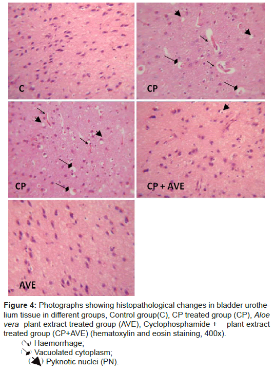
Figure 4: Photographs showing histopathological changes in bladder urothelium tissue in different groups, Control group(C), CP treated group (CP), Aloe vera plant extract treated group (AVE), Cyclophosphamide + plant extract treated group (CP+AVE) (hematoxylin and eosin staining, 400x).
➘Haemorrhage;
➘Vacuolated cytoplasm;
➘Pyknotic nuclei (PN).
Despite its potent antitumor activity, CP-induced urotoxicity remains the major limiting step for its clinical application. Urotoxic effects of CP can be dose limiting and have proven fatal. Since the bladder is the site for the storage of urine, the concentration of CP toxic metabolites is higher in the bladder than in any other organ. However, the mechanisms that determine the individual susceptibility to CP and the mediation of bladder toxicity remain unclear.
Treatment with buthionine sulfoximine (BSO), a potent inhibitor of r-glutamylcysteine synthetase, has been reported to enhance the in vitro antitumor efficacy of a number of drugs, including CP [24]. In fact, it has been shown in several animal model systems to enhance the cytotoxic effects of specific chemotherapeutic agents and radiation therapy [25]. BSO and other agents directed at modulating glutathione levels affect the content of this metabolite in normal tissues as well as in tumors. Several studies have shown that depletion of GSH after BSO treatment result in a variety of toxic effects, including cardiotoxicity, hepatic damage, and respiratory problems [26,27].
Considering the various toxic effects associated with CP treatment used alone or in combination with BSO, special attention has been given in recent research to the search for novel compositions for use as broadspectrum chemoprotectants to help alleviate or prevent such damages. Such novel compositions could serve as adjutants to chemotherapies to protect patients’ normal cells from the toxicity associated with such therapies. In this respect, folk medicine seems to be gaining increasing respect from the medical research community. More succinctly, there is a growing tendency in recent research to look for tumor-therapeutic agents of natural origins. In fact, although neither its active components nor mechanisms of action are fully understood, folk medicine has often been used with enormous success for the treatment of various diseases.
In this context, a number of natural products and compounds have been shown to reduce BSO and CP-induced toxicity mainly due to their antioxidant action [8]. Several extracts from plant origin have been described to exert valuable protective and/or restorative effects on CP-induced decreases of GSH [9]. Of particular interest, the Aloe vera plant has often been reported to possess attractive healing properties. Accordingly, the present study was undertaken to investigate the effects of the oral administration of an Aloe vera plant extract in terms of peroxide hydrogen (H2O2) production, antioxidant and lipid peroxidation (LPO) activities in the urinary bladder of CP and BSO treated mice that were pre-disposed or concomitantly exposed to a GSH reducing agent in the form of either an infection or antibiotic use.
The findings revealed that throughout the treatment no mortalities were recorded for the different experimental groups of animals, and that significant changes were observed in terms of their body weights. In fact, when the values pertaining to GSH reduction were compared, BSO was noted to exhibit more significant effects than CP. A difference of 42% was recorded between both agents, which, as previously reported in the literature, confirm that BSO is a more potent depletor of GSH than CP [28]. CP-induced depletion of GSH is primarily mediated by the interaction of its reactive metabolite, acrolein, with GSH (Kehrer and Biswal 2000). Acrolein not only interacts with GSH but also with cysteine, which is one of the constituent amino acids of GSH (Kehrer and Biswal 2000). Several reports in the literature have indicated that compounds containing free sulfhydryl groups may protect from the urotoxic effects of cyclophosphamide. A number of sulfhydryl (-SH) compounds, and cysteine itself; have been observed to protect experimental animals from the toxic effects of CP [28].
The intra-peritoneal administration of CP was noted to significantly induce LPO in the bladder. In fact, lipid peroxidation is widely used as an indicator to reflect oxidative stress and cell membrane damage. Free radicals, such as superoxide anion and hydroxyl radical, exert their toxic effect by acting on DNA, membrane proteins, and lipids. CP-induced LPO has been reported in different tissues of exposed animals. In CPinduced LPO, the role of acrolein has also been implied [29]. It has been suggested that by binding to nucleophilic amino acids, acrolein could directly affect transcription as well as modulate this process through its ability to deplete GSH (Kehrer and Biswal 2000).
As far as BSO treatment was concerned, the findings revealed that it resulted in the depletion of GSH and the increase of LPO in the urinary bladder. The depletion of GSH is also reported to increase the susceptibility of cells to apoptosis [30]. A depletion of intracellular GSH has been described in a number of different apoptotic systems, with several studies showing that GSH loss in cells undergoing apoptosis is the result of accelerated efflux rather than depletion by oxidation [31].
Moreover, when BSO and CP were administered together, an additive effect was observed in case of GSH and LPO among several other parameters. The purpose of using BSO along with CP was to study a likely scenario where the host is exposed to a combination of GSH depleting agents, including pathogens, and to assess whether the herbal extract treatment of Aloe vera has any modulatory effect on their commutative/additive effect.
In this study, pre-treatment with Aloe vera extract not only showed a marked protective effect with regards to CP urotoxicity but also played an efficient protective role for the animals treated with the CP + BSO combination (Group VII).
The pre-treatment with the Aloe vera extract was noted to restore the depleted GSH and other antioxidants and, at the same time, reduced the LPO levels in the bladder. In fact, CP-induced immunosuppression is likely to increase the incidence of infections that may deplete GSH, as many infectious agents are reported to deplete GSH [10]. Overall, the findings indicate that the herbal extract presented in the current study is a promising immunomodulatory herbal extract with powerful GSH restoring effects. This extract could, therefore, open new opportunities for the reduction of the adverse effects of CP and BSO cancer treatments.
In fact, the immunomodulatory effect of Aloe vera has previously been demonstrated in mice [32]. Aloe-Emodin (AE) was found to alter the expression of a number of proteins involved in oxidative stress, cell-cycle arrest, anti-metastasis, and apoptosis [33]. Moreover, AE was capable of enhancing the intracellular level of reactive oxygen species (ROS). This may not unexpected since AE has a quinone structure that is highly redox active in nature and that can form a redox cycle with their semiquinone radicals, thus leading to the formation of ROS. AE was recently found to be able to induce DNA damage through excessive production of ROS in human lung carcinoma cells [34]. This anticancer specificity further suggests that AE could be a potent chemotherapeutic agent or chemo-preventive compound. It is noteworthy that endothelial cells have recently been reported to be sensitive to AE [35]. This property was suggested to be useful for the modulation of angiogenesis as well as antitumor effects [35].
Moreover, the findings presented in the current work suggest that Aloe vera has a potent chemo-preventive potential for the inhibition of the toxicity processes by modulating lipid peroxidation and cellular antioxidant environments. The findings of the present study demonstrate that Aloe vera extract pre-treatment prevented CP urotoxicity that was primarily mediated by LPO and depleted GSH by reversing those effects.
In the present study, we have reported that exposure of urothelial cell line T24 to CP (200 and 800μM) increased significantly H2O2 production in extracellular medium indicating the role of ROS generation as primary mechanism for CP-induced toxicity. The ability of Aloe vera extract to exert great effect on CP-induced cellular injury is consistent with its increased potency in reducing ROS (e.g. H2O2) generation. Our results are consistent with those of Fu et al. [36] who have found that plant extract, has an antioxidative activity and free radical scavenging properties in vitro, which can scavenge various oxidizing radicals such as OH•, NO2•, O2•, RNS•.
Furthermore, Histopathological examination of the bladder urothelium tissue reveals that CP treatment causes abnormal cellular arrangement with few pyknotic nuclei, vacuolated spaces and hemorrhage. However, co-treatment with AVE prevents these changes and also maintains normal architecture with less number of pyknotic nuclei.
In short, the present work is the first attempt to focus on the biological activities and bladder urotoxicity in Tunisia. In fact, the plant extract presented here showed a significant antioxidant potential in different assays in vivo. The findings provided ample support for the efficiency of the Aloe vera extract as a natural antioxidant agent. Due to its widespread availability in nature and relative lack of toxicity, this herbal extract could be considered a potential strong candidate for future applications as an adjutant to cancer chemotherapies. Accordingly, further studies are currently underway in our laboratories to further explore this extract in terms of cell cycle regulatory activity and antioxidant activity, to further validate its efficacy as an antitumor agent, and to make its use suitable for potential pharmaceutical applications as a therapeutic agent.
This study was supported by the Tunisian Ministry of Higher Education and Scientific Research and Technology and the Tunisian Ministry of Public Health. The authors would like to express their sincere gratitude to Prof. ANOUAR Smaoui from the English Section at the Sfax Faculty of Science for his valuable language polishing and proofreading services.