Reproductive System & Sexual Disorders: Current Research
Open Access
ISSN: 2161-038X
ISSN: 2161-038X
Research Article - (2022)Volume 11, Issue 3
Background: The etiology of embryonic demise is multifactorial, with chromosomal abnormalities being the most common (60%). The purpose of this study is to evaluate the correlation between a serum biomarker, Placental Growth Factor (PlGF) and an ultrasonographic parameter, the distance between Yolk Sac (YS) and embryo (DYSE), in assessing the prognosis of first trimester pregnancies outcome.
Materials and findings: The study is a case-control prospective analysis that includes two groups of patients: 81 patients with viable first-trimester pregnancy and 89 patients with embryonic demise, all of the patients having amenorrhea between 6 and 11 weeks. The endovaginal ultrasonographic exploration was performed to evaluate the distance between the lower pole of the embryo and the yolk sac. From each subject enrolled in the study, 20 ml of blood was collected for PlGF dosing.
Results: Regarding the DYSE in the case group, lower values were observed compared to the control group, the difference being statistically significant. In the statistical analysis of serum PlGF values, statistically significant differences were observed between the two groups (p<0.0001).
Conclusions: DYSE has a high positive predictive value in identifying pregnancies with potentially reserved outcome, this study demonstrating that a DYSE<3 mm may lead to an unfavorable pregnancy outcome. The low serum level of PlGF is associated with an increased rate of non-viable embryos.
Automatic gantry; MIG; Welding system; PLC; Weld quality
Undeveloped embryos are an element with an expanded rate, with over 25% of the 21st century female populace being impacted by this pathology [1]. In over 40% of cases, the etiology of undeveloped pregnancies is the presence of chromosomal anomalies [2]. Fruitful blastocyst implantation requires exact synchronization between the embryo and the uterine climate. The endometrium is a particular and hormonal tissue, which doesn't permit incipient embryos to stick all through the monthly cycle [3].
Ultrasound assessment is the gold standard evaluation for monitoring the early pregnancy outcome [4]. Antenatal assessment looks to expand the adequacy of screening techniques and work on strategies for diagnosing first-trimester pregnancies whose potential outcome can be reserved. At first, the undeveloped embryo is identified promptly near the Yolk Sac (YS). Assuming the undeveloped embryo is isolated from it, detachment is because of its growth. As the Crown-Rump Length (CRL) increases, the distance between the embryo and YS likewise becomes larger. For embryos with a CRL of 5 mm or less, there should be no division of the YS from the embryo or a tiny one (<2 mm). A little distance (thought about 3 mm in the most recent reviews) between an embryo and YS with CRL larger than 5 mm is a negative prognostic marker. A number of sonographic characteristics can indicate that a pregnancy is not viable. The entity known as the yolk stalk sign is one of them. The yolk stalk is also named the vitelline duct or the omphalomesenteric duct, has an endodermal origin and serves the connection between the yolk sac and the midgut. It is very well known the positive predictive value of the "yolk stalk sign" in determining early pregnancy failure for an embryo with a CRL of 5 mm or less and no visible heartbeat. The yolk stalk sign is important for evaluating pregnancy outcome and can be utilized autonomously or in mix with other atipical signs [5]. Notwithstanding this strategy we added a touchy and explicit biomarker to decide the rate of viables embryos in the first trimester of pregnancy. PlGF is an angiogenic protein delivered by the placenta and is involved in trophoblastic invasion of the maternal spiral arteries [6,7]. Pregnancy is a physiological interaction wherein a fine harmony among inflammatory and antiinflammatory substances is necessary to fight pregnancy failure [8-10]. Among the few inflammatory substances present in the uterus, PlGF plays a transcendent part [11-17]. PlGF is upregulated by various triggers, for example, hypoxia, cytokines, development factors and hormones, all processes being present during implantation and placenta development [18-21].
The significance of PlGF, an angiogenic factor very notable as marker of typical vascularization of the first trimester pregnancy, was featured by the new studies of critical contrasts in its appearance in tissues of a physiological pregnancy contrasted with a failed one [22,23]. PlGF is the most plentiful angiogenic factor present in physiological first trimester pregnancy, with a vital job in pregnancy failure [24]. The point of the current study is to assess the relationship between's a serum biomarker (PlGF) and an ultrasonographic marker, the Distance between Yolk Sac and Embryo (DYSE), in monitoring the result of the first trimester pregnancies.
This prospective, observational study was performed between April 2016 and December 2017 and it included pregnant women who got antenatal care at “Dominic Stanca” Obstetrics and Gynaecology Clinic, Cluj-Napoca, Romania. All subjects gave their informed consent for inclusion before they took part in the study. The study was directed in accordance with the Declaration of Helsinki, and the protocol was approved by the Ethics Committee of the University of Medicine and Pharmacy “Iuliu Hatieganu” Cluj-Napoca no 85/24.03.2016.
Patients were splited in 2 groups-the control group with 81 firsttrimester viable pregnancies with DYSE >4 mm and the case group with 95 poor viability markers pregnant patients with DYSE ≤ 3 mm, both groups having amenorrhea between 6-11 weeks. As far as the number of patients included in the study of each group, homogeneous lots were made in every amenorrhea week. For 6 patients of the case group, the forecast of the considered ultrasonographic parameter to a bad outcome was not affirmed by repeated evaluations, being accordingly rejected from the study. In the end, the control group was formed by 89 patients.
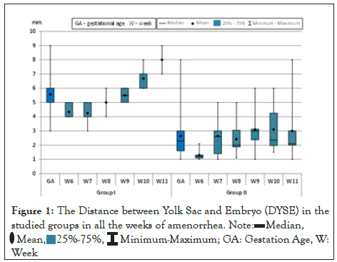
Ultrasound assessment was performed with a 6.5 MHz nominal frequency endovaginal probe by the same expert to decrease interobserver variety, using a Toshiba Aplio 300 machine. To asses DYSE, it was additionally important to independently assess the parameters included: Crown-Rump Length (CRL) and appearance and size of YS. There were on average 2-3 sequential assessments, performed at standard time of every 3-5 days, until the last conclusion of pregnancy failure. The estimations of DYSE were performed utilizing the sagittal section and assesing 3 unique distances from the inferior pole of the embryo and YS, the smallest of the 3 distances being considered for examination (Figure 1). The patients required an alternate number of assessments until affirmation of the absence of embryonic cardiac activity. During their first visit, from each subject signed up for the study, 20 ml of blood were collected by venipuncture into anticoagulant-free tubes for PlGF dosing. Blood tests were centrifuged for 15 minutes at 300 g. The serum was collected and stored at -20°C for up to 4 days, and afterward it was put away at -70°C. To get the information we utilized the Human PlGF ELISA Kit, with range of detection between 15.625-1000 pg/ml, with a sensitivity of under 9.375 pg/ml.

Figure 1:The Distance between Yolk Sac and Embryo (DYSE) in the
studied groups in all the weeks of amenorrhea. Note:

The Shapiro-Wilk test was utilised to test the normal distribution. On account of information with normal distribution the t (Student) test was utilised and on account of values with uneven distribution or ranks the non-parametric Mann-Whitney (U) test was used. The Pearson correlation coefficient (r) was used to seek the correlation between two continuous quantitative variables with uniform distribution. In the case of variables with uneven distribution, the correlation coefficient of the Spearman ranks (ρ) was used. The examinations of the correlation coefficients were performed utilizing Colton's rule. The Odds Ratio (OR) of PlGF was obtained using logistic regression analysis.
No statistically significant difference between the 2 groups (p>0.05) was found in the statistical analysis of the Gestational Age (GA) (Table 1).
| Indicator | Group | Mean ± SD | Statistical significance (p) |
|---|---|---|---|
| GA | I | 8.49 ± 1.59 | 0.779 |
| II | 8.56 ± 1.62 |
Abbrevation: GA:Gestational age,
Note: Results are expressed as mean (weeks) ± standard deviation
Table 1: Gestational age: Comparative analysis and statistical significance.
No statistically significant difference between the two groups (p>0.05) was found in the statistical analysis of the CRL values.
From the 170 patients, 81 patients (47.65%) had viable embryos and with the CRL situated between 6.7 mm and 44.1 mm, and 89 patients (52.35%) had non-viable embryos with the CRL ranging between 6.6 mm and 33.4 mm (Table 2). It is worth mentioning that the normal CRL values between these gestational ages are between 5.5 mm and 60 mm.
| Week | Group | Mean ± SD | Statistical significance (p) |
|---|---|---|---|
| DYSE | I | 5.57 ± 1.33 | <0.0001 |
| II | 2.6 ± 1.43 | ||
| CRL | I | 20.8 ± 1.10 | 0.49 |
| II | 19.5 ± 1.01 |
Abbrevations: DYSE: distance between yolk sac and embryo; CRL: Crown-Rump Length
Note: Results are expressed as mean (mm) ± standard deviation
Table 2: Ultrasonographic indicators: Comparative analysis and statistical significance.
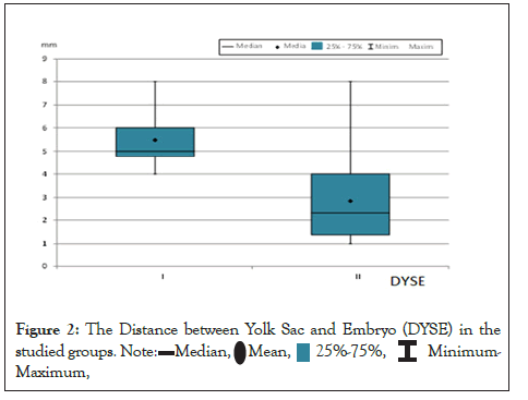
In the statistical analysis of DYSE values, statistically significant differences were found between the 2 groups (p<0.001) in all of the weeks of amenorrhea examined (Figure 1). It was found that a DYSE of <3 mm is correlated with a negative outcome of pregnancy viability (Figure 2).

Figure 2: The Distance between Yolk Sac and Embryo (DYSE) in the
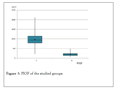
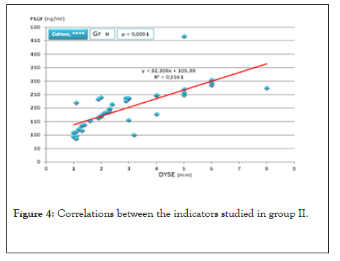
In the statistical analysis of the values of Placental Growth factor (PlGF), statistically significant differences were found between the two groups (p<0.001) (Figures 3 and 4). It is known that the normal ranges of PlGF at those gestational ages are situated between 750-1300 ng/ml.

Figure 3: PlGF of the studied groups.

Figure 4: Correlations between the indicators studied in group II.
After we examined a comparative analysis of the studied indicators we detected that there is a correlation between the serum level of PlGF and the negative outcome of the first trimester pregnancy but also that a DYSE value smaller than 3 mm correlates with a low level of PlGF (Tables 3 and 4).
| Indicators | Group | Media | ES | Median | SD | Min | Max | Statistical significance |
|---|---|---|---|---|---|---|---|---|
| DYSE | I | 5,48 | 0.1961 | 5 | 12,401 | 4 | 8 | <0.0001 |
| II | 2,85 | 0.2747 | 2,3 | 17,372 | 1 | 8 | ||
| PlGF | I | 970,81 | 5,08,447 | 9,42,013 | 32,15,703 | 22,638 | 2100,88 | <0.0001 |
| II | 197,88 | 1,19,001 | 189,85 | 7,52,629 | 86,45 | 465,91 |
Table 3: Comparative analysis and statistical significance of the studied indicators.
| Indicators | Group I | Group II | ||||
|---|---|---|---|---|---|---|
| r/ρ | Colton | p | r/ρ | Colton | p | |
| PlGF | 0.1192 | 0.462 | 0.8247 | <0.0001 | ||
Table 4: Statistical analysis of correlation between the values of the studied indicators.
In this study we showed a correlation between the serum level of PlGF and the negative outcome of the first trimester pregnancy yet in addition that a DYSE value of under 3 mm correspond with a low serum level of PlGF. Although the PlGF purpose in the first-trimester pregnancy pathology is known, as far as anyone is concerned, this is the first study in the literature to exhibit the relationship between an ultrasound marker (DYSE) and this serum parameter [23].
Transvaginal ultrasound has turned into a significant technique for evaluating the first trimester pregnancy and is assisting with distinguishing new parameters of viability [25]. The information from our study affirmed that DYSE is an ultrasonographic marker that shows the risk of early pregnancy failure. For an early and precise analysis, we thought about that the correlation of DYSE with PlGF may be helpful and could bring important data in regards to the early development of the pregnancy.
Additionally, it is known that PlGF has synergistic impacts as far as angiogenesis with vascular endothelial factor, but vessels induced by PlGF are more mature and stable than vessels induced by VEGF [26,27]. PlGF is plentifully spread in human placenta and might be a significant paracrine controller of decidual angiogenesis and an autocrine mediator of trophoblast function [28].
This study uncovered that the vascular variation to implantation is obviously significant in the development of a solid pregnancy. We demonstrated that vascular transformation happens, potentially affected by numerous pregnancy-induced hormones by upgrading the vascular surface and managing the outflow of angiogenic factors. PlGF seem, by all accounts, the key regulator of the vascular adaptive process and may be partly responsible for the vascular changes at the site of early stage implantation.
Shanti Muttukrishna et al observed that PlGF levels were lower in Threatening Miscarriage (TM) patients with a subsequent pregnancy failure (44% decrease, p= 0.001) compared to TM patients who subsequently had a term live birth and TM patients with subsequent miscarriage had lower levels of PlGF (42% decrease, p= 0.002) in contrast to asymptomatic pregnant women.
In this study, we have shown that PlGF could be another delicate indicator of a subsequent pregnancy failure in patients with early pregnancy. The majority of the past investigations that have shown the capacity of serum biomarkers to expect early stage pregnancy failure have been performed on groups of patients previously encountering symptoms of spontaneous abortion (bleeding and abdominal pain) [29].
The information from our study features the significance of the markers utilized for early identification of a negative outcome of first trimester pregnancy, when the embryo is still with cardiac activity and the patients have symptoms.
Then again, the significant limitation of this study comprises of the way that the study was led on a small number of patients, chosen from the same geographical region.
Further larger studies are expected to evaluate the predictive efficiency of these markers. The patients were randomly chosen and they were not evaluated for explicit specific associated pathologies, for instance thrombophilia or antiphospholipidic syndrome, which may sometimes influence the outcome of pregnancy.
The DYSE has a high positive predictive value in recognizing potentially reserved outcome of pregnancies, demonstrating in the current study that a DYSE<3 mm leads to a negative outcome of the pregnancy. Likewise, correlating this ultrasound parameter with a serologic one, that is, with the PlGF serum level, achieves significant data about the viability of this pregnancy in the first trimester. Low PlGF serum levels are correlated with an increased rate of nonviable embryos.
All of the authors contributed with the project and data interpretation, the writing of the article, the critical review of the intellectual content and with the final approval of the version to be published.
The authors have no confiicts of interests to declare.
[Cross Ref] [Google Scholar] [Pub Med]
[Cross Ref] [Google Scholar] [Pub Med]
[Cross Ref] [Google Scholar] [Pub Med]
[Cross Ref] [Google Scholar] [Pub Med]
[Cross Ref] [Google Scholar] [Pub Med]
[Cross Ref] [Google Scholar] [Pub Med]
[Cross Ref] [Google Scholar] [Pub Med]
[Cross Ref] [Google Scholar] [Pub Med]
[Cross Ref] [Google Scholar] [Pub Med]
[Cross Ref] [Google Scholar] [Pub Med]
[Cross Ref] [Google Scholar] [Pub Med]
[Cross Ref] [Google Scholar] [Pub Med]
[Cross Ref] [Google Scholar] [Pub Med]
[Cross Ref] [Google Scholar] [Pub Med]
[Cross Ref] [Google Scholar] [Pub Med]
[Cross Ref] [Google Scholar] [Pub Med]
[Cross Ref] [Google Scholar] [Pub Med]
[Cross Ref] [Google Scholar] [Pub Med]
[Cross Ref] [Google Scholar] [Pub Med]
[Cross Ref] [Google Scholar] [Pub Med]
[Cross Ref] [Google Scholar] (All versions) [Pub Med]
[Cross Ref] [Google Scholar] [Pub Med]
[Cross Ref] [Google Scholar] [Pub Med]
[Cross Ref] [Google Scholar] [Pub Med]
[Cross Ref] [Google Scholar] [Pub Med]
[Cross Ref] [Google Scholar] [Pub Med]
[Cross Ref] [Google Scholar] [Pub Med]
Citation: Bucuri CE, Ciortea R, Oprea VAC, Malutan AM, Nicula R, Oancea MD, et al. (2022) The Distance between the Embryo and the Yolk Sac in Correlation to the Serum Level of Placental Growth Factor: How Reliable Is It? Reproductive Sys Sexual Disord. 11:311.
Received: 21-Mar-2022, Manuscript No. RSSD-22-16310; Editor assigned: 23-Mar-2022, Pre QC No. RSSD-22-16310(PQ); Reviewed: 12-Apr-2022, QC No. RSSD-22-16310; Revised: 21-Apr-2022, Manuscript No. RSSD-22-16310(R); Published: 28-Apr-2022 , DOI: 10.35248/2161-038X.22.11.311
Copyright: © 2022 Bucuri CE, et al. This is an open-access article distributed under the terms of the Creative Commons Attribution License, which permits unrestricted use, distribution, and reproduction in any medium, provided the original author and source are credited.