Anesthesia & Clinical Research
Open Access
ISSN: 2155-6148

ISSN: 2155-6148
Case Report - (2022)Volume 13, Issue 6
Rigid bronchoscopy, which is done both as a diagnostic or therapeutic intervention can cause rare but life-threatening complications like tension pneumothorax and can lead to cardiac arrest if not identified and treated immediately. A 26-days old preterm male baby who developed tension pneumothorax and cardiac arrest during rigid bronchoscopy for the diagnosis of non-resolving right upper lobe collapse, and survived successfully because of the active interventions which were made promptly during the intraoperative period. An integrated team effort with effective communication prevented devastating neurological sequelae from hypoxic-ischemic encephalopathy.
Rigid bronchoscopy; Respiratory distress; Papilloma; Cardiopulmonary resuscitation
Rigid bronchoscopy is done for the diagnosis and treatment of intra and/or extraluminal obstruction in the airway for adults and children. Performing these procedures in paediatric patients is a special challenge to both the anesthesiologist and the surgeon. Pneumothorax is one of the catastrophic complications resulting from lower airway manipulation during rigid bronchoscopy [1]. We herein report a challenging case of rigid bronchoscopy to diagnose persistent right upper lobe collapse and respiratory distress in a preterm neonate. Where the patient developed cardiac arrest due to pneumothorax, pneumomediastinum and subcutaneous emphysema, and was successfully revived with efficient cardiopulmonary resuscitation.
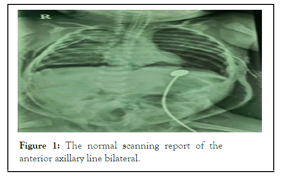
A 26-days old neonate with a history of postnatal apnoea/ aspiration pneumonia who has been on mechanical ventilation for 3 days for the same and later weaned off from ventilator and referred to our institution for the further management given persistent non-resolving right upper lobe collapse. After theinitial evaluation, right upper lobe collapse was confirmed with CT thorax. Hence planned for diagnostic bronchoscopy to rule out any foreign body, vascular rings/congenital tumours or mucous plug [2]. A detailed pre-anaesthetic evaluation was done. This baby was one among the twins and was born out of a nonconsanguineous marriage and emergency LSCS was done at 34 weeks in a local hospital. The immediate postnatal period was uneventful and on the 18th postnatal day, the baby developed respiratory distress and was managed initially suspecting aspiration pneumonia. Higher antibiotics were given and mechanically ventilated for 3 days because of impending respiratory failure. The baby was extubated after 3 days and referred to us. Examination revealed a 26-day old male baby weighing 2.3 kg feeding on an orogastric tube, active and afebrile with subcostal indrawing. Heart rate was 140/min regular, no stridor, Respiratory rate 54/min, SPO2 98% in 6 l of oxygen. No pallor, cyanosis or oedema. On auscultation CVS S1 S2heard, no murmurs but there were a minimal reduction in air entry on the right side. Patent intravenous access noted. High risk written informed consent was taken. Preoperative advice regarding NPO was instructed. Laboratory investigations were within normal limits and a negative anterior axillary line bilaterally shown in Figure 1.

Figure 1: The normal scanning report of the anterior axillary line bilateral.
Reverse transcription-polymerase chain reaction (RTPCR) for COVID-19 was obtained. Chest X-ray revealed right upper lobe non-homogenous opacity with no mediastinal shift. A cautious intravenous induction of general anaesthesia with intermittent positive pressure ventilation through the side port of the bronchoscope was planned. OT was kept ready for the baby. Baby was premedicated with IV ketamine 0.5 mg/kg, IV atropine 0.02 mg/kg and IV fentanyl 2 μ/kg. Preoxygenated for 3 minutes, the Child was then induced with IV propofol 2 mg/kg and oxygen in Sevoflurane 2% by face mask [3]. For muscle relaxation IV succinylcholine 1.5 mg/kg was administered and topical lidocaine 3-4 mg/kg was sprayed in the larynx and tracheobronchial tree to prevent laryngospasm. IV dexamethasone was given at a dose of 0.04 mg/kg. Once the child was apnoeic, the surgeon introduced an appropriately sized bronchoscope and intermittent positive pressure ventilation was continued through the side port of the bronchoscope. Anaesthesia was maintained with repeat doses of ketamine and intermittent oxygen. Pedunculated Papillomatosis lesion noted at the right apical bronchus obstructing the same. Bronchoalveolar lavage and excision was attempted and suddenly the baby developed subcutaneous emphysema extending from the face, neck, and upper chest up to the scrotum. The baby became cyanosed and bloated. Heart rate dropped to 98/min and IV atropine of 0.02 mg/kg was administered, SPO2 declined to 74%. Bronchoscope removed at once and bag and mask ventilation with 100% oxygen attempted. The reservoir bag was tight, and ventilation was found to be difficult. ECG showed bradycardia culminating in asystole. Chest compression began according to PALS (Paediatric Advanced Life Support) protocol, IV adrenaline 1 in 10,000 dilution 0.5 ml given. Successful endotracheal intubation was done in the second attempt. Confirmed by the appearance of the EtCO2 waveform. Bilateral air entry was diminished [4]. Needle thoracostomy was performed bilaterally using a 24 G needle in the 2nd intercostal space at the midclavicular line and CPCR continued.
Emergency intercostal drainage was done in the fifth intercostal space in the anterior axillary line bilaterally. SPO2 improved to 94% and heart rate increased to 164/min with a BP of 70/60 mmHg. The stomach was decompressed using an 8Fr feeding tube [5]. Parents were informed about the events and the need for postoperative ventilation. IV fentanyl 5 μg, IV atracurium 1 mg were given and the baby was shifted to NICU with the monitor attached. Serial Chest X-rays were taken and the baby became conscious, active and alert on the next postoperative day. Vitals remained stable and weaned off from ventilator on 3rd postoperative day. Bilateral ICDs were removed after full expansion of the lungs. Shifted from NICU after 2 weeks with stable vitals and no residual respiratory or neurological deficits [6].
Rigid Bronchoscopy is a simple but challenging procedure, especially in paediatric patients. Various complications can happen such as laryngospasm, bronchospasm, slippage of the foreign body to the opposite side, bleeding, infections, laryngeal oedema, pneumomediastinum and pneumothorax. Tension pneumothorax is a rare but life-threatening complication if not dealt with on time. The possibility of developing tension pneumothorax during bronchoscopy for foreign body removal is approximately 1% as reported by Rothmann [7]. Once there is a suspicion of tension pneumothorax, immediate intervention should be performed to prevent life-threatening events [8]. An immediate intercostal drainage tube can revert the situation if done promptly on time. The addition of positive pressure converts pneumothorax into a tension pneumothorax. This occurs when a one-way valve mechanism develops because of tracheobronchial instrumentation leading to inadvertent rent in the airway, thereby allowing the air to enter the pleural cavity during positive pressure ventilation and preventing exit during the expiratory phase [9]. As the air builds up in the pleural space, the ipsilateral lung is compressed followed by mediastinal shift and compression of the contralateral lung and intrathoracic vasculature leading to severe hypoxemia and cardiovascular compromise [10].
A high index of suspicion must be maintained for pneumothorax during rigid bronchoscopy whenever sudden deterioration of hemodynamic or hypoxia ensues. Intraoperative catastrophe from which a child cannot be revived can lead to irreversible neurological damage. Paediatric bronchoscopy needs practice and understanding of instrument handling in small tracheobronchial airways. A small breach in the airway can lead to a life-threatening complication. Proper training in instrument insertion and constant patient monitoring is very crucial to prevent such events. Communication between the anaesthesiologist, the otolaryngologist, the paediatric pulmonologist and the paediatric intensivist is very important for a better outcome in such cases.
[Crossref] [Google Scholar] [Pubmed]
[Crossref] [Google Scholar] [Pubmed]
[Crossref] [Google Scholar] [Pubmed]
[Crossref] [Google Scholar] [Pubmed]
[Crossref] [Google Scholar] [Pubmed]
[Crossref] [Google Scholar] [Pubmed]
[Crossref] [Google Scholar] [Pubmed]
[Crossref] [Google Scholar]
[Crossref] [Google Scholar] [Pubmed]
Citation: Vidya AS (2022) Tension Pneumothorax and Surgical Emphysema in a Neonate during Rigid Bronchoscopy: A Case Report. J Anesth Clin Res. 13: 1066
Received: 02-Jun-2022, Manuscript No. JACR-22-18521; Editor assigned: 06-Jun-2022, Pre QC No. JACR-22-18521(PQ); Reviewed: 22-Jun-2022, QC No. JACR-22-18521; Revised: 28-Jun-2022, Manuscript No. JACR-22-18521(R); Accepted: 05-Jul-2022 Published: 05-Jul-2022 , DOI: 10.35248/2155-6148.22.13.1066
Copyright: © Vidya AS. This is an open-access article distributed under the terms of the Creative Commons Attribution License, which permits unrestricted use, distribution, and reproduction in any medium, provided the original author and source are credited.