Journal of Applied Pharmacy
Open Access
ISSN: 1920-4159
ISSN: 1920-4159
Research Article - (2017) Volume 9, Issue 3
Curcumin is the yellow pigment of turmeric. In addition to its positive safety profile, curcumin is reported to have beneficial pharmacologic effects as an antioxidant, antitumor, and anti-inflammatory agent, along with other promising pharmacologic effects on the cardiovascular and digestive systems. Curcumin is poorly absorbed, which limits its value in clinical application. In order to improve the poor bioavailability and enhance the pharmacologic action of curcumin, we studied its absorption mechanisms in an animal model in vivo and in a Caco-2 cell model in vitro. The absorption rates of curcumin at different concentrations in blank intestinal juice were not the same. The absorption rate of curcumin solution at a concentration of 5 μg/mL was the highest, followed by 10 μg/mL, and the minimum absorption occurred at 20 μg/mL. The absorption rate in the ileum decreased as the concentration of curcumin increased, which reminds us that absorption in the ileum does not result from simple passive diffusion but rather shows the characteristics of active transport. Curcumin may be a P-glycoprotein (P-gp) substrate and thus may be affected by P-gp efflux, and thus the addition of a P-gp inhibitor such as verapamil can promote the intestinal absorption of curcumin. A Caco-2 cell model was established to accurately study curcumin’s absorption mechanisms. We found that, for curcumin in a 5 μg/mL solution, the Caco-2 cell monolayer transport was passive, and when the concentration was increased to 10 μg/mL efflux influenced the transport but not extensively. The transport mode of curcuminthe appears to be passive diffusion at concentrations <10 μg/mL, but at concentrations >10 μg/mL active transport is involved. In summary, curcumin is transported by a combination of passive diffusion and active transport, curcumin is a substrate for the intestinal transporter P-gp, and intestinal absorption of curcumin is regulated by intestinal P-gp transport.
<Keywords: Curcumin, Intestinal absorption, P-glycoprotein, Caco-2 cell
Curcumin, the yellow pigment of turmeric, is a natural active ingredient extracted from the root of turmeric or curcuma. It is soluble in methanol, ethanol, alkali, acetic acid, acetone, chloroform, and other organic solvents (Figure 1). Numerous studies have demonstrated its positive safety profile and beneficial pharmacologic effects as an antioxidant, its antitumor and anti-inflammatory activities, its free radical scavenging and antimicrobial effects, and many other pharmacologic effects on the cardiovascular and digestive systems [1]. In particular, recent findings suggest that curcumin is potentially a powerful tool to reverse cisplatin-induced toxicity, and it has attracted considerable interest over the last decades due to its beneficial effects for human health [2]. Studies of the absorption and metabolism of curcumin have been conducted since at least the 1980s [3]. Ravindranath studied the absorption and tissue distribution of curcumin in rats that were orally given 400 mg of curcumin. At 15 min to 24 h after oral administration of curcumin, only trace amounts of 5 mg·L–1 were detected in the hepatic portal vein [4]. Eight volunteers were given 2 g curcumin, after which the content of curcumin in human serum was very low or undetectable; the peak concentration was only 0.006 ± 0.005 mg·L–1; and at 3 h after administration, the serum concentration reached zero. Overall, curcumin is poorly absorbed and has low bioavailability, both of which limit its clinical applications and therapeutic efficacy [5]. At present, the mechanism by which curcumin is absorbed in vivo is not clear. In order to find suitable methods to improve poor bioavailability and enhance its pharmacologic action, we studied the absorption mechanisms of curcumin.

Figure 1: Structure of curcumin.
In this research, we used an animal model in vivo and a Caco-2 cell model in vitro in order to study the absorption process and the characteristics of curcumin. At the same time, we wanted to provide a theoretical basis to solve the problem of the low absorption rate of curcumin via the oral route and to find a new way to improve the biological use of some drugs that are poorly absorbed in the gastrointestinal tract.
Drug and chemicals
Reference-grade curcummin (purity 99.8%) were purchased from Nanjing Zelang Medical Technology Co., Ltd. (ZL20090515A). Hank’s buffered salt solution (HBSS) was prepared as follows: anhydrous calcium chloride (0.40 g), magnesium chloride hexahydrate (0.10 g), and magnesium sulfate heptahydrate (0.10 g) were dissolved in 1000 mL of distilled water, and then potassium chloride (0.40 g), potassium dihydrogen phosphate (0.06 g), sodium bicarbonate (0.35 g), water (1000 mL), disodium hydrogen phosphate (0.12 g), sodium chloride (8.00 g), and glucose (1.00 g) were added and mixed until completely dissolved. All other reagents were of High-performance Liquid Chromatography (HPLC) grade or were of the highest purity commercially available.
Experimental animals
Animal experiments in this study were used in compliance with standard ethical guidelines and were approved by Nanjing University of Chinese Medicine Animal Care and Use Committee. Forty male SD rats, weighing 220 ± 20 g, were purchased from Shanghai Silaike Experiment Animal Co., Ltd. (SCXK2007-0005; Shanghai, China) and were housed in a 23°C room with a 12 h/12 h light–dark cycle and were allowed to acclimate to the laboratory for 1 week before experiments.
Instrumentation and analytical conditions
The concentration of curcumin was analyzed with an LC-10A HPLC (Shimadzu Corporation, Kyoto, Japan) equipped with an SPD-10Avp UV detector, a quaternary pump, an online degasser, and an automatic plate sampler. Chromatographic separations were performed with a Dimma ODS-2 C18 column (150 × 4.6 mm5 μm packing) with a column temperature of 35°C. The mobile phase was methanol: water (70:30). The flow-rate was 0.2 mL/min. The detection wavelength was 425 nm. Aliquots of 20 μL were injected into the HPLC system for analysis.
Reverted gut sac experiments in vitro
Precise amounts of curcumin were added to a 0.1% Tween 80 solution containing HBSS in order to make concentrations of 5, 10, and 20 g/mL HBSS solution. The rats were fasted 24 h (water was freely available) and were anesthetized by intraperitoneal injection of 20% urethane anaesthesia. First we fixed the animals’ backs on the operating table, and then we made a 3-cm incision along the ventral midline and quickly removed a 20-cm-long segment of the ileum. Next, we removed the small intestine and immediately placed it in saline solution, washed the small intestine, and then transferred the intestinal segment to the HBSS fluid; surface fat and blood vessels were removed by stripping.
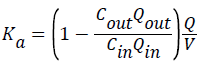
Then the ileum was divided into 2 sections, each portion of which was sealed at one end with a silk thread ligation. A fine glass rod with a diameter <5 mm of fine glass rod for outer intestine, the small intestinal mucosa of lateral outside, and the other end is tied to one end of the glass tube bulge. Blank HBSS buffer 0.5 mL injection; the gut sacs thus prepared were placed into the containers with HBSS buffer maintained at 37°C. HBSS buffer was placed in a 100-mL beaker, and 95% O2 and 5% CO2 were aspirated for 5 min, and then the gut sac was placed into the oxygenated HBSS solution 100 mL containing 5, 10, or 20 g/mL of drugs of HBSS solution. 0.5 mL was taken out of the gut sac at 30 min, 60 min, 90 min, and 120 min; then we added and equivalent amount of fresh blank HBSS solution. The cumulative absorption amount per unit area (Q) was determined by the following formula:
Qn=C × V/S
Where n=the number; V=the solution volume (0.5 mL) in the intestine; C=sample concentration (g/mL), and S=intestinal segment area (cm2).
In situ intestinal perfusion experiment
Rats were fasted 16 h overnight. After anaesthesia, animals were placed for surgery, and the ileum was removed. Then glass tubes were inserted into both ends of the intestinal segments. The segments were fixed and ligated, cleaned with a 37°C saline flush, and the lengths of the intestinal segments were measured at the end of the test.
HBSS solutions containing 0.1% Tween 80 buffer solution were prepared to concentrations of 5 g/mL, 10 g/mL, and 20 g/mL curcumin solution as the test group. The ahead solution served as the blank control. The amount of 50 mL containing curcumin intestinal perfusate, adding a 100 mL flask circulation device. Researchers placed the flask in a 37°C water bath and opened the constant-flow pump.
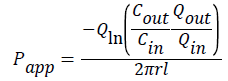
The flow rate was adjusted to 0.2 mL/min, replaced the infusion vial and liquid vial and weighing every 15 min, then calculated the perfusate volume. Drug concentrations at each time point were determined by HPLC. The experiment lasted 120 min, when all the vials were collected for determination, and the rats were killed. The ileum cannula along the cut ends, gently on the droplet adding 37°C Krebs–Ringer solution, measuring intestinal length, measuring inner diameter with thread. Calculation of relevant data was calculated as follows:
 (1)
(1)
 (2)
(2)
Where Ka is absorption rate, Papp is apparent permeability coefficient, Qin and Qout are the volumes of intestinal fluid imported and exported (mL), respectively. Cin and Cout were the concentrations of curcumin in the intestinal fluid imported and exported (μg/mL), respectively. L and R were the length (cm) and the cross-section radius (cm) of the intestinal segment, Q was the perfusion rate (0.2 mL/min), t was the perfusion time (0.25 h), and V was the perfusion volume of the intestinal segment.
Effect of the P-gp inhibitor verapamil on the transport of curcumin
A solution of 5 μg/mL of curcumin was used in the control group, and a 5 μg/mL curcumin solution containing 500 μmoL/L verapamil hydrochloride was used in the experimental group. We conducted rat ileum perfusion experiments using the method just described, and both Ka and Papp values were calculated.
Study of the mechanism of curcumin activity in Caco-2 cell culture
Caco-2 cells from the Shanghai Cell Bank, Chinese Academy of Sciences were grown to <90% confluency in DMEM containing 10% FBS, 100 U/mL penicillin, 0.1 mg/mL streptomycin in a humidified atmosphere of 5% CO2 in air maintained at 37°C. The cells were used from passage 50 to 90. After trypsinization, Caco-2 cells were seeded onto Millicell hanging cell culture inserts (Supplier, City, Country) in 24-well plastic plates at a density of 1 × 10⁵ cells/cm2 for approximately 21 days.
The integrity of the cell monolayer was checked before and after each experiment by measuring transepithelial electrical resistance (TEER) using a volt-ohmmeter (EVOM; Millipore, Tokyo, Japan). Only monolayers with a TEER value >350 Ω·cm2 were considered intact and suitable for transport studies. To obtain Caco-2 monolayers with various TEER values, the monolayers were treated with cytochalasin D (0.1 mM) from the apical side.
Transepithelial transport experiments
Curcumin was diluted to working concentrations of 5, 10, and 20 μg/mL, and we prepared a 500 M verapamil with HBSS solution containing 0.1% Tween 80. All experiments were performed in HBSS buffer. After washing the inserts 3 times with warm HBSS buffer for 30 min, researchers added transport medium containing curcumin (5, 10, or 20 μg/mL with 500 M verapamil) either to the apical or basolateral compartment (0.4 or 0.6 mL, respectively). After shaking at 55 rpm at 37°C, 100-μL samples were collected after 30, 60, 90, and 120 min from the receiver compartment then immediately replaced with an equal volume of prewarmed fresh HBSS (n=4). Simultaneous determinations of TEER values were made during the process of the experiments. Papp (apparent permeability coefficient) and ER (efflux ratios) were calculated:
Papp=Q/(t·A·C0);
Q: Transport quantity in t (μg);
A: Surface area of the carbon film (cm2);
C0: Initial concentration of the drug (μg/mL).
ER=PappB→A/PappA→B
PappB→A Apparent permeability coefficient from B side to A side (cm/s);
PappA→B Apparent permeability coefficient from A side to B side (cm/s).
Data analysis
The differences between the groups were analyzed using Student’s t-test when only 2 groups were compared or one-way analysis of variance (ANOVA) when >2 groups were compared. P values <0.05 indicate statistical significance. All statistical analyses were performed using SPSS 17.0 software (SPSS, Chicago, IL, USA).
HPLC method for curcumin assay
HPLC mapping was used to measure the intestinal absorption of curcumin in different concentrations. The curcumin standard curve is shown by: y =31648x–22253, R=0.9996. In the range of (0.5 to approximately 50 g/mL), the concentration of curcumin was in good linear relationship with the peak area. Compared with the initial value at 0 h, different concentrations of curcumin at different time points were not significantly different (P>0.05). Compared with the initial concentration, the concentration at 2 h was 98%. The intestinal solution can maintain a steady state of curcumin, and the HPLC assay can quickly and accurately determine the concentrations of curcumin, which can be used for quantitative analysis.
Intestinal absorption rates of different concentrations of curcumin in reverted gut sac experiments
The absorption rates of curcumin at different concentrations were not the same. Results are shown in Figure 2. The absorption rate of the curcumin solution with a concentration of 5 μg/mL was the highest, followed by the second-highest absorption of the 10 μg/mL solutions, but the minimum absorption occurred with the 20 μg/mL solutions. The absorption rate of the ileum decreased as the concentration of curcumin increased. Absorption in the ileum is not the result of simple passive diffusion and instead shows the characteristics of active transport.

Figure 2: Absorption rate of curcumin in the ileum segment.
In situ intestinal perfusion experiment of different concentrations of curcumin in the presence of a P-gp substrate
When the concentration of curcumin increased from 5 μg/mL to 10 μg/mL, the Ka value decreased, and the Ka value continued to decrease at a concentration of 20 μg/mL. Similarly, the Papp value also decreased. The concentration continued to increase to 20 μg/mL when the Papp decreased slightly (Table 1).
| Dose group | Ka | Papp |
|---|---|---|
| (×10-2 min-1) | (×10-3 cm × min-1) | |
| 20 μg/mL curcumin | 11.92 ± 1.10 | 8.23 ± 0.29 |
| 10 μg/mL curcumin | 15.99 ± 0.63 | 12.29 ± 3.51 |
| 5 μg/mL curcumin | 20.22 ± 0.07 | 21.09 ± 0.42 |
Table 1: Absorption rate and apparent permeability coefficient of various concentrations of curcumin.
The experimental results show that the absorption rate (Ka) and apparent permeability coefficient (Papp) decreased when the concentration of curcumin was increased. We observed a high concentration of saturation phenomenon. When 5 μg/mL of curcumin solution was added to the 5 M P-gp inhibitor verapamil, the Ka value was increased from 20.22 ± 0.07 × 10⁻² min-1 to 41.61 ± 5.71 × 10⁻² min-1 (P<0.01), and the Papp value increased from 21.09 ± 0.42 × 10⁻³ cm × min-1 to 29.14 ± 4.00 × 10⁻³ cm × min-1 (P<0.05). Results are shown in Figures 3 and 4. The effect of P-gp on the intestinal absorption of curcumin in the presence of verapamil is relatively large, and curcumin may be a substrate of P-gp and may be affected by the P-gp efflux function.

Figure 3: Absorption rate of various concentrations of curcumin.

Figure 4: Apparent permeability coefficient of various concentrations of curcumin.
Transport across Caco-2 cells
The experimental results are shown in Tables 2 and 3. As the concentration of curcumin increased, TEER values gradually increased in a concentration-dependent manner: Following addition of 500 μM verapamil, the curcumin PappA→B in the 10-μg/mL and 20-μg/mL solutions increased. There was no significant decrease in TEER. After verapamil was added to the 500-μM solution, the PappA→B values between the 2 concentrations were almost equal and were quite close to the PappA→B of 5 μg/mL. After adding P-gp inhibitors, the apparent permeability coefficient of curcumin PappA→B value remained constant, indicating that passive diffusion was the main transport mechanism. For curcumin in a 5 μg/mL solution, the Caco-2 cell monolayer transport mechanism is still passive transport; when the concentration rose to 10 μg/mL, the efflux exerted an influence on the transport, but the efflux effect was not very strong. The transport mode of curcumin involves passive diffusion at concentrations <10 μg/mL, but when the concentration is>10 μg/mL, active transport is involved. Overall, the transport mechanism of curcumin involves a combination of active and passive transport, and curcumin is a P glycoprotein substrate.
| Sample | Papp (×10–6 cm/s) | Efflux ratio | |
|---|---|---|---|
| A→B | B→A | ||
| 5 µg/mL curcumin | 1.50 ± 0.43 | 1.17 ± 0.12 | 0.78 |
| 10 µg/mL curcumin | 1.23 ± 0.21 | 1.44 ± 0.09 | 1.17 |
| 20 µg/mL curcumin | 0.97 ± 0.23 | 1.56 ± 0.26 | 1.62 |
| 10 µg/mL curcumin+500 µM verapamil | 1.59 ± 0.17 | - | - |
| 20 µg/mL curcumin+500 µM verapamil | 1.59 ± 0.10 | - | - |
Table 2: Apparent permeability coefficient (Papp) and efflux ratios of curcumin transport across Caco-2 cell monolayers (n=4).
| Time(min) | 0 | 60 | 120 | After 24 h of cultivation |
|---|---|---|---|---|
| A→B | ||||
| 5 µg/mL curcumin | 407 ± 56 | 460 ± 16 | 678 ± 33 | 848 ± 36 |
| 10 µg/mL curcumin | 435 ± 14 | 447 ± 6 | 665 ± 2 | 776 ± 183 |
| 20 µg/mL curcumin | 394 ± 11 | 450 ± 8 | 773 ± 83 | 544 ± 226 |
| 10 µg/mL curcumin+500 µM verapamil | 422 ± 9 | 434 ± 12 | 653 ± 26 | 728 ± 219 |
| 20 µg/mL curcumin+500 µM verapamil | 444 ± 16 | 454 ± 6 | 722 ± 27 | 980 ± 432 |
| B→A | ||||
| 5 µg/mL curcumin | 437 ± 29 | 444 ± 6 | 655 ± 11 | 870 ± 212 |
| 10 µg/mL curcumin | 427 ± 21 | 437 ± 4 | 661 ± 14 | 804 ± 118 |
| 20 µg/mL curcumin | 407 ± 13 | 446 ± 14 | 755 ± 11 | 675 ± 18 |
Table 3: TEER of the Caco-2 cells monolayers in the Caco-2 transport study (n=4).
Everted gut sacs are commonly used to study the intestinal absorption of drugs. The rate and amount of curcumin absorption can be studied by determining the composition of the intestinal absorption solution. Because of the lack of intestinal blood circulation and nerve control in this model, long-term in vitro intestinal mucosal functions can easily change, so an improved method was used in this experiment. The culture medium with HBSS was treated with 95% O2/5% CO2 so the exposed mucosal cells received an adequate supply of O2 to ensure the continuing intestinal functions during the experiment. The everted sac model retains the characteristics of intact tissue and mucosa and can be used to simulate the absorption of drugs in different intestinal segments in vitro. Our study of the absorption mechanism in the ileum is perhaps the most helpful and persuasive portion of this experiment. On the basis of the experiment, we further investigated the mechanism of intestinal absorption of curcumin by using in vivo intestinal perfusion model. In situ intestinal perfusion is a technique to determine the absorption rate of a drug in situ in rats, which has recently become one of the main research models of drug absorption [6-8].
P-gp is a membrane transport protein that pumps the drug out of the cell, thereby reducing the bioavailability of drugs. P-gp is an ATP-dependent efflux pump that is highly expressed in multi-drug resistant tumor cells and the blood–brain barrier [9]. P-gp inhibitors can slow the drug efflux function of P-gp, thereby reversing drug resistance and increasing the amount of drug in the tissues. In order to improve the bioavailability of drugs, the combination of curcumin and P-gp inhibitors such as verapamil and cyclosporine can be considered. The key factor to ensure the effect of an oral medicine is that it must be absorbed in the digestive tract, and the intestinal transport protein P-gp plays a very important role in the transmembrane transport (including absorption and secretion) of lipid-soluble drugs. Therefore, it is necessary to strengthen the research on P-gp and its substrates [10].
The Caco-2 cell model has been widely used to study the absorption and transport mechanisms of drugs in the small intestine. Caco-2 cells were first isolated from human colon cancer cells in the 1970s, and the model was originally proposed by Markowska et al. [11]. Caco-2 cells possess the same cell polarity and compact single-cell layer as do human intestinal epithelial cells. The morphology and function of cells were similar to those of human intestinal epithelial cells. Because most of the absorption processes and mechanisms of action of Chinese herbal medicines are not yet elucidated, the Caco-2 cell model has wide applications in the study of the oral absorption of Chinese medicines [12-14].
The Papp value reflects the ability of drug molecules to penetrate the intestinal tract, and ER reflects the role of the efflux protein to expel drugs—the greater the ER, the more powerful the efflux effect. Based on our findings using a Caco-2 cell model to study curcumin, we determined that the transmembrane transport of curcumin can be enhanced by the presence of a P-gp inhibitor, and the Papp value was increased after the addition of verapamil. From the results, we see that in a monolayer of Caco-2 cells, the transport mode of curcuminthe is passive diffusion at concentrations <10 μg/mL and when the concentration is >10 μg/mL, active transport is involved. The transport mechanism for curcumin is a combination of active and passive processes. This is confirmed in the experiments by the addition of an inhibitor, and the value is close to PappA→B of 5 μg/mL.
Curcumin is present in many foods and traditional Chinese medicines and is an important material basis for food nutrition and traditional Chinese medicine. In this paper, the experimental results reveal that, when the content of curcumin in food and medicinal materials is low, curcumin can be absorbed into the body at low dose levels by passive diffusion and is a substrate for the drug intestinal transporter P-gp. Careful use of other P-gp components can improve a drug’s competitive transport into the gastrointestinal tract. The results of this study can also be used to explain some of the reasons for the poor performance of certain formulations of curcumin as a high-purity compound. Chinese foods and Chinese herbal medicines containing curcumin can still play a role in nutrition and health.
In summary, curcumin transport results from a combination of passive diffusion and active transport. Curcumin is a substrate of the intestinal transporter P-gp, and the mechanism by which intestinal absorption of curcumin takes place is related to the mechanism of intestinal P-gp transport.
The authors declare that they have no competing interests in this work.
This thesis is sponsored by Jiangsu Province and the Opening Foundation of Key Laboratory of acupuncture and moxibustion (No. KJA200802)