Journal of Plant Biochemistry & Physiology
Open Access
ISSN: 2329-9029
ISSN: 2329-9029
Research Article - (2015) Volume 3, Issue 4
Natural products are often a source for bioactive compounds which have great potential for developing novel therapeutic agents. The present study was to evaluate anti-inflammatory potential of six compounds A: Raffinose, B: Sucrose, C: Benzoic acid, D: Trehalose, E: Quinic Acid and F: Ferulic acid on bacterial lipopolysaccharide (LPS) induced inflammation in RAW 264.7 macrophages by employing in vitro methods: nitric oxide (NO) and Nitro blue tetrazolium (NBT) assays. These compounds are found in many plants, including palm, date palm, pomegranate, custard apple, wood apple, water chestnut and Indian plum, among others and it has been found that many of these compounds are effective anti-inflammatory agents, with anti-oxidant activities. The study revealed that compound C exhibited maximum anti-inflammatory activity at a concentration as low as one ng/ml. Compounds A, B and E also showed anti-inflammatory activity but at a higher concentration but compound D and F did not show antiinflammatory activity.
Keywords: Oxidative pathways; Therapeutic molecule; Prophylaxis; Functional food; Cell based assays
Inflammation is part of a complex biological response that takes place in vascular tissues when exposed to harmful stimuli, like pathogens, damaged cells or irritants [1,2]. It is an organism’s protective mechanism, by which it can eliminate the damaging stimuli and start the process of healing. Inflammation is part of an organism’s innate immunity [3,4], and is characterized by five cardinal signs: pain (dolor), heat (calor), redness (rubor), swelling (tumor) and loss of function (functio laesa) [5]. Acute inflammation occurs initially, when the body is exposed to the damaging stimulus. The body’s initial response involves increased movement of plasma and leukocytes from the blood to the site of injury or inflammation. Acute inflammation is initiated by cells that are already present at the site, like resident macrophages, dendritic cells, Kupffer cells and mast cells. At the start of an infection, these cells get activated and release inflammatory mediators, which are responsible for the clinical signs of inflammation [6]. Prolonged, or chronic, inflammation leads to a change in the type of cells at the site of inflammation, and is characterized by a simultaneous destruction and healing of the tissue. Inflammation leads to increased production of reactive species like ROS (reactive oxygen species), NOS (nitric oxide synthase) and their product peroxy nitrite (ONO2 -) by activated macrophages. This increase in oxidative stress leads to decrease in effectiveness of oxidant defenses, that is, reduction in antioxidants.
Due to the various side effects and other complications of modern medicine, the use of traditional medicines and natural products is gaining popularity. Phytotherapy involves use of phytochemicals from fruits as possible sources of therapeutic agents. Different biological activities of these chemicals, including their anti-oxidant properties and their anti-inflammatory properties, have been tested in vitro, as well as in vivo [7-9].
A natural product is a chemical compound or substance produced by a living organism that is, the product is found in nature [10,11]. Natural productshave been used as medicines for a very long time, the earliest written document being a 4000 year old Sumerian clay tablet, recording remedies for various illnesses. Natural products sometimes have pharmacological or biological activity that can be of therapeutic benefit in treating diseases. As such, natural products are the active components not only of most traditional medicines but also many modern medicines. The identification of the molecular structures of many natural products has allowed chemists to synthesize the products, rather than isolating them from natural sources [12]. This has markedly reduced the cost of drug production, and allowed the drugs to be of improved potency.
Plants are a major source of complex and highly structurally diverse chemical compounds (phytochemicals), this structural diversity attributed in part to the natural selection of organisms producing potent compounds to deter herb ivory (feeding deterrents). Major classes of phytochemicals include phenols, polyphenols, tannins, terpenes, and alkaloids. Though the number of plants that have been extensively studied is relatively small, many pharmacologically active natural products have already been identified [13].
Here we have assessed the effects of six compounds (A: Raffinose, B: Sucrose, C: Benzoic acid, D: Trehalose, E: Quinic Acid and F: Ferulic acid) on bacterial lipopolysaccharide (LPS) induced inflammation in RAW 264.7 macrophages by performing cell based assays, namely NO and NBT assay. LPS activates macrophages and production of pro-inflammatory cytokines. NO is a diatomic free radical that is extremely short lived in biological systems. It is used as a biomarker of inflammation in many biological samples. NO is involved in many physiological processes including regulation of blood pressure, immune response and neural communication. Therefore its accurate detection and quantification is critical to understanding health and disease. Inhibition of NO-production in LPS stimulated RAW cells is an effective screening method. In addition, NBT is a chemical compound composed of two tetrazole moieties. This assay is used to determine the production of superoxide anion (O2 -) in various phagocytic cells. Inflammation leads to an increased production of O2 - radicals, which in turn, leads to higher reduction of NBT, and greater colour in the colorimetric reaction. Thus, inhibition of NBT reduction can be used as a screening method as well.
The compounds which we are using in our studies are found in many plants, including palm, date palm, pomegranate, custard apple, wood apple, water chestnut and Indian plum, among others. In many Indian cultures, these plants have been traditionally used as medicinal plants to combat various diseases. Over the years, the active ingredients of these plants have been identified, and it has been found that many of these compounds are effective anti-inflammatory agents, with antioxidant activities.
Compound A: raffinose
Raffinose (C18H32O16.5H2O) is a trisaccharide, with a molecular weight of 594.51 gm/mol, composed of galactose, glucose and fructose. Also called (α-D-Galactosylsucrose, it is found in beans, cabbage, Brussels sprout, broccoli and whole grains. It can be hydrolyzed to D-galactose and sucrose by the enzyme α-galactosidase (α-GAL), which is not present in the human digestive tract. Raffinoses, among other sugars from the raffinose family of oligosaccharides (RFOs), pass undigested into the lower intestine, where gas producing bacteria, which produce α-GAL, ferment these sugars. The gases released in this process of fermentation (carbon dioxide, methane or hydrogen) lead to flatulence. However, it has been found that raffinose increases the population of indigenous gut micro flora, especially Bifidobacterium population, in humans and rats, and also suppresses Th2 mediated immune response in mice [14].
`Compound B: sucrose
Sucrose (C12H22O11) is a disaccharide, with a molecular weight of 392.3 gm/mol, composed of a glucose unit and a fructose unit. Commonly called table sugar, cane sugar, beet sugar, or just sugar it is a white, odorless, crystalline compound with a sweet taste. Sucrose is a non-reducing sugar, and its hydrolysis is a very slow process. However, the gastric enzymes are capable of breaking it down into glucose and fructose. Sucrose is a natural product found in sugarcane, sugar beet and date palm, but it can be synthesized chemically. It was first synthesized by Ref. [15].
Compound C: benzoic acid
Benzoic acid (C6H5COOH) is a simple aromatic carboxylic acid, of molecular weight 122.12 gm/mol. It is a colorless, crystalline solid, found naturally in many plants, especially in berries, and it serves as an intermediate in the biosynthesis of many secondary metabolites. Benzoic acid is relatively nontoxic [16]. Benzoic acid is used as a preservative, as it can inhibit the growth of moulds, yeast and some bacteria [17] Benzoic acid is a major constituent of a number of topical ointments, used in the treatment of fungal diseases, and has also been used as an inhalant decongestant, expectorant, analgesic and antiseptic [18,19].
Compound D: trehalose
Trehalose (C12H22O11) is a disaccharide, with a molecular weight of 342.3 gm/mol, formed of two glucose units joined by a (α, α-1, 1) glucoside bond. It is a non-reducing sugar that can be hydrolysed by the enzyme trehalase, present in the intestinal mucosa. It is nutritionally equivalent to glucose, because it is broken into two glucose molecules. Trehalose is found in many plants, like sunflower seeds and moonwort, and animals and microorganisms. It has been implicated in the ability of plants and animals to withstand prolonged periods of desiccation. It is also an anti-oxidant. Trehalose is a stress response factor, made when cells face environmental stresses like heat, cold, oxidation and desiccation, where trehalose helps cells maintain their structural integrity by preventing protein denaturation [20]. Trehalose has been found to inhibit growth of melanoma cells [21].
Compound E: quinic acid
Quinic acid (C7H12O6) is a cyclitol and a cyclohexane carboxylic acid, of molecular weight 192.2 gm/mol. It is a crystalline acid, found in the barks of the cinchona tree, the coffee plant and in some other plants. Quinic acid is used as an astringent. Quinic acid and its compounds are known for their anti-inflammatory effects through mechanisms involving the inhibition of the pro-inflammatory transcription factor, nuclear factor kappa-B (NF-κB) [22,23].
Compound F: ferulic acid
Ferulic acid (C10H10O4) is a hydroxycinnamic acid of molecular weight 194.2 gm/mol. It is a phenolic phytochemical, found abundantly in plant cell wall components. It is a component of asafoetida, and is also found in the seeds of coffee, apples, peanuts and oranges. It has antibacterial [24] and antioxidant [25] properties. In vitro studies have shown that ferulic acid may have anti-tumor activity in cancer [25,26], and can also reduce oxidative stress and formation of thymine dimers if applied topically with ascorbic acid and vitamin E.
Rationale
The various side effects and complications of medicines have urged scientists to look for less harmful alternatives. These alternatives can be found in natural products, which have been exploited for centuries in the treatment of various diseases. These products are obtained from plants, and can also be synthesized chemically at a much lower cost. The active ingredients of a number of natural products have been isolated. Our study aims to assess the anti-inflammatory activity of some of these chemical compounds isolated from several plants, and to determine the lowest concentration at which the compounds act.
RAW 264.7 macrophage cells were cultured in our laboratory. Dulbecco’s Modified Eagle Medium (DMEM), fetal bovine serum (FBS), Penicillin Streptomycin (Pen-Strep), and Nitro blue Tetrazolium chloride (NBT) were bought from Himedia, India. Sulfanilamide, N-1-naphthyl ethylene diamine dihydrochloride (NED) and dimethyl sulfoxide (DMSO) were obtained from Sisco research Laboratories (SRL), India. Ortho phosphoric acid, sodium nitrite (NaNO2), methanol and potassium hydroxide (KOH) were bought from Merck, India. Bacterial lipopolysaccharide (LPS) was obtained from Sigma Aldrich. 1X phosphate buffered saline (PBS; pH 7.4) was prepared using 137 mM sodium chloride (NaCl; Merck, India), 2.7 mM potassium chloride (KCl; Himedia India), 10 mM disodium hydrogen phosphate (Na2HPO4; Qualigens, India), and 2 mM potassium dihydrogen phosphate (KH2PO4; Himedia India). 96 well plates were obtained from Nest Biotech Co. Ltd., China, and were incubated in a CO2 incubator (Thermo Fisher) at 37°C, with 5% CO2. Cells scrapers from Himedia, India were used to detach cells. Absorbance readings were taken in a multiplate reader (Thermo Fisher Multiskan EX). All cell-culture work was done inside the biosafety cabinet.
Culture of RAW 264.7 macrophages
RAW 264.7 macrophages were cultured in DMEM, with 10% FBS and 1% Pen strep, and incubated at 37°C with 5% CO2, till confluent. The cells were scraped off using scrapers, collected, washed in 1X PBS, and resuspended in DMEM. Approximately 100 μl of 1 × 10⁵ cells/ml were plated in the wells of a 96 well plate, so there were 104 cells per well. The wells were marked as Negative Control (only cell), Positive Control (cell+LPS), 1.00 mg/ml, 0.10 mg/ml, 0.01 mg/ml, 1.00 μg/ml, 0.10 μg/ml, 0.01 μg/ml and 1.00 ng/ml.
Treatment of cells: The cells in the wells marked Positive Control, 1.00 mg/ml, 0.10 mg/ml, 0.01 mg/ml, 1.00 μg/ml, 0.10 μg/ml, 0.01 μg/ ml and 1.00 ng/ml were treated with 1 μg/ml LPS, and incubated for 3 hours at 5% CO2, 37°C. After 3 hours, the cells in the wells marked 1.0 mg/ml, 0.10 mg/ml, 0.01 mg/ml, 1.00 μg/ml, 0.10 μg/ml, 0.01 μg/ml and 1.00 ng/ml were treated with the respective concentrations of the compounds A-F. They were then incubated for 1 hour at 5% CO2, 37°C. NO and NBT assays were done with these cells.
NO assay: The reaction was standardized using different concentrations of NaNO2 (100 μM, 50 μM, 25 μM, 12.5 μM, 6.25 μM, 3.13 μM, 1.56 μM and 0 μM), using the method in Promega User Guide (Product G2930). 100 μl of sulfanilamide solution (1% Sulfanilamide in 5% ortho phosphoric acid) was added to each well, and incubated at room temperature for 5 minutes, in dark. 100 μl of NED solution (0.1% NED in distilled water) was then added, and incubated at room temperature for 5 minutes, in dark. Absorbance was measured in a plate reader at 540 nm. Using the standard curve prepared, the absorbance values of the samples were plotted to get the concentrations of NO produced (in μM). The concentrations of NO were plotted against each sample. The experiment was repeated twice (Table 1).
| NO Assay | Fold changes in concentration of pure compounds with respect to +ve control | ||||||
|---|---|---|---|---|---|---|---|
| A | B | C | D | E | F | ||
| +ve control Wrt –ve control | (+) 1.40 | (+) 1.40 | (+) 1.40 | (+) 1.61 | (+) 1.61 | (+) 1.61 | |
| Pure compounds with respect to +ve control |
1.00 mg/ml | (-) 2.35 | (-) 2.53* | (+) 1.20 | (+) 1.19 | (+) 1.01 | (+) 1.20 |
| 0.10 mg/ml | (-) 1.60 | (+) 1.06 | (-) 1.60 | (+) 1.18 | (-) 1.05 | (+) 1.28 | |
| 0.01 mg/ml | (-) 1.76 | (-) 4.47* | (-) 1.40 | (+) 1.14 | (+) 1.06 | (+) 1.23 | |
| 1.00 µg/ml | (-) 1.13 | (-) 8.11* | (-) 1.17 | (+) 1.26 | (+) 1.16 | (+) 1.08 | |
| 0.10 µg/ml | (-) 1.25 | (-) 12.51* | (+) 1.06 | (+) 1.21 | (+) 1.02 | (+) 1.14 | |
| 0.01 µg/ml | (-) 1.03 | (-) 7.29 | (-) 2.72 | (+) 1.11 | (-) 1.12 | (+) 1.32 | |
| 1.00 ng/ml | (+) 1.17 | (-) 1.25 | (-) 3.96 | (+) 1.73* | (+) 1.14 | (+) 1.62 | |
Table 1: Fold changes in the concentrations of nitric oxide produced by RAW macrophages in presence of pure compounds, assessed by NO assay using sulfanilamide and NED. Compound B shows a maximum decrease (12.51 fold; p<0.05) in NO concentration, at 0.10 μg/ml.
NBT assay: 40 μl of NBT solution (0.5 mg/ml NBT in 1X PBS) was added to the cells in the 96 well plate, and incubated for 1 hour in a CO2 incubator at 5% CO2, 37°C. After 1 hour, the cells were washed twice with warm, 1X PBS, then with methanol, and air dried. 40 μl of 2M KOH was added, followed by 60 μl of DMSO, and incubated at room temperature for 10 mins, with shaking. Absorbance was measured immediately in a plate reader at 620 nm. The absorbance at 620 nm was plotted. Taking the absorbance of the samples to be directly correlated to the amount of intracellular O2 - produced, the changes in O2 - were calculated. The experiment was repeated twice in Table 2.
| NBT Assay | Fold changes in concentration of pure compounds with respect to +ve control | ||||||
|---|---|---|---|---|---|---|---|
| A | B | C | D | E | F | ||
| Pure compounds with respect to +ve control |
+ve control wrt -ve control | (+) 1.83 | (+) 1.83 | (+) 1.83 | (+) 2.13 | (+) 2.13 | (+) 2.13 |
| 1.00 mg/ml | (-) 2.00 | (-) 1.83 | (-) 1.83* | (-) 1.55 | (-) 2.13 | (-) 2.43 | |
| 0.10 mg/ml | (-) 1.83 | (-) 1.10 | (-) 1.69 | (+) 1.53 | (+) 1.53 | (-) 1.31 | |
| 0.01 mg/ml | (-) 2.75* | (+) 1.36 | (+) 1.18 | (-) 2.13 | (-) 1.89 | (-) 2.43 | |
| 1.00 µg/ml | (-) 1.29 | (-) 1.47 | (-) 1.69 | (+) 1.35 | (-) 1.21 | (-) 1.70 | |
| 0.10 µg/ml | (-) 1.83 | (-) 1.69 | (-) 2.44 | (-) 1.06 | (-) 1.89 | (-) 2.43 | |
| 0.01 µg/ml | (-) 1.57 | (-) 1.69 | (-) 2.00 | (-) 1.21 | (-) 1.70 | (-) 2.43 | |
| 1.00 ng/ml | (-) 1.16 | (-) 2.44* | (-) 2.00 | (-) 2.43 | (-) 2.43 | (-) 2.43 | |
Table 2: NBT assay of RAW macrophages in presence of pure compounds, correlating to the production of superoxide radicals by the cells. Compound E shows a maximum increase of 3.43 fold (p<0.05), at a concentration of 0.10 mg/ml.
NO assay
The effect of six compounds (A: Raffinose, B: Sucrose, C: Benzoic acid, D: Trehalose, E: Quinic Acid and F: Ferulic acid) on bacterial lipopolysaccharide (LPS) induced inflammation in RAW 264.7 macrophages was checked by NO assay. This assay gives the concentration of nitric oxide produced by macrophages in response to inflammation. The NO content of cells treated with LPS increased in comparison to the untreated negative control. In contrast, compounds A-C showed a reduced NO content, compared to the positive control. In our study, we found that the compound B showed high antiinflammatory activity, as it lowered the NO content the most. It showed a 12.51 fold decrease (p<0.05) at a concentration as low as 0.10 μg/ml. Compound A inhibits the nitric oxide production at higher concentrations (1 mg/ml). Compounds D and F, however, show higher production of NO, as compared to positive control in Figures 1 and 2.
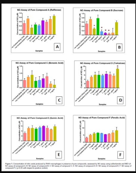
Figure 1: Concentration of nitric oxide produced by RAW macrophages in presence of pure compounds, assessed by NO assay using sulfanilamide and NED (A: NO assay of compound A; B: NO assay of compound B; C: NO assay of compound C; D: NO assay of compound D; E: NO assay of compound E; F: NO assay of compound F) (*p<0.05 with respect to positive control).
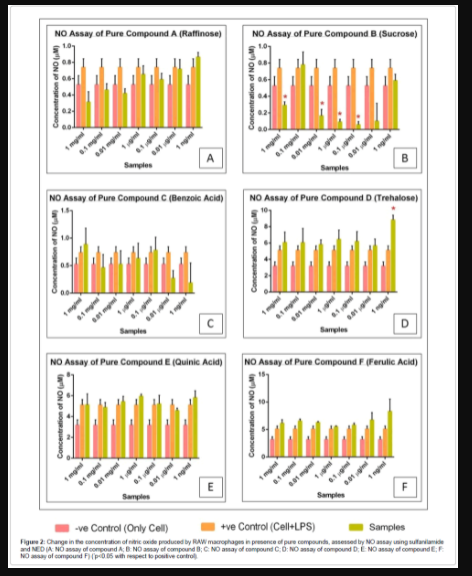
Figure 2: Change in the concentration of nitric oxide produced by RAW macrophages in presence of pure compounds, assessed by NO assay using sulfanilamide and NED (A: NO assay of compound A; B: NO assay of compound B; C: NO assay of compound C; D: NO assay of compound D; E: NO assay of compound E; F: NO assay of compound F) (*p<0.05 with respect to positive control).
NBT assay
The effect of six compounds (A: Raffinose, B: Sucrose, C: Benzoic acid, D: Trehalose, E: Quinic Acid and F: Ferulic acid) on bacterial lipopolysaccharide (LPS) induced inflammation in RAW 264.7 macrophages was also performed by NBT assay. This assay quantitates the superoxide produced by the enzyme NADPH oxidase during the phagocytosis due to inflammation. NBT reacts with the superoxide radicals to give mono and di-formazan, which has purple colour. The formazan, deposited in the phagocytic cells can be extracted and the extent of colour is indicative of the superoxide production. In our study, we found that treatment with LPS led to an increase in the absorbance at 620 nm. This indicates that formazan production increased with LPS, and thus, superoxide production increased with inflammation. Treatment with pure compounds led to a decrease in the superoxide production, with the maximum decrease seen in compound A, at a concentration of 0.01 mg/ml in Figures 3 and 4.
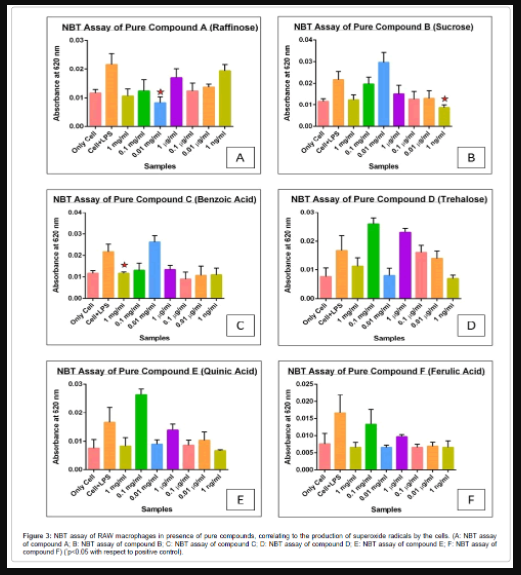
Figure 3: NBT assay of RAW macrophages in presence of pure compounds, correlating to the production of superoxide radicals by the cells. (A: NBT assay of compound A; B: NBT assay of compound B; C: NBT assay of compound C; D: NBT assay of compound D; E: NBT assay of compound E; F: NBT assay of compound F) (*p<0.05 with respect to positive control).
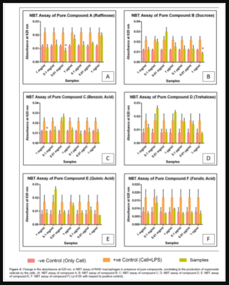
Figure 4: Change in the absorbance at 620 nm, in NBT assay of RAW macrophages in presence of pure compounds, correlating to the production of superoxide radicals by the cells. (A: NBT assay of compound A; B: NBT assay of compound B; C: NBT assay of compound C; D: NBT assay of compound D; E: NBT assay of compound E; F: NBT assay of compound F) (*p<0.05 with respect to positive control).
Inflammation is a response of living tissue to injuries that involve activation of various enzyme, mediators release, cell migration, tissue breakdown and repair. A number of inflammatory stimuli, such as LPS and pro-inflammatory cytokines (e.g., TNF-α), activate immune cells to up regulate inflammatory states. Therefore, they represent useful targets for developing new anti-inflammatory constituents and exploring their molecular. In addition, there are free radicals which are constantly generated and they can cause extensive damage to tissues and biomolecules leading to various disease conditions, especially degenerative diseases and extensive lysis. The commonly used drugs for management of inflammatory conditions are non-steroidal antiinflammatory drugs, which have several adverse side effects. Therefore, search for natural antioxidants with anti-inflammatory activity has been greatly increased in the recent years. An alternative solution to the problem is to consume natural antioxidants and anti-inflammatory products from food supplements and traditional medicine. In our study, we have selected two types of cell based assays, namely NO and NBT assay. Nitric oxide endows macrophages with cytostatic or cytotoxic activity against viruses, fungi, bacteria, protozoa and tumor cells. Suppression in the production of nitric oxide is usually observed with immunosuppressive drugs, for example glucocorticoids, and excessive production of NO is known to play important role in inflammation and arthritis. Therefore, effect on NO production should be carefully assessed for enhancement and suppression effects. This assay gives the concentration of nitric oxide produced by macrophages in response to inflammation. NBT assay gives an idea about the superoxide radicals produced by cells during inflammation. Due to inflammation, cells produce higher superoxide, which leads to greater reduction of NBT, and greater production of purple formazan.
In our present study, we found that all the compounds A, B, C, D, E and F, have, to some extent, anti-inflammatory activity, on bacterial lipopolysaccharide (LPS) induced inflammation RAW 264.7 macrophage cells. Compound A shows maximum anti-inflammatory activity at a concentration of 1 mg/ml. Compound B shows dose dependent anti-inflammatory activity between 0.01 mg/ml (10 μg/ml) and 1 ng/ml. Compound C is active at concentrations below 1 mg/ ml. Compound E shows activity below 0.0 μg/ml. No solid conclusion could be drawn for Compounds D and F, since the readings for the two assays did not tally. The results of the two assays show that compounds A, B, C and E do have some anti-inflammatory effect against LPS induced inflammation.
In this experiment, six pure compounds extracted from plant sources were screened for their anti-inflammatory activity against LPS induced inflammation in RAW macrophages, by NO and NBT assays. It could be concluded that compounds A (Raffinose), B (Sucrose), C (Benzoic acid) and E (Quinic Acid) could be used as anti-inflammatory agents, at low concentrations. However, compounds D (Trehalose) and F (Ferulic acid) did not give satisfactory results. Further assays, both in vitro and in vivo, have to be done to verify and validate these effects, before they can be considered for clinical use.
In this experiment, six pure compounds extracted from plant sources were screened for their anti-inflammatory activity against LPS induced inflammation in RAW macrophages, by NO and NBT assays. It could be concluded that compounds A (Raffinose), B (Sucrose), C (Benzoic acid) and E (Quinic Acid) could be used as anti-inflammatory agents, at low concentrations. However, compounds D (Trehalose) and F (Ferulic acid) did not give satisfactory results. Further assays, both in vitro and in vivo, have to be done to verify and validate these effects, before they can be considered for clinical use.
SM and SK performed all experiments and SM drafted the manuscript. ERB conceptualized the project, designed all experiments and analyzed the data and wrote this manuscript. The suggestion for initiating this screening and providing the pure compounds came from BD.