Journal of Clinical & Experimental Dermatology Research
Open Access
ISSN: 2155-9554
ISSN: 2155-9554
Research Article - (2021)
The association with the intake of thiazide medication and an increased photosensitivity was investigated by testing 15 patients on thiazide medication and 15 controls with stepwise UVA-, UVB- and UVB 311 nm-irradiation. Erythema and blood flow 24 h after irradiation were quantified by visual assessment (MED) and objective methods (Dermacatch, Minolta Chromameter, laser doppler flowmetry). A higher UVA-photosensitivity in patients taking thiazides was found by visual assessment. The sensitivity and specifity of the different devices was compared; objective measurement of erythema in all chosen wavelengths generally showed the best results with laser doppler flowmetry closely followed by colorimetry. Phototherapy in patients on thiazide medication should be carried out with caution since many factors of pharmacologically increased photosensitivity are still not explored, but it can be assumed that for the frequently used UVB 311 nm phototherapy no special precautions need to be taken.
Photosensitivity; Hydrochlorothiazide; Phototherapy
Hydrochlorothiazide (HCT) is one of the most frequently prescribed active ingredients in the world [1]. Shortly after the introduction the first cases with photosensitive reactions to HCT were published [2] and a review from the year 2014 identified 62 published cases with photosensitivity [3]: The most common presentations were eczematous lesions in a photodistributed pattern. Photosensitivity can generally occur in the context of natural sun exposure but also of artificial ultraviolet (UV) exposure. General recommendations for the implementation of UV phototherapy and photochemotherapy include special precautions such as close monitoring and modified treatment regimens in patients requiring photosensitizing drugs [4]. Photosensitivity is usually analysed by performing phototests such as determination of the Minimal Erythema Dose (MED). The visual scoring can be objectified by measuring the erythema by colorimetry and laser doppler flowmetry in each irradiation field [5]. It was the aim of this study to investigate the photosensitivity of patients on thiazide medication in comparison to control patients using visual assessment and objective methods including colorimetric measurements.
Study group
30 patients (17 men, 13 women; age 44 to 87 years; thiazide group: mean=66.4 ± 9.6 years, controls: mean=64.2 ± 8.6 years) with an indication for phototherapy (mainly for eczema and psoriasis) were included in the study. 15 patients were on thiazide medication (≥ 3 months; duration of the intake 1.5 to 18 years) and 15 without thiazide medication (controls). 14 of 15 patients of the control group were taking hydrochlorothiazide (dose: n=10: 12.5 mg/d, n=3: 25 mg/d, n=1: 6.25 mg) and one patient was taking indapamide (1.25 mg/d). Most of the patients were classified as skin type II according to Fitzpatrick (n= 27: skin type II, n= 3: skin type III). Exclusion criteria were diseases with a priori increased photosensitivity as for example SLE or Porphyria cutanea tarda, the intake of other photosensitizing medication (as for example furosemide, tetracyclines, phytotherapeuticals) and a recent or continuing topical (at the area of UV-testing) or systemic steroid medication.
UV-testing
Stepwise UVB, UVB 311 nm and UVA exposure on UV-unexposed and non-inflammatory skin at the lower back was performed. UVB irradiation was performed with TL20W/12 Philips (Hamburg, Germany) bulbs (285 nm-350 nm, maximum between 310 and 315 nm) at doses of 25, 50, 75, 100, 125 and 150 mJ/cm2; UVB- 311 nm irradiation was performed with a Phototest 300 (Medilux Medizintechnik, Korntal-Münchingen, Germany) device (310 nm- 315 nm, maximum at 311 nm) at doses of 0.03, 0.07, 0.18, 0.30, 0.37, 0.56, 0.75 J/cm2 and UVA irradiation with a Waldmann UVA 700 (Waldmann, Villingen-Schwenningen, Germany) device (330 nm-450 nm, maximum between 360 nm-370 nm) at doses of 5, 10, 15, 20, 25, 30 J/cm2.
Evaluation of erythema
24 ± 2 hours after UV-exposure the erythema in each irradiation field was visually determined (minimal erythema dose, MED). An objective evaluation was done by laser doppler flowmetry (Laser Doppler Imager moorLDI, Lawrenz, Sulzbach, Germany) and colorimetry using two different devices (Minolta Chromameter CR-400, Konica Minolta, Langenhagen, Germany; Dermacatch, Colorix, Neuchâtel, Switzerland).
Statistics
Poisson regression models were used to compare the photosensitivity between the two patient groups in the respective UV range (UVA, UVB, UVB 311 nm) on the basis of the results of the visual and instrumental evaluation using Dermacatch. Therefore the integer number of the first erythematous irradiation field per subject served as the outcome while the factor variables “group” and “gender”, as well as the continuous variable “age” were used as covariates. The presented average values per group are marginal means computed from the models averaged across gender and age. Linear regression models were used to explain the redness intensity by the factor variables “irradiation level”, “group” and the interaction thereof. The latter was used to infer on different group specific effects. The robust Huber-White method was used to compute the model’s covariance matrix to account for repeated measurements per subjects. ROC (Receiver Operating Characteristics) analyses for clustered data were used to compare the performance of the instrumental measurement methods as a diagnostic test of the visual evaluation (gold standard) of the multiple irradiation fields per patient [6]. Statistical hypothesis testing has been performed on exploratory two-sided 5% significance levels. Analyses were conducted with R 3.5.0 (The R Foundation for Statistical Computing, Vienna, Austria).
Patients’ characteristics
27 patients were classified as Fitzpatrick skin-type II, 3 as Fitzpatrick skin-type III. The mean age of the patients taking thiazides (8 men, 7 women) was 66.4 ± 9.6 years, of the controls (9 men, 6 women) 64.2 ± 8.6 years.
Evaluation of minimal erythema dose (MED)
Visual assessment: On average there were less erythematous irradiation fields in the group taking HCT compared to the control group after UVB (2.74 vs. 2.92, p-0.773) and UVB 311 nm (1.15 vs. 1.33, p-0.655) irradiation. In the group taking HCT the mean number of erythematous irradiation fields was significantly higher than in the control group after UVA irradiation (1.33 vs. 0.6, p-0.047).
Colorimetric measurement (Dermacatch): There were slight, statistically Not significant differences with respect to the number of erythematous irradiation fields between the group taking HCT and the control group after UVA (3.4 vs. 3.02, p-0.557), UVB (3.67 vs. 3.7, p-0.966) and UVB 311 nm (3.31 vs. 3.24, p-0.917) irradiation.
Evaluation of Erythema intensity
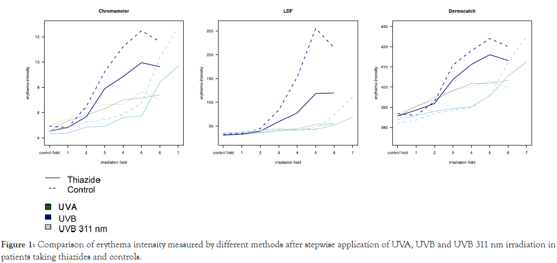
Laser doppler flowmetry: The regression models of blood flow intensity by UV doses showed overall increasing mean blood flow values with increasing irradiation doses (p<0.0001). There were minor differences between the two groups for UVB and UVA irradiation. For UVB 311 nm irradiation the patients taking HCT showed a significant lower course of the curve (p-0.014) than the controls.
Colorimetric measurement: The regression models of erythema intensity by UV doses showed overall significantly increasing erythema values with increasing irradiation doses (p<0.0001). There were minor differences between the curves of the two groups for UVB, UVB 311 nm and UVA irradiation for both devices.
An overview of these results can be found in Tables 1 and 2, Figure 1.

Figure 1: Comparison of erythema intensity measured by different methods after stepwise application of UVA, UVB and UVB 311 nm irradiation in patients taking thiazides and controls.
| Test method | Wavelength | Mean thiazide group | Mean control group | Ratio | p-value |
|---|---|---|---|---|---|
| Visual | UVA | 1.33 | 0.6 | 2.23 ± 1.49 | 0.047 |
| UVB | 2.74 | 2.92 | 0.94 ± 1.25 | 0.773 | |
| UVB 311 nm | 1.15 | 1.33 | 0.86 ± 1.39 | 0.655 | |
| Colorimetry (Dermacatch) | UVA | 3.02 | 3.4 | 1.13 ± 1.22 | 0.557 |
| UVB | 3.67 | 3.7 | 0.99 ± 1.21 | 0.966 | |
| UVB 311 nm | 3.31 | 3.24 | 1.02 ± 1.22 | 0.917 |
Table 1: Comparison of minimal erythema dose by visual determination and colorimetric measurement (Dermacatch) after stepwise application of UVA, UVB and UVB 311 nm irradiation in patients taking thiazides and controls (marginal means computed from the poisson regression models averaged across gender and age).
| Laser doppler flowmetry | ||
|---|---|---|
| UVA | Patients taking thiazides | Controls |
| Dose (J/cm2) | Mean ± SD | Mean ± SD |
| 5 | 32.51 ± 7.40 | 33.01 ± 5.35 |
| 10 | 36.02 ± 9.63 | 36.68 ± 4.61 |
| 15 | 40.05 ± 10.01 | 40.81 ± 7.58 |
| 20 | 44.52 ± 13.12 | 40.59 ± 6.78 |
| 25 | 53.63 ± 19.70 | 48.66 ± 19.90 |
| 30 | 55.41 ± 35.32 | 53.68 ± 35.43 |
| UVB | Patients taking thiazides | Controls |
| Dose (mJ/cm2) | Mean ± SD | Mean ± SD |
| 25 | 33.14 ± 5.84 | 34.45 ± 4.83 |
| 50 | 39.25 ± 7.52 | 44.90 ± 11.11 |
| 75 | 59.43 ± 22.47 | 83.85 ± 50.84 |
| 100 | 77.75 ± 46.18 | 153.08 ± 104.84 |
| 125 | 118.58 ± 64.30 | 255.18 ± 192.42 |
| 150 | 119.52 ± 74.14 | 214.82 ± 161.45 |
| UVB 311 nm | Patients taking thiazides | Controls |
| Dose (J/cm2) | Mean ± SD | Mean ± SD |
| 0.03 | 36.22 ± 8.03 | 37.38 ± 6.34 |
| 0.07 | 40.58 ± 11.41 | 42.92 ± 6.49 |
| 0.18 | 44.40 ± 10.59 | 42.67 ± 8.07 |
| 0.30 | 41.65 ± 9.09 | 44.61 ± 8.42 |
| 0.37 | 43.15 ± 9.92 | 43.71 ± 10.27 |
| 0.56 | 51.88 ± 19.46 | 73.25 ± 37.34 |
| 0.75 | 68.88 ± 31.14 | 111.65 ± 55.53 |
| Minolta Chromameter | ||
| UVA | Patients taking thiazides | Controls |
| Dose (J/cm2) | Mean ± SD | Mean ± SD |
| 5 | 5.37 ± 2.06 | 5.53 ± 2.85 |
| 10 | 5.81 ± 1.99 | 6.31 ± 2.65 |
| 15 | 6.31 ± 2.23 | 6.65 ± 2.55 |
| 20 | 7.00 ± 2.67 | 6.65 ± 1.93 |
| 25 | 7.14 ± 2.41 | 7.66 ± 2.83 |
| 30 | 7.41 ± 2.38 | 7.03 ± 1.89 |
| UVB | Patients taking thiazides | Controls |
| Dose (mJ/cm2) | Mean ± SD | Mean ± SD |
| 25 | 4.83 ± 1.79 | 4.82 ± 2.62 |
| 50 | 5.67 ± 1.79 | 6.49 ± 2.44 |
| 75 | 7.89 ± 2.72 | 9.22 ± 4.29 |
| 100 | 8.86 ± 2.66 | 11.22 ± 4.02 |
| 125 | 9.95 ± 3.40 | 12.47 ± 4.75 |
| 150 | 9.63 ± 3.63 | 11.61 ± 4.35 |
| UVB 311 nm | Patients taking thiazides | Controls |
| Dose (J/cm2) | Mean ± SD | Mean ± SD |
| 0.03 | 4.38 ± 1.60 | 4.89 ± 2.53 |
| 0.07 | 4.86 ± 1.76 | 5.24 ± 2.63 |
| 0.18 | 6.31 ± 5.78 | 5.46 ± 2.52 |
| 0.30 | 5.61 ± 1.90 | 5.86 ± 2.56 |
| 0.37 | 5.73 ± 2.01 | 6.73 ± 2.68 |
| 0.56 | 8.40 ± 2.94 | 10.39 ± 3.69 |
| 0.75 | 9.68 ± 3.06 | 12.71 ± 3.35 |
| Dermacatch | ||
| UVA | Patients taking thiazides | Controls |
| Dose (J/cm2) | Mean ± SD | Mean ± SD |
| 5 | 390.51 ± 14.67 | 390.55 ± 13.83 |
| 10 | 393.67 ± 12.38 | 394.89 ± 15.49 |
| 15 | 398.24 ± 13.54 | 398.49 ± 15.71 |
| 20 | 401.62 ± 14.76 | 400.25 ± 13.76 |
| 25 | 401.89 ± 13.07 | 403.02 ± 16.98 |
| 30 | 403.36 ± 13.33 | 399.91 ± 13.13 |
| UVB | Patients taking thiazides | Controls |
| Dose (mJ/cm2) | Mean ± SD | Mean ± SD |
| 25 | 388.60 ± 13.70 | 385.96 ± 13.80 |
| 50 | 391.89 ± 13.87 | 392.82 ± 12.18 |
| 75 | 403.78 ± 16.18 | 410.93 ± 22.88 |
| 100 | 411.24 ± 16.56 | 418.02 ± 21.34 |
| 125 | 416.05 ± 19.02 | 424.07 ± 26.18 |
| 150 | 413.22 ± 19.57 | 420.09 ± 24.85 |
| UVB 311 nm | Patients taking thiazides | Controls |
| Dose (J/cm2) | Mean ± SD | Mean ± SD |
| 0.03 | 386.00 ± 16.10 | 383.62 ± 16.34 |
| 0.07 | 388.00 ± 17.27 | 386.89 ± 16.85 |
| 0.18 | 389.33 ± 16.26 | 388.60 ± 14.95 |
| 0.30 | 390.60 ± 13.58 | 389.78 ± 17.03 |
| 0.37 | 395.27 ± 12.58 | 395.89 ± 15.96 |
| 0.56 | 405.58 ± 15.43 | 412.15 ± 18.37 |
| 0.75 | 412.53 ± 14.88 | 425.07 ± 16.57 |
Table 2: Trend and evaluation of blood flow (laser doppler flowmetry) and erythema intensity (Minolta Chromameter, Dermacatch).
Comparison of objective measurements and visual assessment of erythema
For UVA irradiation the laser doppler flowmeter showed a higher area under the curve than colorimetic measurements with the Minolta Chromameter (p<0.004) and Dermacatch (p<0.001). Comparison of the Minolta Chromameter with the Dermacatch showed a higher area under the curve for the Minolta Chromameter (p<0.008). For UVB irradiation the area under the curve between laser doppler flowmetry and the Minolta Chromameter as well as between the Minolta Chromameter and Dermacatch showed similar values without significant differences. The laser doppler flowmeter showed a higher area under the curve than the Dermacatch (p<0.011). For UVB 311 nm irradiation the area under the curve between all devices was similar and showed no significant differences.
An overview of these results can be found in Table 3.
| Laser doppler flowmetry (1) | Minolta Chromameter (2) | Dermacatch (3) | p-value (1 vs. 2) | p-value (1 vs. 3) | p-value (2 vs. 3) | |
|---|---|---|---|---|---|---|
| UVA | 0.98 ± 0.01 | 0.80 ± 0.06 | 0.65 ± 0.08 | 0.004 | <0.001 | 0.008 |
| UVB | 0.96 ± 0.02 | 0.91 ± 0.03 | 0.88 ± 0.03 | 0.090 | 0.011 | 0.080 |
| UVB 311 nm |
0.83 ± 0.06 | 0.95 ± 0.02 | 0.89 ± 0.04 | 0.086 | 0.376 | 0.066 |
Table 3: Comparison of the diagnostic ability of the objective measurements (Dermacatch, Minolta Chromameter, laser doppler flowmetry) using visual assessment of erythema as gold standard.
This study with a special focus on patients with an indication for phototherapy showed that there was not an increased UVBphotosensitivity in the group taking HCT compared to the control group. Therefore it can be assumed that especially for the frequently used UVB 311 nm phototherapy no special precautions need to be taken in patients with no history of UV-distributed skin diseases. The higher UVA-photosensitivity in patients taking HCT versus controls found by visual assessment was less expressed for objective laser doppler or colorimetric measurements of erythema. In most published studies in-vitro or in-vivo photosensitivity to HCT was due to UVA alone or to both UVA and UVB suggesting an action spectrum mainly in the UVA range [3,7,8]. Recently an association between high use of HCT and the development of nonmelanoma skin cancer was published [9]. According to in vitro and animal studies also UVA irradiation might be responsible for photocarcinogenic effects due to HCT [10].
Objective measurement of erythema in all chosen wavelengths generally showed the best results measuring the blood flow with laser doppler flowmetry closely followed by evaluation of the color by colorimetry. The higher sensitivity of the laser doppler flowmeter might be due to the earlier detection of relative changes in the Blood flow compared to the color changes. Nevertheless in daily clinical practice measurements with laser doppler flowmetry are time-consuming, less practical and reserved for special investigations where minimal changes need to be registered. Previous studies could show the positive correlation between visual, colorimetric and laser doppler flowmetric assessment of skin erythema [11,12]. Overall colorimetric measurements as well as laser doppler flowmetry are reliable tools for such purposes [5]. Our results confirm that also small and easy to handle devices as the Minolta Chromameter and the Dermacatch are suitable for the objective evaluation of skin erythema in daily clinical practice.
Citation: Balzer C, Hapfelmeier A, Zink A, Biedermann T, Eberlein B (2020) Quantification of UV-Induced Skin Erythema before Phototherapy in Individuals taking Hydrochlorothiazide and Controls. J Clin Exp Dermatol Res. 12:538.
Received: 21-Dec-2020 Accepted: 04-Jan-2021 Published: 11-Jan-2021 , DOI: 10.35248/2155-9554.21.s8.538
Copyright: © 2021 Balzer C, et al. This is an open-access article distributed under the terms of the Creative Commons Attribution License, which permits unrestricted use, distribution, and reproduction in any medium, provided the original author and source are credited.