Journal of Glycobiology
Open Access
ISSN: 2168-958X
ISSN: 2168-958X
Research Article - (2013) Volume 0, Issue 0
Keywords: Photodynamic therapy; Photosensitizer; Squaraine dye; Biodistribution; Skin tumor; Oxidative stress; Lipid peroxidatin; Antioxidant enzymes
PDT: Photodynamic Therapy; GPx: Glutathione Peroxidase; GR: Glutathione Reductase; NADP+: Nicotinamide Adenine Dinucleotide Phosphate; NADPH: Nicotinamide Adenine Dinucleotide Phosphate Reduced; SOD: Superoxide Dismutase; TBARS: Thio Barbituric Acid Reactive Substances; TCA: Trichloroacetic Acid; H2O2: Hydrogen Peroxide; NaOH: Sodium Hydroxide; ANOVA: Analysis of Variance
Photodynamic therapy (PDT) is emerging as a promising noninvasive treatment for cancers [1]. It involves three key components; a photosensitizer, light and tissue oxygen.
Photodynamic therapy involves administration of a tumor localizing photosensitizing agent, followed by the activation of the agent by light of a specific wavelength. This therapy results in a sequence of photochemical and photo biological processes that cause irreversible photo damage to tumor tissue. Photodynamic therapy (PDT) has emerged as an alternative strategy for treating cancer. PDT consists of three main components: a photosensitizer, light, and oxygen. PDT takes advantage of an appropriate wavelength of light that excites a photosensitizer to a triplet energy state [2-4]. In the presence of molecular oxygen, energy is transferred to relax the excited state of the photosensitizer. This energy transfer in turn excites molecular oxygen to form excited, singlet state oxygen. Singlet oxygen induces cell death via damaging oxidation or redox-sensitive cellular signaling pathways, thus mediating the effects of PDT [2,5,6]. Intriguingly, PDT has also been shown to regulate processes beyond tumor cell death including tumor angiogenesis and modulation of the immune system [2,5,7,8]. Each photosensitizer is activated by light of a specific wavelength [1]. This wavelength determines how far the light can travel into the body [9,10].
An ideal photosensitizer should meet some of the following criteria that are clinically relevant: a commercially available pure chemical, low dark toxicity but strong photocytotoxicity, good selectivity towards target cells, long-wavelength absorbing, rapid removal from the body and ease of administration though various routes. These criteria provide a general guideline for comparison. Although some photosensitizers satisfy all of or some of these criteria, there are currently only a few photosensitizers that have received official approval around the world. However, PDT with the currently FDA-approved photosensitizers is not without adverse effects. For example, Photofrin®, the first systemic drug to be approved, is well known for causing an intense inflammatory and necrotic reaction at the treated site and prolonged widespread photosensitivity for up to several weeks post-PDT, thereby imposing severe limitations on the lifestyle [11]. Because of this and other drawbacks of Photofrin®, many additional photosensitizers have been synthesized, and a few of them have developed into FDA-approved drugs or are in clinical trials.
Interest in the synthesis and evaluation of new photosensitizers for use in PDT has grown as a result of both the encouraging initial clinical results of this therapy and the documented need to improve upon the limitations of currently available photosensitizers. The dye selected in our study- Symmetric diiodinated squaraine- is one of the newly developed photosensitizers. We have done the in vivo biodistribution of the dye on normal and skin tumor induced animal models to check the retention time of the dye. Before analyzing the therapeutic efficacy of the compound as a photosensitizer, it should be confirmed that it does not elicit any toxic manifestations in the body, when not illuminated. One among the most important criteria of an ideal photosensitizer is that it should induce no dark-toxicity; i.e., it should be non-toxic in the dark [6]. Thus we have checked whether symmetrical diiodinated squaraine is an ideal photosensitizer in this aspect by checking the antioxidant status of dye administered mice.
Chemicals
The photosensitizer Symmetrical diiodinated squaraine was synthesized and supplied by the Photochemistry Research Unit, National Institute of Interdisciplinary Science and Technology (NIISTCSIR), Thiruvananthapuram, India. All reagents used were of analytical grade. All biochemicals used were obtained from M/s. Sigma, St. Louis, MO, USA.
Animal models
The animal models (Swiss albino mice-Male, weighing 20-25g) were from the departmental animal house. The animals were housed in polypropylene cages in rooms maintained at 25 ± 1oC. Drinking water was given ad libitum. Mice were fed with standard laboratory diet supplied by Lipton India Ltd. For maintaining the experimental animals, the Institutional Ethical guidelines were absolutely followed as per CPCSEA rules [Sanction No: IAEC- KU- 4/2010-11-BC-AA (15)].
Studies on cellular uptake and distribution
Swiss albino mice (Male, 20-25 g) were used for the in vivo studies. The dye was administered to the mice by intraperitoneal injection at different concentrations (12.5 mg/kg body weight dissolved in phosphate buffered saline). Thereafter, the animals were kept in dark until the termination of the experiment to eliminate any possible tissue damage due to the exposure to sunlight. After injection, the animals were sacrificed at different intervals of time (1, 2, 3, 4, 5, 6, 10, 20 and 24 h post injection). Serum and other tissue samples like liver, kidney, skin, heart, and spleen were collected for the estimation of the dye. The concentration of the dye in different tissues after intraperitoneal administration was determined by the method described by Moan et al. [12] with some modifications.
For the induction of skin tumors the method of Ewing et al. [13] was followed. Dorsal hair between the cervical and caudal portion of mice was removed using a surgical clipper, two days prior to the initiation of the experiment. DMBA (100 µg dissolved in 50 µl acetone) was used as a tumor initiator and applied to the skins of mice. After fourteen days, the tumor initiation by DMBA was promoted by the topical application of croton seed oil (1% v/v in acetone) thrice a week for the next 20 weeks [13,14]. The biodistribution studies were done as described above.
For the extraction process, the tissues were washed free of blood. Approximately 500 mg of each tissue was disrupted in a homogenizer, dissolved in methanol (0.5 g/ml) and centrifuged at 20,000 rpm. The supernatant was analyzed with a spectrofluorimeter (excitation wavelength: 660 nm and emission wavelength: 678 nm) and the concentrations of the extractable dye were determined by comparing with the standard curve. The dye was directly extracted into methanol from serum and the concentration was estimated.
Analysis of oxidative stress in vivo
To determine whether there is any oxidative stress produced by the dye in the body without illumination, the following experiment was done.
Female Swiss Albino mice were divided into three groups containing six animals in each as:
Group I: Normal mice (Control).
Group II: Treated with squaraine dye (Sacrificed at 4 h after the administration of the dye).
Group III: Treated with squaraine dye (Sacrificed at 24 h after the administration of the dye).
The dye was administered to the mice of Group II and Group III by intraperitoneal injection at a concentration of 12.5 mg/kg body weight dissolved in PBS. The animals were sacrificed at 4 h (when the amount of dye in the body of Group II mice is found to be maximum, according to the distribution studies) and 24 h (when the dye is completely removed from the body) after the injection and the tissues were collected. The biochemical parameters marking oxidative profile: the levels of lipid peroxidation products (Malondialdehyde and Conjugated diene), the activities of the antioxidant enzymes - superoxide dismutase (SOD), catalase, glutathione peroxidase (GPx) and glutathione reductase (GR) were determined.
Estimation of lipid peroxide – TBARS
TBARS was estimated by the thiobarbituric acid assay method of Niehaus and Samuelson [15].
The tissue homogenate was prepared on 0.1 M Tris-HCl buffer, pH 7.5. 1 ml of the homogenate was combined with 2 ml of TCATBA -HCl and mixed thoroughly. The solution was heated for 15 min in a boiling water bath. After cooling, the flocculent precipitate was removed by centrifugation at 1000 g for 10 min. The absorbance of the sample was read at 535 nm against a blank that contained no tissue homogenate. The molar extinction coefficient of malondialdehyde is 1.56×10⁵ mol/cm.
Estimation of the levels of conjugated diene
Levels of conjugated diene in lens were estimated by the method of Reckangal and Ghoshall [16]. Tissue homogenate prepared in Tris - HCl (0.1 M, pH 7.5) was centrifuged and 1 ml supernatant was mixed with 5.0 ml of chloroform: ethanol mixture (2:1). The content was centrifuged at 1000 x g for 5 min and the upper layer removed by aspiration. 3.0 ml of the lower chloroform layer was taken in a test tube and evaporated to dryness at 45°C in a water bath. The residue was dissolved in 1.5 ml cyclohexane and absorbance noted at 233 nm against a cyclohexane blank. The concentration of conjugated dienes was calculated from its extinction coefficient (2.52 × 10 m-1 cm-1).
Assay of glutathione peroxidase (EC 1. 11 .1.9)
Activity of GPx was determined by the procedure of Brien and Little [17]. The assay system contained 2ml phosphate buffer, 0.3 ml sodium azide, 0.2 ml EDTA, 1ml reduced Glutathione, 0.1 ml H2O2, 0.1 ml NADPH, 0.5 ml water and 0.2 ml enzyme source. The oxidation of glutathione was measured at 340 nm at an interval of 15 seconds for two min. Values have been expressed as – mg of GSH oxidized/min/ mg tissue.
Assay of glutathione reductase (EC 1.6.4.2)
The method is as described by Bergmeyer [18]. The assay system contained 0.067 mM phosphate buffer, pH 7.6, 7.5 mM oxidised glutathione, 0.3 mM NADPH and the enzyme in a final volume of 1ml .The decreased in OD was followed at 340nm. Activity is expressed as µmoles of NADPH oxidised/min/g tissue.
Assay of Superoxide Dismutase (SOD, EC 1.15. 1. 1)
SOD activity was determined by the method described by Kakkar et al. [19]. The assay mixture contained 1.2 ml sodium pyrophosphate buffer (0.052 mM pH 8.3), 0.1 ml 186 mM Phenazine methosulphate, 0.3 ml 30 µM Nitro blue tetrazolium, appropriately diluted enzyme preparation (0.2 ml) and water in a total volume of 3 ml. The reaction was started by the addition of 0.2 ml 780 µM NADH. After incubation at 30°C for 90 seconds, the reaction was stopped by the addition of 1 ml glacial acetic acid. The reaction mixture was stirred vigorously and shaken with 4 ml of n-butanol. The mixture was allowed to stand for 10 min. Centrifuged and butanol layer was taken out. Colour intensity of chromogen in the butanol was measured at 560 nm against butanol (Stable up to 48 h). A system devoid of enzyme served as control. One unit of enzyme activity is defined as the enzyme concentration required to inhibit the OD at 560 nm of chromogen produced by 50% in one minute under the assay condition and expressed as specific activity in milli units/mg protein.
Assay of catalase (EC 1. 11. 1. 6)
Catalase was assayed by the method of Machly and Chance [20]. The tissue was homogenized in 2 ml phosphate buffer at 1-4°C and centrifuge at 5000 rpm. The estimation was done spectrophotometrically following the decrease in absorbance at 230 nm. 3 ml H2O2 – phosphate buffer and about 0.01 – 0.04 ml of enzyme solution was pipetted into experimental cuvette. Read after every 15 seconds for about 1-2 min. Read against control cuvette containing enzyme solution as in the experimental cuvette but containing H2O2 free buffer.
Estimation of protein
The estimation of protein was done by the method of Lowry et al. [21]. 1 ml 10% TCA to 0.1 ml of the tissue homogenate was added. The tube was kept in ice for 10 min and centrifuged for 10 min and the supernatant was decanted. Then the precipitate was dissolved in 1 ml 0.1 N NaOH.
0.1 ml of the above solution was taken as test solution. Tubes were made up to 1 ml with 0.1N NaOH, mixed well. Then 5 ml of alkaline copper reagent was added and kept for 10 min. 1 ml of diluted Folin’s reagent was added to all the tubes. A standard as well as a blank were also treated same way. The blue colour was obtained. The optical density was read spectrophotometrically at 670 nm within 30 min.
Statistical analysis
The data were statistically analyzed using analysis of variance (ANOVA) and significant difference of means was determined using Duncan’s multiple range test [22] at the level of p<0.05.
Biodistribution studies
The distribution of the photosensitizer among the various organs in the body represents a key factor for the efficacy of photodynamic therapy. The simplest method to measure the drug concentration is to detect the fluorescence emitted by the drug.
The dye (12.5 mg/kg body weight dissolved in phosphate buffered saline) was administered to different groups of mice by intraperitoneal injection. Thereafter, the animals were kept in dark until the termination of the experiment to eliminate any possible tissue damage due to the exposure to sunlight. After injection, the animals were sacrificed at different intervals of time (1, 2, 3, 4, 5, 6, 10, 20 and 24 h post injection). Serum and other tissue samples like liver, kidney, heart, spleen, lungs, and skin were collected for the estimation of the dye.
The concentration of the squaraine dye in different tissues except serum of the animals was maximal after 4 h of injection. In serum, the highest concentration was obtained 1h after the administration of dye and it was progressively reduced with time. At 24 h after injection, the concentration of the dye observed in the tissues was zero. Thus it can be assumed that it has been cleared out from the system by this time interval (Figure 1).
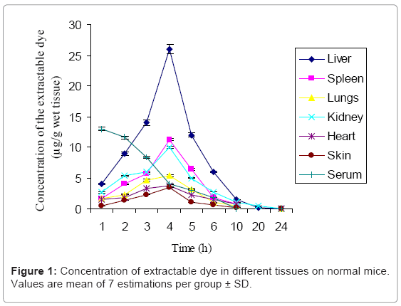
Figure 1: Concentration of extractable dye in different tissues on normal mice. Values are mean of 7 estimations per group ± SD.
Photosensitizing compounds are thought to be taken up in the malignant tissue both by active and by passive paths. The preferential accumulation of the photosensitizer in tumor tissue compared with the normal tissues may be a result of the greater proliferative rates, poorer lymphatic drainage, leaky vasculature and large interstitial space of neoplastic cells, physiological differences between tumor and normal tissues or some more specific interaction between the photosensitizer and marker molecules on neoplastic cells [23,24]. In general, the fast angiogenesis of tumor tissue results in vessels of a lower quality that have a tendency to leak, allowing macromolecules to pass into the interstitial compartment, which often is larger in malignancies. Since there is a poor lymphatic drainage system for the malignant cells, the photosensitizer once get entered into the tumor cells are retained there for a long time. One suggested specific interaction is between the lowdensity lipoprotein (LDL) receptor and photosensitizer leading to increased photosensitizer concentrations in neoplastic tissue. Tissues with high mitotic activity (like malignant tumor tissue) express an increased amount of low-density lipoprotein (LDL) receptors on the cell surface. The increased expression of LDL receptors in malignant cells may be caused by either an increased rate of cell proliferation or an increased rate of membrane turnover without proliferation. The uptake of sensitizer molecules associated with the receptormediated endocytosis of LDL can therefore be of importance. LDLassociated photosensitizer is then targeted to cellular or vascular components of the tumor. Tumors contain many macrophages that can ingest and monomerize aggregated photosensitizers as well as lipoprotein bound drugs [23]. This may be another reason for the tumor specificity of photosensitizers. Differences in water content and in other physiological parameters between tumors and normal tissue play roles for tumor localization of drugs. A higher content of collagen seems to be present in several tumors than in normal tissues [25]. Most of the photosensitizers have a high affinity towards collagen. These conclusions are based largely on photosensitizer pharmacokinetics and tissue distribution studies with a number of photosensitizers.
Skin tumor was induced in Swiss albino mice (Male, 20-25 g) using DMBA and croton seed oil. After the completion of the study period (20 weeks), the tumor bearing mice with a tumor radius greater than 2 mm were divided into ten groups with six animals each. The dye was administered to the different sets of mice except the control by intraperitoneal (i.p) injection at a concentration of 12.5 mg/kg body weight dissolved in phosphate buffered saline. Thereafter, the animals were kept in dark until the termination of the experiment to eliminate any possible tissue damage due to the exposure to sunlight. After injection, the animals were sacrificed at different intervals of time (1, 2, 3, 4, 5, 6, 10, 20 and 24 h post injection). Serum and other tissue samples like liver, kidney, heart, spleen and lungs were collected for the estimation of the dye. Skin flaps and tumor tissues from the back of mice were excised. All samples were washed free of blood by rinsing with physiological saline, blotted dry and protected from light. Skin/tumor tissues were immediately frozen at -80°C until used for estimations.
The concentration of extractable dye from different tissues was determined fluorimetrically. The compound is tumor specific since after 24 h of injection, it was retained only in the tumor site and in the immediate surroundings of the tumor. This shows the selectivity of the dye to tumor tissue compared to the normal nonmalignant tissues. There is a slight diffusion of the dye to the surrounding normal tissue as well. Thus recurrence of tumor in the pre existed- tumor margins can be prevented in the case of treatments with squaraine PDT.
Biochemical parameters marking oxidative profile
The biochemical parameters marking oxidative profile like the levels of lipid peroxidation products (Thiobarbituric acid reacting substances-TBARS) and conjugated dienes, the activities of the antioxidant enzymes - superoxide dismutase (SOD), catalase, glutathione peroxidase (GPx) and glutathione reductase (GR) were determined 4 h and 24 h after the injection of the squaraine dye. The results obtained are given in the following figures (Figures 2-8).
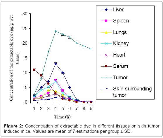
Figure 2: Concentration of extractable dye in different tissues on skin tumor induced mice. Values are mean of 7 estimations per group ± SD.
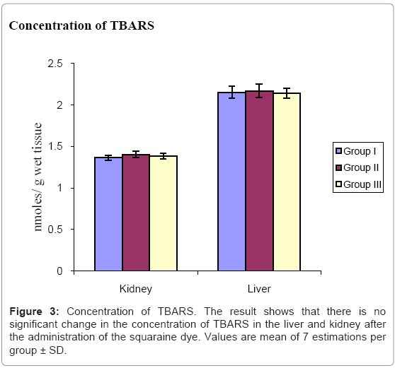
Figure 3: Concentration of TBARS. The result shows that there is no significant change in the concentration of TBARS in the liver and kidney after the administration of the squaraine dye. Values are mean of 7 estimations per group ± SD.
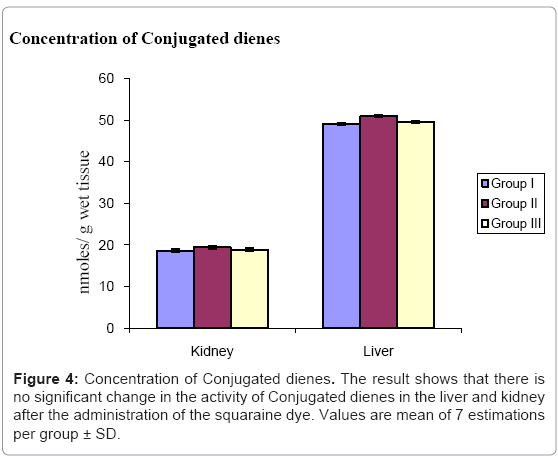
Figure 4: Concentration of Conjugated dienes. The result shows that there is no significant change in the activity of Conjugated dienes in the liver and kidney after the administration of the squaraine dye. Values are mean of 7 estimations per group ± SD.
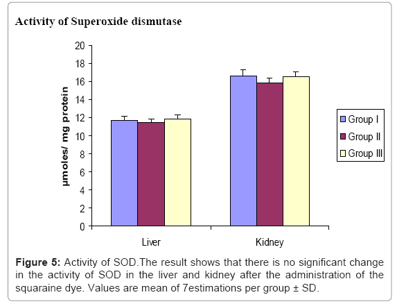
Figure 5: Activity of SOD.The result shows that there is no significant change in the activity of SOD in the liver and kidney after the administration of the squaraine dye. Values are mean of 7estimations per group ± SD.
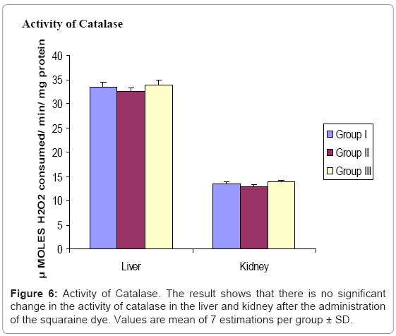
Figure 6: Activity of Catalase. The result shows that there is no significant change in the activity of catalase in the liver and kidney after the administration of the squaraine dye. Values are mean of 7 estimations per group ± SD.
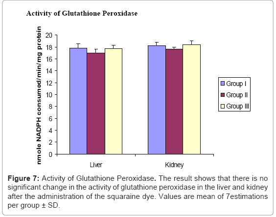
Figure 7: Activity of Glutathione Peroxidase. The result shows that there is no significant change in the activity of glutathione peroxidase in the liver and kidney after the administration of the squaraine dye. Values are mean of 7estimations per group ± SD.
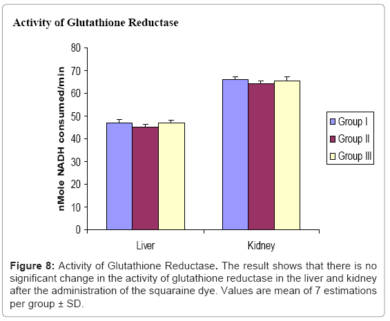
Figure 8: Activity of Glutathione Reductase. The result shows that there is no significant change in the activity of glutathione reductase in the liver and kidney after the administration of the squaraine dye. Values are mean of 7 estimations per group ± SD.
Concentration of TBARS
Before analyzing the therapeutic efficacy of the compound as a photosensitizer, it should be confirmed that it does not elicit any toxic manifestations in the body, when not illuminated. One among the most important criteria of an ideal photosensitizer is that it should induce no dark-toxicity; i.e., it should be non-toxic in the dark.
It has been reported that PDT induces tumor damage through the generation of oxidative stress in the target tissue. But an ideal photosensitizer is one which does not lead to any change in the oxidative status of the accumulated tissues, when not gets illuminated. Free radicals are constantly generated and eliminated in the biological system and play important role in a variety of normal biochemical functions and abnormal pathological processes. The products of lipid peroxidation include malondialdehyde (TBARS), hydroperoxides, conjugated dienes etc. An increase in lipid peroxidation indicates serious damage to cell membranes and their fluidity, inhibition of several enzymes and oxidation of proteins. The analysis of oxidative profile of different groups establishes no evidence of oxidative damage, which was measured in terms of the level of TBARS in the dye administered animals compared to the control animals.
In photodynamic therapy, the combination of photosensitizer and visible light causes generation of singlet oxygen and other free radicals which are extremely reactive. Since photodynamic therapy associated tumor destruction is mediated through the generation of oxidative stress, the study examines whether the photosensitizer alone can alter the oxidative profile of the normal system. For this the levels of lipid peroxidation products (Malondialdehyde and Conjugated dienes) and the markers of oxidative stress (superoxide dismutase (SOD), catalase, glutathione peroxidase (GPx) and glutathione reductase (GR) were analyzed and no significant change was observed in the levels of these parameters in the dye administered group when compared to control (Figures 3-8).
Altogether, in the present study we have studied the in vivo biochemical effects of Symmetrical diiodinated squaraine on experimental models. In the normal mice, the concentration of the dye in liver, kidney, spleen, heart, lungs and skin were maximal at 4 h after the injection whereas in serum maximum concentration was found at 1 h after the administration of the dye and was completely removed from the tissues by 24 h. This result shows that the dye is not retained in the normal tissues after 24 h. The biodistribution studies on skin tumor induced mice shows that the dye is retained in the tumor area after 24 h whereas it is completely removed from the normal tissues by that time and so the dye is efficient for PDT applications. The administration of the squaraine dye in the system induces no significant change in the level of lipid peroxidation products and activity of catalase, superoxide dismutase, glutathione peroxidase and glutathione reductase when compared to the control mice. This indicates that the administration of the squaraine dye does not alter the antioxidant status of the body. Thus, from the present study, it can be concluded that the dye selected for our study (Symmetrical diiodinated squaraine) is a promising agent for the application in photodynamic therapy, without any significant toxicity to the normal tissues.
Financial assistance in the form of Junior Research Fellowship to Soumya M.S. from CSIR, Government of India, and New Delhi is gratefully acknowledged. We are also thankful to Dr. Suresh Das, Director, NIIST, Trivandrum for providing the material for the study.