Journal of Clinical & Experimental Dermatology Research
Open Access
ISSN: 2155-9554
ISSN: 2155-9554
Research Article - (2014) Volume 5, Issue 5
Background: Low-fluence 1064 nm Q-switched Nd:YAG laser has recently been popular in the treatment of melasma but with dissatisfaction of a high recurrence and adverse effects in our practice. The optimal regime of 1,064- nm QS-Nd:YAG laser in the treatment of melasma is not established. It raises a question as to whether we can explore the possibility of a new treatment model.
Objectives: To establish a new laser regime of treatment using 1064 nm Q-switched Nd:YAG laser of melasma in Chinese patients, which on the hypothesis to decrease the melanosome without destroying the normal balance of production and transportation of melanin.
Materials and Methods: 35 Chinese with melasma were treated with the 1064 nm Q-switched Nd:YAG laser (4-mm spot size, 4.5 J/cm² fluence, one pass) at 4-week interval for an average of 24 months to the entire lesion. Two independent investigators evaluated the results by Modified Melanin Area and Severity Index (mMASI), Derma- Spectrometer of melanin index (MI) and erythema index (EI) at baseline, each session and 8,16 weeks after the completion of the treatment. Patients were asked to rate satisfaction at last visit. All possible side effects were recorded.
Results: The effective rate of patients at 8, 16 weeks follow up was 85.71% (30/35), 88.57% (31/35) compared with the baseline (P<0.01). The mean mMASI score decreased from 17.56 ± 10.74 at baseline to 3.11 ± 2.73 at the last visit. Correspondingly, the mean MI data lowered from (49.04 ± 10.32)% to (34.82 ± 7.70)% at 16-week follow up and showed a statistically decrease (P<0.01). There was no liner correlation between MI and EI. No severe adverse events were observed.
Conclusions: The new approach of 1064 nm Q-switched Nd:YAG laser (4.5 J/cm2, 4.0 mm, one pass) can be effectively and safely used in the treatment of melasma and achieved a marked curative effect. It can supply a good alternative option compared with low fluence laser therapy.
<Keywords: Melasma, 064 nm Q-switched Nd:YAG laser, mMASI, Spectrometer
Melasma is a common pigmentary disorder in middle-aged Chinese women. Topical bleaching creams using chemical peels such as hydroquinone, Kligman’s formula, trichloroacetic acid (TCA) have been popular [1]. However, in darker skin types of Asian women, the choice of the peeling agent becomes limited because dermal or mixed melasma does not respond well to conventional topical therapies [2,3].
Therefore, the importance of an approach that physically removes melanin or destroys melanosomes such as laser treatment is magnified. 1,064 nm Q-Switched Nd:YAG laser is considered to be the most effective till now and has increasingly been used as laser toning for the treatment of melasma in Asian. It involes multiple passes of low fluence (e.g., 1.6-3.5 J/cm²), a large spot size (e.g., 6-8 mm spot size) to achieve the clinical endpoint of mild to moderate erythema, and can reduce the size, appearance and homogeneity of melasma lesions [4-6]. Goldberg and Whitworth were the first to suggest the concept of ‘laser toning’ in 1999 [7]. Several studies have since shown that multiple passes of QS 1,064 nm Nd:YAG laser with a low-fluence could lead to dermal remodeling and rhytides improvement with histological evidence of collagen remodeling and regeneration [8]. In recent years, 1,064 nm Q-switched Nd:YAG laser as laser toning gained much popularity with variable efficacy in the Asian patients in the melasma treatment. The procedure is often performed every 1-2 weeks for a course of several weeks to even months. Polnikorn reported two case treatments of refractory dermal melasma using the 1,064-nm QS-Nd:YAG laser at sub-threshold photothermolytic fluences and got reduction of epidermal and dermal pigmentation [9].
Although laser toning is commonly performed, its efficacy and safety, time-limited clinical improvement for melasma has only been documented in parts of published reports [10]. Wattanakrai et al. treated dermal or mixed-type melasma using 1,064-nm QS-Nd:YAG laser (6-mm spot size, 3.0-3.8J/cm2) combined with 2% hydroquinone for five sessions at 1-week interval [11]. Seven of 22 patients developed rebound hyperpigmentation and mottled hypopigmentation, and all patients had recurrence of melasma during 12-week follow-up. Furthermore, some more recent articles have reported that undesirable fibrosis in the papillary dermis is induced by Q-switched Nd:YAG laser toning [12]. Hypopigmentation, depigmentation or punctate leucoderma has also been reported in a series of Asian patients treated with laser toning. A study of adult zebra fish found that there is recovery of pigmentation following selective photothermolysis of laser toning in the treatment of melisma [13]. As a result, laser toning does not prevent melanosome regeneration.
We also had experienced limited improvement and uncontrolled side effects in the practice of melasma therapy using 1,064-nm QSNd: YAG laser (6-mm spot size, 2.0-3.5 J/cm2). Common complications were hypopigmentation, rebound hyperpigmentation, depigmentation, and melasma recurrence. What is more, one of patient in our clinic experienced that the multiple hypopigmented-depigmented macules interspersed with mottled hyperpigmented macules after receiving 12 weekly Q-switched Nd:YAG laser treatments for melasma, which is difficult to repair (Figure 1). It has been proposed that 1,064 nm QSNd: YAG laser caused dermal and epidermal melanosome rupture in melanocytes, and destruction of dermal melanophages.

Figure 1: Fgure shows a female, 46 years old, has suffered from middle to severe melasmaon the whole face for 10 years, after receiving 12 weekly Q-switchedNd:YAG laser treatments (3.5 J/cm2, 6 mm spot size, 2-3 passes) for melasma, the multiple hypopigmented–depigmented macules interspersed with mottled hyperpigmented macules appeared, which is difficult to repair at 2 years’ follow up.
The optimal regime of 1,064-nm QS-Nd:YAG in the treatment of melasma is not established. More recently, Chan et al. further demonstrated in an animal study that repeated treatment with high-energy laser and IPL radiation produced elevations of p16 and proliferating cell nuclear antigen expression, indicating DNA damage [14].
The production and transportation of melanin in melasma is a complex dynamic process that is not easily modulated by lasers. S. B. Cho combined two treatment pattern (2.5 J/cm2, 6-mm spot size for two passes with appropriate overlapping; 4-5 J/cm2, 4-mm spot size for two passes with appropriate overlapping) and got 72% marked or near-total clinical improvement. It raises questions as to whether we can explore a more proper fluence for the treatment of melasma, which is to avoid deeper cellular destruction and heat accumulation among melanocytes [15]. Meanwhile, based on the current histological experiment reports, we designed a new treatment model (4-mm spot size, 4.5 J/cm² fluence, one pass) for melasma using 1064 nm Q-switched Nd:YAG laser in Chinese patients on the hypothesis to decrease the melanosome without destroying the normal balance of production and transportation of melanin. It may supply an alternative option in the melasma treatment [16,17].
To our knowledge, this new approach has not been reported previously.
35 Chinese patients (all female; mean ± SD age 42.7 ± 2.1 years, range 36-52 years, Fitzpatrick skin types III-IV)with different distribution of melasma on the face were enrolled in the study. All patients were suffering from middle to severe melasma on sun-exposed face for average of 5.57 years (SD 2.19, range 1.5-12). They did not receive any other laser therapies or chemical peels on their face at least 3 months before and during the treatment. Those patients with systemic illness or psychological disorder within the last 6 months, pregnancy, poor compliance or unrealistic expectation were excluded at the same time. An explanation and informed consent including the course, benefits, risks and potential complications were provided to the patients.
Patients wiped off their make up before the procedure. The subjects’ eyes were protected with external ocular shields. No anesthesia was required before the treatment. No patients used hydroquinone or any other topical agents containing a-hydroxy acid, retinoic acid and any bleaching agents during the treatments. Patients were advised to avoid sun exposure during and after the treatment.
A 1064-nm Q-Switched Nd:YAG laser (MedLite_C6; HOYA ConBio, CA, USA) with fluence of 4.5 J/cm2, spot size of 4 mm, one pass and 10% overlap, a repetition rate of 10 Hz was used in the treatment of melasma. The endpoint was adjusted individually where mild erythema developed. An average of course was 24 times at 1-month interval. All the treatment was performed by a single physician using a collimation hand piece each session.
High-quality digital photographs (Canon 50D, 17-40 mm; Tokyo, Japan) of the patients were taken prior to treatment each time, using frontal, 45° oblique and lateral photographs encompassing the treated areas.
The MASI is a reliable measure of melasma severity, while area of involvement and darkness are sufficient for accurate measurement [18]. The mMASI is calculated:
mMASI=0.3Df Af+0.3Dmr Amr+0.3Dml Aml+0.1DcAc
f:forhead, mr: right malar, ml: left malar, c:chin, A is the area of involvement (0-6 points: 0=absent, 1 ≤ 10%, 2=10%-29%, 3=30% -49%, 4=50%-69%, 5=7 0%-8 9%, and 6=90-100%), D is the pigment darkness (0-4 points: 0=absent, 1=slight, 2=mild, 3=marked, and 4=severe). Two blinded experienced physicians assessed the modified Melasma Area and Severity Index (mMASI) at the same time using photographs that were taken under standardized conditions, at the baseline, the 6th session, the 12th session, the 18th session, the final session and 8 weeks and 16 weeks follow up.
At the last visit, patients were asked to complete a questionnaire to rate their satisfaction about the overall improvement on a fourpoint scale (1: no satisfied, 2: barely satisfied, 3: somewhat satisfied, 4: satisfied).
Derma Spectrometer (X Derma Spectrometer; Cortex Technology, Hadsund, Denmark) was used to evaluate the melanin and vessel distribution. The same region of melasma lesion and normal skin respectively in every patient was evaluated three times repeated with a standard black-white colorimetric board contrast every pre-treatment. The detector of spectrometer touched skin softly and got its mean melanin index (MI) and erythema index (EI), which represented the severity of melasma and telangiectasia on the face.
Statistical Package for Social Sciences (SPSS software, version 11.0; SPSS Inc, Chicago, Illinois) was used to all of statistical analysis. mMASI scores, MI scores and EI scores were analyzed using a paired t-test and linear correlation. The effective rate in different groups was compared by Chi-square test. Significance level was set at 0.05.
The clearance rate of skin lesions (%)=(the mMASI before treatmentthe mMASI after treatment)/ the mMASI before treatment×100%.
Clinical assessment was conducted based on the following criteria:
Cure- ≥ 90% clearance,
Excellent response-60-89% clearance,
Good response-30-59% clearance, and
Poor response-<29% clearance or no significant response
the effective rate(%)=(the cases of cure + the cases of excellent response)/total cases×100%
At each visit, adverse effects of laser therapy were assessed. Patients were examined for presence of any possible complications and side effects such as persistent erythema, blistering, edema, crusting, bleeding, post-inflammatory hyperpigmentation (PIH), confetti-like pigmentation, depigmentation, mottled hypopigmentation, Punctate leucoderma, atrophy, scars and so on.
A total of 35 Chinese women which were enrolled in the study completed treatments without any significant adverse reactions, the specific session was depended on the type and severity of melasma seen at baseline, which was an average of 24 sessions (Table 1).
| mMASI (baseline) | No.(n) | Treatment session(mean) | mMASI (8-week, mean) | mMASI (16week, mean) | effective rate |
|---|---|---|---|---|---|
| >20 | 10 | 24.8 | 4.23 ± 2.11* | 4.12 ± 2.07* | 80% (8/10) |
| 10~20 | 16 | 22.6 | 3.46 ± 2.77* | 3.44 ± 2.81* | 87.50% (14/16) |
| <10 | 9 | 21.9 | 2.56 ± 1.98* | 2.39 ± 2.10* | 88.88% (8/9) |
Table 1: Patients Outcomes of Different mMASI at 8-week and 16-week follow up.
There was not obvious difference in the mean mMASI between the baseline and the 6th session (P>0.5). At the 12th session, the mean mMASI began to decrease and appear distinct decrease at 18th session. At final treatment, the mean mMASI decreased from 17.56 ± 10.74 at baseline to 3.12 ± 2 .68 (P<0.01), and to 3.06 ± 2.44 (P<0.01) at 8-week follow up and 3.11 ± 2.73 (P<0.01) at 16 weeks follow up, achieving a 58.9%, 60.0% and 58.9% reduction compared with the baseline, respectively. There was not statistically difference between 8 weeks and 16 weeks follow up of the mean mMASI (P>0.5) (Figure 2).

Figure 2: Histogram showing the decrease of mean mMASI scores from baseline to the final treatment and 8 weeks and 16 weeks follow up. Meanwhile, mean mMASI score showed no significant decrease after six sessions of treatment.
The effective rate of patients at 8, 16 weeks after the final treatment was 85.71% (30/35), 88.57% (31/35) compared with the baseline (P<0.01). 42.85% (15/35) of patients in the category got almost total clearance and distinct clearance at the 16 weeks follow up.
The mean MI was significantly different between melasma lesion (49.04 ± 10.32)% and normal skin (32.69 ± 9.86)% (P>0.5) at baseline. At the 6th month, the mean MI scores of treated region did not change statistically compared to the baseline (P>0.5), which corresponds to the development of mean mMASI. At 12th session, 16th session and the last session, the mean MI of treated region lowered to (42.62 ± 8.40)%, (37.82 ± 7.98)% and (34.82 ± 7.70)%, which showed a statistically decrease (P<0.05) compared with the baseline and no obvious increase afterwards (Figure 3).

Figure 3: Histogram showing the decrease of mean MI score from baseline to the final treatment and 8 weeks and 16 weeks follow up, compared to the mean MI of normal skin in melasma patients, which has a high correlation with mean mMASI.
The mean EI exhibited a slight decrease from (1.82 ± 1.43)% at baseline to (1.69 ± 1.51)% at the final treatment (P>0.5). There was not obvious change of mean EI in the whole treatment (Figure 4). What is more, no liner correlation between MI and EI was found.

Figure 4: Histogram showing the change of mean EI score in melasma lesion from baseline to the final treatment and 8 weeks and 16 weeks follow up, which shows no linear correlation with mean MI, telling telangiectasia exist after all the sessions.
At the last visit, patients were asked to assess their treatment results and satisfaction on a four-point scale, 74.28% (26/35) of patients rated their treatment results as satisfied, 20.00% (7/35) of patients got somewhat satisfied, while 5.71% (2/35) of patients were barely satisfied, no one was aggravated (Figures 5-7).

Figure 5: 50-year-old female, Fitzpatrick IV. (A and D) Clinical photography of melasma before the laser treatment. Large, bilateral, brown discrete patches on both cheeks and partial periorbital area; (B and E) Clinical appearance after 12 sessions over the course of the study. Sporadic regression of melasma was seen on both cheeks; (C and F) At the final treatment, melasma on both cheeks disappeared almost.

Figure 6: 42-year-old female,Fitzpatrick III. (A and D) Serial photographs of representative melasma lesion; (B and E) More than 90% clearance of melasma was seen after 23 sessions; (C and F) At 16 weeks follow up, no relapse was observed.

Figure 7: 40-year-old female, Fitzpatrick III. (A, B, C) representative melasma lesion before the treatment; (D, E, F) After 24 treatment sessions, more than 95% clearance of melasma was seen at 16 weeks follow up.She was also observed the benefits of improvement in skin texture and lightened skin color on the laser-treated region.
The main adverse effects were transient pain, erythema during the treatment, which were tolerated and disappeared within 1 hr. Most of the melasma lesions unchanged or slightly aggravated at the 6th session and began to fade away at the following sessions. Mild linear depigmentation was observed in four patients (4/35) during the study period, but was transient (disappeared naturally within subsequent 8 months), and no extra interference was required.
Mechanism of action for the treatment of melasma can be grouped as follows: reduction and control of epidermal melanin synthesis; increased melanin transportation and shedding [19,20]. Traditional approaches to melasma have met with partial and temporary success. Epidermal types usually respond well to topical treatment such as hydroquinone, tretinoin, glycolic acid and so on, whereas dermal and mixed types respond poorly and have high recurrence rates, and thus, treatment methods should be tailored to the melasma type [3,4,20]. However, the mixed and dermal type of melasma is often seen in the majority Chinese. The theory of laser treatment for melasma is subcellular-selective photothermolysis (SSP) [21]. Wattanakrai et al. [11] used these low energy model and found 13.6% patients developed faint, spotty hypopigmentation after the five laser treatments. Most of patients sought additional once-weekly laser treatments to treat their recurrent melasma after completing the study session, which represented the recovery of melanosome, and a majority of patients came back with side effects of disfiguring hypopigmented macules intermingled with mottling hyperpigmented macules in areas of melasma. Alternatively, multiple sub threshold exposures to the 1,064-nm wavelength may stimulate melanogenesis in some areas and produce rebound hyperpigmentation similar to the guinea pig model [16]. Therefore, to avoid these serious complications, we caution the use of too frequent QS- Nd:YAG laser sessions.
Till now, the mechanism of laser toning is not understood sufficiently. Mun [22] found that treatment of melasma with Q-switched Nd:YAG laser resulted in the decreased number of melanocyte dendrites and altered ultrastructure of mature melanosome using 3D structure. Tan et al. [23] reported two cases of biopsy-proven hydroquinoneinduced exogenous ochronosis. One of them had also undergone 1,064-nm QS-Nd:YAG laser therapy for worsening melasma, and the clinical examination described mottled, lace-like hyperpigmented macules of ochronosis with confetti-like hypopigmented macules. The melanogenisis cannot be prevented by laser toning treatment of melasma in adult zebrafish [13]. As a result, laser toning does not stop melanosome regeneration.
The long-term safety of large-spot-size, low-fluence QS-Nd:YAG laser in the treatment of melasma should be further studied [10].
In our practice, multiple treatments with a 1,064-nm QS-Nd:YAG laser at lower sub-photothermolytic fluence for treatment of dermal and mixed melasma produced only temporary improvement of melasma. However, complications of recurrent melasma, rebound hyperpigmentation, and mottled hypopigmentation were common, making this a discouraging treatment for melasma. Meanwhile, the total cumulative dose after multiple subthreshold scanning models and a relative shorter interval could be higher than used in standard energy treatment and cause direct photo toxicity damage and cellular destruction of melanocytes, which has been suggested as the most likely cause depigmentation and leucoderma punctate. As the performance of laser toning has increased, an approximately 10% risk of hypopigmentation and a higher chance of hyperpigmentation of the surrounding areas in Asian patients with melisma [23-25]. For patients whose hypopigmentation persists, treatment is often difficult [26].
Anderson and colleagues conducted a study demonstrating melanosome rupture within melanocytes and keratinocytes [27]. Only 532 and 1,064 nm at threshold and suprathreshold exposures produced permanent leukotrichia due to follicular depigmentation. At subthreshold exposures, they stimulated melanogenesis and prominence of dendritic melanocytes in guinea pig skin. In a cultured PIG cells trial, Low fluence of 1064-nm Q-switched Nd:YAG laser could stimulate tyrosinase to enhance melanogenesis, a proper fluence 3.0 J/cm² could suppress melanogenisis while no decreased cell viability [16]. Meanwhile, Melanosomes in melanophores were almost completely destroyed at 0.4 J/cm² in adult zebrafish melanophores model. However, at a fluence of 0.5 J/cm² or more, apoptotic cells became widespread in the hypodermis and muscle layer [17].
Melasma is often recalcitrant to many therapies, which may be caused by the presence of biologically active melanocytes, rather than an increase in melanocytes. Compared to the traditional energy parameter, fluence of 4.5 J/cm² and beam size of 4 mm spot was the first time to be used in the treatment of melasma. The subcellular-specific melanin lyse without cellular damage or cell death at this dose, in which melanin granules will be fragmented and dispersed into cytoplasm gradually [22,23,28].
The most notable finding of this study was the mean mMASI score significantly decreased from 17.56 ± 10.74 at baseline to 3.11 ± 2.73 at the last visit (P<0.01), showing 58.9% improvement after an average of 24 sessions of treatment, which had a high correlation with MI. 74.28% (26/35) of patients rated their treatment results as satisfied, part of them approached almost total clearance at the follow up visit. After the completion of the treatment, the production of melanin was induced to a normal level, which approached to the index of the normal skin. Both mMASI scores and MI scores had not statistically increase at the follow up visit (P>0.5). There was no linear correlation between MI and EI, as a result of facial telangiectasia persisting even if melasma lesion had disappeared.
From our results, we postulate that the lightening of melasma was due to melanin granule dispersion and fragmentation [22,29]. The ultimate aim of treatment is to regulate the production of melanin to a normal level. The laser treatment may also produce nonspecific dermal wound and non-inflammation, which facilitates migration of melanophages. There was no epidermal disruption and inflammation when a proper fluence was used and no increased epidermal turnover. Repeated laser treatments may reduce or exhaust hyperactive melanocytes, which may be the cause of confetti-like hypopigmented macules [24,30]. We caution that even slight hypopigmentation should contraindicate treatment with sub-threshold fluence, 1,064-nm QSNd: YAG lasers. Treatment of hypopigmentation is difficult and may lead to worsening of the melisma [31]. Mild linear depigmentation was observed in four patients during the study period, but was transient and reversible, which disappeared at 16-week follow up and no extra interference was required.
The severity of melasma at baseline is variable; meanwhile variability alters the response to treatment. Since multiple linear regression analysis showed a correlation between mMASI score at baseline and treatment outcome, such that higher initial mMASI scores (worse melasma) exhibited less clinical response, which accord with our study results. Furthermore, minimal improvement was seen in our study while response happened only after at least 12 cumulative treatments.
The penetration and energy of laser can be calculated by the mathematical formula [32]:
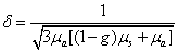
μα is the absorption coefficient of tissue,μs is the scatter coefficient, g is the average scatter cosine.
The endpoint was adjusted individually to the mild erythema developed by single pass with 10% overlap in each session. Negishi [33] Reported aggravation of post-treatment melasma like hyperpigmentation induced by high-fluence IPL irradiation. Actual melasma appeard where erythema lasting for several hours. Highfluence Q-switched 1064 nmNd:YAG laser treatment may activate immature melanocytes and destroy the basal membrane in melasma skin, which is the major risk factor for aggravation of melasma [12,25,34]. It is possible that dermal inflammation induced by a highfluence laser could be associated with the activation of MMPs, but further evidence is needed [35].
Most of the patients also observed the benefits of improvement in skin texture and lightened skin color on the laser-treated side. Textural changes such as this are thought to be the result of collagen remodeling and are the basis of improvement seen with treatment of photorejuvenation effect using nonablative lasers [36]. The nonablative, dermal remodeling effects of the 1,064-nm QS- Nd:YAG laser in the treatment of wrinkles and atrophic acne scars have been clinically and histologically confirmed [37,38].
Interestingly, there was not obvious improvement till the 6th session during the treatment, even slightly aggravation in some patients. Longo C found the presence of dendritic-shaped cells that maybe represent reasonably activated melanocytes upon confocal microscopy in four patients presenting with an early relapse of melasma (revealing the same pattern and severity of melasma seen at baseline) during laser sessions [12]. Naturally, it opens the question whether the laser treatment should be stopped or modulated at this time. Without cellular damage or cell death, the production of melanin could be modulated by melanocytes. An inspired news was clinical improvement was observed in all cases at the 12th session time point. This clinical outcome further supported the hypothesis that this gentle laser therapy began to suppress melanogenisis after an early remodeling phenomenon of melanin related proteins, tyrosine and so on without cellular destruction [13,39,40]. However, further studies on large basis are needed to fully elucidate the dynamic mechanism of melasma.
The study indicated that 1064 nm Q-switched Nd: YAG laser (4.5 J/cm², 4.0 mm, one pass) is duplicable effective and safe. Compared to other laser therapy, a long therapy course, high cost and prolonged effectiveness are its main drawback. What is more, Data of long-term effects are still lacking. Further well designed randomized controlled studies are required to address these issues. Large case number group data and more pathological research study should be carried out to assess the further value of this regimen. It is still a long way to go with optimization of treatment regimes. Our exploration can be a good alternative option in the treatment of melasma in Chinese.