Enzyme Engineering
Open Access
ISSN: 2329-6674
ISSN: 2329-6674
Research Article - (2016) Volume 5, Issue 1
Keywords: Curcumin; Iron(III); Photocytotoxicity; Photodynamic Therapy; Apoptosis
DCFDA: 2',7'- Dichlorofluoresceindiacetate; DMEM: Dulbecco’s Modified Eagle’s Medium; DMF: Dimethylformamide; FACS: Fluorescence Assisted Cell Sorting; HeLa: Human Cervical Carcinoma; MCF-7, Human Breast Adenocarcinoma; MTT: 3-(4,5- Dimethylthiazol-2-yl)-2,5-Diphenyltetrazolium Bromide; PDT: Photodynamic Therapy; PI: Propidium Iodide; ROS: Reactive Oxygen Species; TCM: Traditional Chinese Medicine
Curcumin (abbreviated Hcurc) is a polyphenolic dye found in root turmeric and can be isolated from the rhizome of the herb Curcuma longa (Figure 1). It is used as dietary spice in India and in Traditional Chinese Medicine (TCM) to treat infections, bite, burns and skin diseases [1-3]. Curcumin (1,7-bis(4-hydroxy-3-methoxy phenyl)-1,6- heptadiene-3,5-dione) consists of two feruloyl chromophores connected by a methylene group. The two aryl rings possessing two ortho-methoxy and phenolic hydroxyl groups are conjugated through a β-diketone moiety. After deprotonation of the acidic hydroxyl group by base treatment, the β-diketone moiety with two chemically equivalent oxygen atoms can act as a strong bidentate ligand for chemically hard metal ions such as oxidovanadium(IV), iron(III), cobalt(III) and lanthanide(III). The β-diketone functionality is also quite reactive and responsible for the occurrence of the well known keto-enol tautomerism in curcumin [2]. Curcumin possesses a visible absorption band near 420 nm with a shoulder around 450 nm. This band is responsible for curcumin’s bright orange-yellow colour [2]. Curcumin emits green fluorescence when excited around 430 nm and the position of this emission band is dependent on the nature of the solvent used. For example, the emission maximum (λem) of curcumin is centred at 524 nm in acetonitrile and 549 nm in ethanol [2]. Curcumin has demonstrated negligible intrinsic toxicity and hence it is safe for oral administration to a limit of 10 grams a day [3]. Recent interests in curcumin is attributable to the fact that it shows a wide range of medicinal properties as anticancer, antioxidant, antiinflammatory, antimicrobial, anti-HIV and anti-diabetic agents [4-7]. The wide range of therapeutic activities of curcumin are attributed to the fact that curcumin can interact with a number of molecular targets in the body [3]. Curcumin shows potent anticancer activity against a variety of cancers and has entered into several clinical trials for the treatment of cancer and other diseases [8-11]. The anticancer activity of curcumin stems from its ability to induce apoptosis in cancer cells without being cytotoxic to normal cells [8-11]. Curcumin interferes with the activity of the transcription factor NF-κB (a protein complex that is involved in the transcription of DNA) which has been linked to a number of inflammatory diseases including cancer [11]. However, the clinical applications of curcumin have been severely impeded by its poor bioavailability and pharmacokinetic profiles due to its hydrolytic instability under physiological conditions [5,12]. The presence of the β- diketone moiety in its structure renders curcumin hydrolytically unstable [5]. To improve the aqueous stability and hence therapeutic efficacy of curcumin, several modifications of curcumin have been synthesized and tested. One very recent and successful strategy to address this problem takes advantage of curcumin’s metal binding property [13-28]. Binding of curcumin to a metal ion via its β-diketone moiety significantly reduces the tendency of curcumin to undergo hydrolysis in aqueous medium and hence could result in improved bioavailability and therapeutic efficacy.
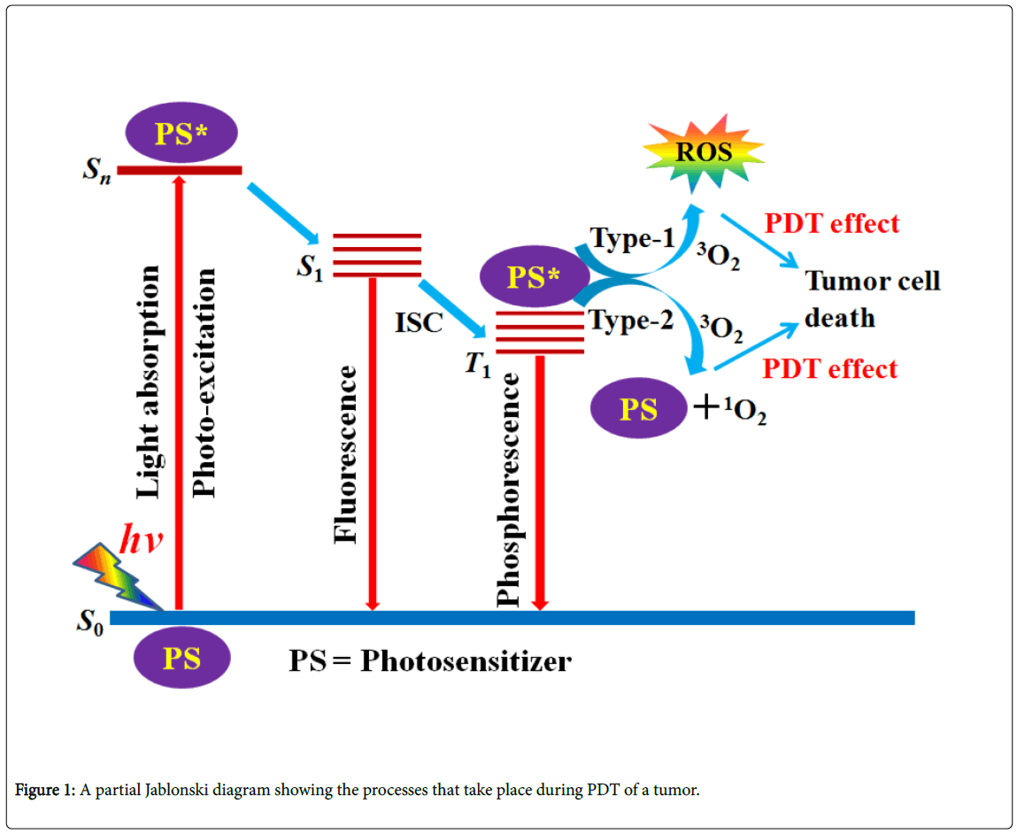
Figure 1: A partial Jablonski diagram showing the processes that take place during PDT of a tumor.
Tissue homogenates from archival breast cancer FFPE sections (6μm) were prepared according to the procedure as described in the QuantiGene Sample Processing Kit for FFPE Tissues (Affymetrix Inc., Santa Clara, CA). Excess paraffin from the slides were removed by soaking the slides in xylene for 5 minutes followed by soaking in 70% ethanol for 5 minutes. The slides were air dried and the tissue from the slides were removed using a clean razor blade and placed into 1.5 mL tube. 300 μL of homogenizing solution supplemented with 2 μL of proteinase K (50 μg/L) were added to the tubes. The tubes were vortexed for 30 seconds and incubated at 65°C for 4 hours. The tissue homogenate was separated from the debris by brief centrifugation, and then transferred to a new tube.
Gene expression analysis was performed using a QuantiGene 10 Plex assay (Affymetrix Inc, Santa Clara, CA). The 12 genes measured in this study included EN1, FOXC1, GABRP, CA12, AGR2, TTF3, GATA3 ESR1 SCNN1A, ERBB2 and the housekeeping genes PPIB and HPRT. Tissue homogenates were transferred to a 96 well hybridization plate containing QuantiGene Plex probe and Luminex beads sets. Each bead type was coated with a different single-strand DNAcapture probe (CP). Several other components of the QuantiGene Probe set are also comprised of single-strand DNA oligonucleotides including the capture extenders (CEs), label extenders (LEs), and blocking probes (BPs). Parts of CE oligonucleotides are complementary to the target mRNA (covering ~500 bases). The QuantiGene Plex Assay was performed according to the manufacturer’s manual with all of the reagents and consumables supplied by the manufacturer. (Quantigene Plex 2.0 Assay, Affymetrix Inc, Santa Clara, CA). For each well of the 96 well hybridization plate, 40 μL of tissue homogenate were transferred to the plate where each well already contained 60 μL of working bead mix (33.3 μL of lysis mixture, 18.5 μL of nuclease free water, 0.2 μL of proteinase K solution, 2 μL of blocking reagent, 5 μL of probe set, 1 μL of magnetic Luminex beads; customised QuantiGene Plex Set Panel). Hybridization was performed overnight for 18 hours at 54°C, with shaking at 600 rpm. The hybridization mixtures were then transferred to a 96-well flat bottom plate. An Affymetrix handheld magnetic bead washer (Affymetrix P/N QP0702) was used to wash the beads, thus removing all unbound materials. 100 μL of preamplifier reagent was added to each assay well. The magnetic separation plate was sealed with adhesive backed foil and incubated for 1 hour at 50°C and 600 rpm. The unbound preamplifier reagent was removed, and beads were washed three times with 100 ml of wash buffer using the handheld magnetic washer. This was followed by similar incubation and washing steps with 100 μL of Amplifier reagent, followed by 100 μL of Label Probe reagent, and finally followed by 100 μL SAPE working reagent. Signals from the beads were measured with a Luminex 200 (Luminex Corp., Austin, TX), after re-suspending the beads in 130 μL of SAPE wash buffer, using dd gate settings of 5,000– 25,000. Per target, 50–100 beads were measured in a sample volume of 100 μL.
Photodynamic Therapy (PDT)
Photodynamic therapy (PDT) is a newly recognized successful chemotherapeutic modality for the treatment of cancer. In PDT, a photoactivatable drug is administered to a cancer patient and the target tissue is exposed to specific light in the presence of molecular oxygen thereby resulting in the production of cytotoxic molecular species [29,30]. PDT has many advantages over conventional cancer therapies such as its low systemic toxicity, selective drug action within the photo-irradiated regions, very low level of invasiveness and ability to overcome drug resistance [29-35]. Light of wavelength in the range 620-850 nm, also known as the PDT window, is ideally required in PDT to activate the drug molecule [36]. Absorption of light by a photoactivatable drug, also called a photosensitizer (PS), causes an electronic transition promoting an electron from a singlet ground state to a singlet excited state. The singlet excited state is relatively shortlived (typically 10-12 to 10-6 s) and there exists several pathways for deactivation of this excited state. Initial excitation of the PS takes it to a higher vibrational level of the first electronic singlet excited state. The molecule then reaches the lowest vibrational level of the first electronic singlet excited state (called S1) by a process known as internal conversion (IC). At this stage, the PS may undergo further deactivation with the emission of a photon (fluorescence) and come back to the ground state. Alternatively, the excited singlet (S1) state may undergo radiationless decay to populate the lower energy triplet excited state (T1) by a process known as intersystem crossing (ISC). This triplet excited state is of importance due to its longer lifetime [36]. In type 1 reaction, the triplet PS can react directly with a substrate, such as the cell membrane or a molecule and in type 2 reaction, the triplet PS can transfer its energy directly to molecular oxygen to form excited state singlet oxygen. Both type 1 and type 2 reactions can result in further reactions resulting in the formation of ROS which are toxic to several vital cellular components and macromolecules such as DNA, lipids, polysaccharides and enzymes. Molecules and organelles that are proximally located within the area ROS production and hence the area of PS localization, are directly influenced by PDT [36]. The various processes that can undergo upon irradiation of a PS are described in a partial Jablonski diagram (Figure 2). Photofrin® is the currently FDA approved organic PDT drug [37]. Unfortunately, the limitations such as skin sensitivity and hepatotoxicity associated with Photofrin® have motivated chemists to search for its alternatives [36-38]. Thus, the photoactivatable metal complexes of non-porphyrinic ligands have emerged as potential candidates in the PDT of cancer [39-48]. Metal complexes possess varying coordination geometries, versatile spectral and redox properties and can induce cell death via photo-redox and/or type-I pathways besides generating cytotoxic singlet oxygen species in a type-II process [39–48].
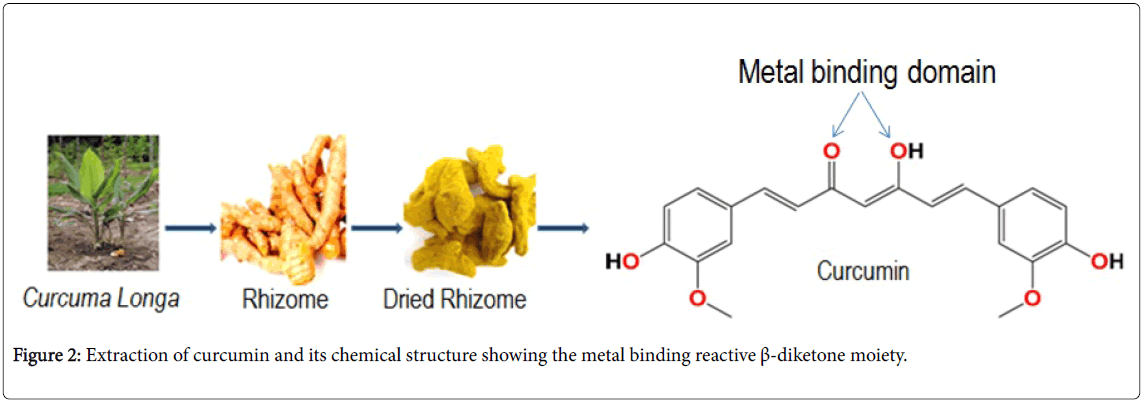
Figure 2: Extraction of curcumin and its chemical structure showing the metal binding reactive β-diketone moiety.
Photocytotoxicity of curcumin
The photophysical and photochemical properties of curcumin have recently been reviewed [2]. In India, turmeric is well known to protect skins from sunburns and used as remedy for skin related diseases such as leprosy and psoriasis [49]. Curcumin was shown to be genotoxic to bacterial systems following photo irradiation with visible light of 420 nm [50]. Dahl and co-workers found that curcumin was toxic to both gram-positive and gram negative bacteria as well as mammalian cells in the presence of light and oxygen [51]. Similarly, the photocytotoxicity of curcumin was observed in NPC/CNE2 cell lines when irradiated with visible light [52]. The mechanism of cell death was shown to be apoptotic thereby indication that curcumin could act as a photochemotherapeutic agent. The phototoxicity of curcumin has been attributed to the generation of singlet oxygen and ROS during photoexcitation [53,54]. Recently, the PDT effect induced by curcumin on cancer cells has also been investigated [55,56]. Very recently, the photocytotoxicity of curcumin has been investigated by Chakravarty and coworkers [25, 26]. Curcumin was found to be photocytotoxic to HeLa cells yielding an IC50 value of 8.2 μM when irradiated for 1 h with visible light (Luzchem photoreactor, 400-700 nm, 10 J cm-2). It was found to be significantly less toxic (IC50 = 85 μM) to the cell line in the absence of light (4 h incubation in the dark). The dark toxicity of Hcurc was reported to be 18 μM on 48 h incubation in the same cell line. The free curcumin, however, was found to undergo rapid hydrolytic degradation in culture media. The green emission of the curcumin ligand was used to study its cellular localization in HeLa cells. Curcumin was found to accumulate slightly within 2hr of incubation as evidenced from the intensity of the green fluorescence inside the cells. Upon increasing the incubation time to 4 h the fluorescence intensity decreased substantially and the cells were no longer seen, possibly due to the degradation of free curcumin molecule in culture media. When the curcumin ligand was allowed to bound to an oxidovanadium(IV) moiety, not only the photocytotoxicity but also the fluorescence intensity increased significantly as a result of increased hydrolytic stability of curcumin on complexation to the VO2+ moiety.
As discussed above, free curcumin rapidly decays hydrolytically to its degradation products in aqueous buffer and this degradation of curcumin can be cleverly arrested if the cheating β-diketone moiety is engaged in bonding to a metal centre such as iron(III). While numerous studies on the anticancer and other biological properties of curcumin and its metal complexes are reported, the photodynamic effect of metal complexes of curcumin in cancer cells remains unexplored despite curcumin’s rich photophysical and photochemical properties [2,13-28]. Earlier reports on cobalt (III), oxidovandium(IV), lanthanide(III), copper(II), zinc(II) and Ir(III) complexes have also shown that the hydrolysis of curcumin gets suppressed upon binding to a metal ion [13-28]. Using this strategy, we have recently reported an oxo-bridged iron(III) complex of curcumin that shows photocytotoxicity in HeLa and MCF-7 cancer cells in visible light (Luzchem photoreactor, 400-700 nm, 10 J cm-2) [57]. In the reported work, two complexes of formulation [{Fe(phdpa)(acac)}2(μ-O)](ClO4)2 (1) and [{Fe(phdpa)(curc)}2(μ-O)](ClO4)2 (2), where phdpa is bis-(2- pyridylmethyl)-benzylamine, acac is acetylacetonate and curc is the monoanion of curcumin (bis(4-hydroxy-3-methoxyphenyl)-1,6- diene-3,5-dione) were prepared in good overall yield and characterized by a number of physico-chemical techniques such as elemental analysis, IR, conductivity, electrochemical, mass spectral, UV-Visible, emission and magnetic measurements (Figure 3). The crystal structure of complex 1 was established by single crystal X-ray diffraction method. The complex was found to be a dinuclear species with a distorted octahedral FeN3O3 core formed by the tridentate N,N,Ndonor phdpa, bidentate O,O-donor acac and the bridging oxo ligands. Absorption spectral study on complex 2 in Dulbecco's Modified Eagle's medium (DMEM) showed that the complexes were stable even after 48 hours [57].
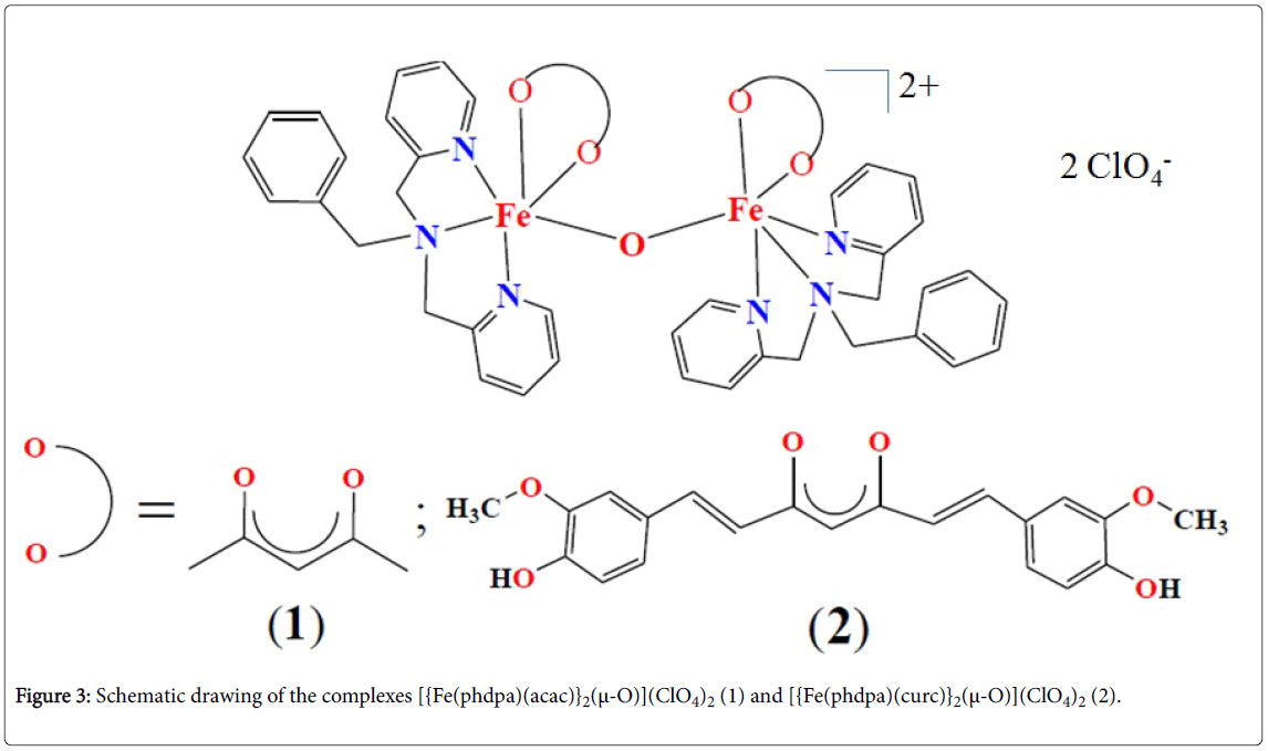
Figure 3: Schematic drawing of the complexes [{Fe(phdpa)(acac)}2(μ-O)](ClO4)2 (1) and [{Fe(phdpa)(curc)}2(μ-O)](ClO4)2 (2).
The photocytotoxicity and hence the anticancer activity of the complexes was studied by the well-known MTT assay in two human cancer cell lines, viz., HeLa and MCF-7 in the presence of visible light. The photoactive complex 2 gave an IC50 value of 3.1 μM when irradiated with visible light of 400–700 nm (10 J cm-2) in HeLa cells (Table 1). Under identical conditions, the same complex gave an IC50 value of 4.9 μM in MCF-7 cells. Interestingly, the complex was negligibly toxic to both the cell lines in the dark (IC50 > 50 μM). The control complex 1, lacking a curcumin unit, was neither photocytotoxic nor dark toxic in these cell lines under similar experimental conditions (Table 1). This result indicates that curcumin is responsible for the observed photocytotoxicity of complex 2. The free curcumin ligand is known to give IC50 values of 8.2 μM and 19.9 μM, respectively, in HeLa and MCF-7 cells in visible light [20,25]. Hence, an increase in activity of curcumin with respect to free curcumin was observed in both the cell lines on binding to iron(III). The photocytotoxicity but negligible dark toxicity displayed by complex 2 is interesting considering the chemistry of iron based PDT agents [57].
| Compound | HeLa | MCF-7 | ||
| Light / µM | Dark / µM | Light / µM | Dark / µM | |
| [{Fe(phdpa)(acac)}2(μ-O)](ClO4)2 (1) | 58.1 ± 1.1 | 79.4 ± 1.3 | 70.4 ± 1.7 | >100 |
| [{Fe(phdpa)(curc)}2(μ-O)](ClO4)2 (2) | 3.1 ± 0.4 | >50 | 4.9 ± 0.5 | >50 |
| Curcumin | 8.2 ± 0.2 | 85.4 ± 0.6 | 19.9 ± 1.4 | 90.3 ± 4.9 |
a References 25 and 57.
Table 1: Cytotoxicity (IC50 values) of the complexes and curcumin in HeLa and MCF-7 cells in the presence of visible light (400-700 nm, 10 J cm-2) and in darka.
To know about the mechanism of cell death, the authors carried out Annexin V-FITC/PI assay using HeLa cells treated with complex 2 under illuminated conditions. The results showed induction of apoptosis in ~52% of the cell populations upon irradiation with a slight occurrence of necrosis (~4% of the cells). No effect of the complex on the cells was seen in the absence of light. To further confirm the above result, the authors carried out propidium iodide (PI) staining experiment with complex 2. Propidium iodide (PI) stains only the cell’s nucleus. HeLa cells treated with complex 2 and incubated 4 hours followed by photo-exposure to visible light of 400-700 nm for 1 h exhibited irregular nuclear morphology on staining with propidium iodide as revealed from the fluorescence microscopy images. Chromatin condensation of nuclei, which is a characteristic feature of cells undergoing apoptosis, was observed. No such changes were seen in the complex untreated and photo unexposed cells suggesting that the complex remained harmless inside the cells in the absence of light. Intracellular ROS are the key players in inducing apoptosis by redox active metal complexes [58, 59]. To detect ROS, 2',7'- dichlorofluoresceindiacetate (DCFDA) assay was performed by FACS analysis. The results showed generation of cytotoxic apoptosis inducing ROS only on exposure to visible light by complex 2 and not in the dark [57].
The green fluorescence of the free curcumin ligand retained upon binding to Fe(III) and this property was exploited by the authors to study the cellular uptake and localization of the complex in the HeLa and MCF-7 cells using fluorescence microscopy. Uptake of complex 2 could be seen in both the cell lines after 2 h of incubation which increased substantially after 4 hr of incubation. Dual staining experiment with Hoechst 33342, which is known to stain only cell’s nucleus with its characteristic blue emission, showed primarily cytosolic localization of complex 2 as evidenced from the merged fluorescence images [57].
There is currently a surge of research interest in curcumin and its metal complexes as an anticancer agent. Recent work has shown that the main drawback of curcumin, which is its poor bio-availability due to its hydrolytic instability, can be successfully overcome by taking advantage of curcumin’s metal binding property. In most cases an increase in anticancer activity of curcumin has been observed on binding to a metal. Our recent work on iron (III) complex of curcumin has shown that curcumin, when bound to iron(III), is a potential photochemotherapeutic agent. There is lot of future scope to design and develop metal complexes of curcumin with enhanced efficacy and selectivity towards cancer cells.
AH thanks the Department of Biotechnology (DBT), Government of India for the U-Excel grant (BT/401/NE/U-Excel/2013). T.S. thanks the DBT for research fellowship.