Journal of Pharmaceutical Care & Health Systems
Open Access
ISSN: 2376-0419
ISSN: 2376-0419
Research Article - (2023)Volume 10, Issue 6
This study aimed to analyze the pharmacokinetic parameters of 1,3-beta-glucans from Maitake Pro4X in BALBc mice through oral and Intra Venous (IV) administration. The objectives were to determine the following parameters: Tmax, Cmax, t1/2, ta1/2, Ke, Ka, Clearance, Vd, Cp0, and AUC. Additionally, the tissue distribution of 1,3-beta-glucans after oral and IV administration of Maitake Pro4X was examined to identify the organs involved in absorption, elimination, and distribution. The results comparison indicated that certain elimination constants were similar in both administration routes (ke3 IV and ke2 oral). Both routes showed a Tmax of 10 hours. The elimination t1/2 was comparable for both routes (12.93 hours for oral and 12.81 hours for IV). Total systemic clearance values were also similar. However, the IV route exhibited a higher volume of distribution but lower AUC Cp vs. time. The findings suggest that the gastrointestinal organs (stomach, duodenum, and colon) exhibited the highest levels of uptake for both administration routes. Conversely, the liver and kidney showed the lowest uptake for both routes. Comparatively, oral administration resulted in greater gastrointestinal accumulation, while cerebral, pulmonary, renal, and hepatic uptakes were higher after intravenous administration. The in vivo pharmacokinetic studies in murine models led to the conclusion that oral and intravenous administration had similar values for elimination rate, Maximum plasma concentration Time (Tmax), Half-Life (T1/2), total systemic clearance, and bioavailability. Both routes exhibited a Cmax peak and a high volume of distribution, indicating low binding to plasma proteins. Biodistribution studies in murine models revealed greater uptake of the compound in the gastrointestinal tract after both oral and IV administration, with gastric uptake being predominant, along with significant uptake in the duodenum and colon (in the order of millions of pg.h/ml). The presence of β-glucans in the brain suggests the ability of Maitake to cross the blood-brain barrier. The lower relative uptake observed in the hepatorenal region indicates a slower rate of inactivation and excretion of the compound, as evidenced by an extended circulation time in the body after a single administration.
D-fraction pro4X; Pharmacokinetic parameters; Tissular biodistribution; 1,3-β-glucans
Tmax: Maximum time; t1/2: Half-life time; Ke: Elimination constant; IV: Intravenous; Cp0: Initial Plasma Concentration; Cmax: Maximum Concentration; ta1/2: Half Time Absorption; Ka: Absorption Constant; Vd: Distribution Volume; AUC: Area Under The Curve; pg.h/ml: Picograms times hour per milliliter; μl: Microliters; LAL: Limulus Amebocyte Lysate; pNA: p-Nitroaniline; nm: nanometers; h: hour; mg: milligrams; Kg: Kilograms; ml: milliliter; NEDA: N-(1-Napthyl) Ethylenediamine Dihydrochloride; CO2: Carbon Dioxide; °C: Centigrade Degrees; pg/ml: Picograms per milliliters; Clt: Total systemic clearance; mL/g; milliliter per gram; pH: -[Log concentration H+]; NO: Nitric Oxide; TNF-α: Tumor Necrosis Factor α; BBB: Blood-Brain Barrier; TLR2: T-Lymphocyte Receptor 2; CNS: Central Nervous System; AMPA: N-methyl-d-aspartate (NMDA) and α-amino-3-hydroxy-5-methyl-4- isoxazolepropionic acid.
In recent years, several fungi have been found to possess Immunomodulatory and antitumor properties [1,2]. These fungi have been consumed for centuries in Asian countries due to their health-promoting and longevity benefits [3]. Among these fungi, Grifola frondosa has been highly regarded as both an edible and medicinal mushroom in Japan for thousands of years [4]. It is known to exert immunomodulatory effects [5]. The soluble β-glucans found in Fraction D of G. frondosa stimulate innate and adaptive immune responses, which contribute to its antitumor activity [6]. Our research group has reported therapeutic benefits in the treatment of mammary carcinogenesis using β -glucans from Maitake Fraction D [7]. However, to ensure the safety and efficacy of administering these molecules with biological activity to patients, further investigation into the pharmacological and toxicological aspects of the extract is necessary. By delving deeper into these aspects, we can provide a comprehensive understanding of the molecules' properties and their effects, thus ensuring their appropriate and effective use in a clinical setting.
Understanding the pharmacokinetic, pharmacodynamic, and toxicological profile of Maitake Fraction D β-glucans is crucial to support their use as a therapeutic agent and evaluate potential interactions with other compounds. The primary objective of the in vivo tests proposed in this study is to elucidate the pharmacodynamic and pharmacokinetic aspects of the β-glucans found in Maitake Fraction D. By conducting these tests, we aim to gain insights into how the β-glucans interact with the body, their effects on biological processes, and any potential risks or interactions that need to be considered for safe and effective therapeutic use.
1,3 Beta-glucans demonstrate a unique mechanism of absorption in the intestine, specifically in the Peyer's patches, which are lymphatic tissues. Within these tissues, key immune cells possess pattern recognition receptors such as dectin-1, which bind to beta-glucans. Upon recognition, macrophages or dendritic cells phagocytose the beta-glucans, resulting in the release of cytokines and chemokines that facilitate communication and initiate complex immune responses.
The size of beta-glucan molecules, ranging from 2 to 4 microns, provides an additional advantage for their action. Importantly, the (1,3) β-glucans found in Maitake Pro4X remain intact and are not degraded in the stomach or duodenum, as observed with beta-glucans derived from commercial yeasts. This ensures that the full structure and functionality of the beta-glucans are preserved, allowing for their optimal Immunomodulatory effects.
Quantification of 1,3-β-D-glucans in blood plasma and tissues of mice treated with maitake pro4X:
It was determined after oral administration or intravenous administration of a single therapeutic dose of the glycoprotein extract (4 μl of product). After the sacrifice of the animals at pre-established times, they were collected blood and histological samples to determine concentration of 1,3-β-D-glucans by the colorimetric method of the Glucatell kit. This test was carried out in Cape Cod Laboratory Inc, Maryland, USA.
Glucatell® Kit (1→3)-ß-D-glucan detection kit:
The Glucatell kit is specific for detection of (1→3)-ß-D-glucan. (Catalog Number GT003-Associates of Cape Cop Laboratories, Maryland, USA). The assay is based upon a modification of the Limulus Amebocyte Lysate (LAL) pathway, is formulated as a chromogenic lysate by adding Boc-Leu-Gly-Arg-pNA. (1,3)-β-Dglucan in the test sample (or standard) activates Factor G, which then activates the proclotting enzyme. The clotting enzyme cleaves p-Nitroaniline (pNA) from the chromogenic peptide substrate. The free pNA is measured at 405 nm (kinetic assay) or alternatively, the pNA is diazotized to form a compound that absorbs at 540-550 nm (endpoint assay).
Assay endpoint procedure:
Briefly, add 50 μL of sample to the unknown wells, add 50 μL of Glucatell reagent to each well, cover the plate with its lid and shake. Incubate the plate to 37°C ± 1°C for the recommended incubation time. Stop the reaction by adding 50 μL of sodium nitrite, then add in sequence 50 μL of ammonium sulfamate, and then 50 μL of N-(1-Napthyl) Ethylene Diamine dihydrochloride (NEDA) (vial 3). Read the color development in microplate reader at 540 nm- 550 nm. Use the plate reader software to plot a linear standard curve and to calculate the concentration of (1,3)-β-D glucan in the unknowns.
Study in oral administration:
Pharmacokinetic parameters were determined by orally, using two animals of both sexes for each time (n=18). If they administered 4 μl of a single therapeutic dose of Maitake Pro4X/mouse, corresponding to about 5 mg of β-glucans/kg. Subsequently proceeded to sacrifice at 30 min, 1 h, 2 h, 4 h, 7 h, 10 h, 16 h, 24 h and 34 h after oral intake, by CO2 asphyxiation in a euthanasia chamber and subsequent dislocation cervical to ensure death. Peripheral blood was collected from the cavities with a micropipette to which the tip facilitates aspiration, transferring to a sterile tube (Vacutainer 3.5 ml). Plasma was obtained after centrifugation of the vacutainer tubes and collected in a clean Eppendorf tube, labeled, and kept at -20°C until was analyzed.
Equations:
Pharmacokinetics in oral administration
Plasma quantification of 1,3-β-glucans was performed after oral administration of Maitake Pro4X in BALB/c mice. For this, two animals were used for each time (condition), sacrificing them at 0.5 h, 1 h, 2 h, 4 h, 7 h, 10 h, 16 h, 24 h and 34 h. The determination of 1,3-β-glucans was carried out by the colorimetric assay of the Glucatell kit, carried out at the Cape Cod Inc laboratory in Maryland, USA, which allowed obtaining curves of Plasmatic Concentration (pg/ml) vs. time post administration (hours) (Figure 1). The pharmacokinetic parameters that were determined were AUC (Area Under the Curve Cp vs. time), T½ (half-life time), Cpmax (Maximum Plasma Concentration), ke (Elimination Constant), ka (Absorption Constant), Ta½ (Absorption Half-Life), Vd (apparent Volume of Distribution) and Clt (Total Systemic Clearance).

Figure 1: Pharmacokinetic Curve of 5 mg/Kg times-glucans from Maitake Pro4X in BALBc mice after oral or intravenous administration. Note: ( )Oral Treatment; (
 ) IV Treatment.
Maximum plasma concentration (Cpmax) and maximum time (Tmax): The results reported in Figure 1 and Table 1, suggest that the plasma levels of 1,3-β-glucans increased rapidly after oral administration, presenting 3 peaks of maximum plasma concentration.
• 1st peak Cpmax>5000 pg/ml half an hour after oral administration of Maitake.
• 2nd peak Cpmax>5000 pg/ml between 2 h and 10 h after oral administration of Maitake.
• 3rd peak Cpmax>5000 pg/ml 34 hours after oral administration of Maitake.
After the first Cpmax peak, plasma levels of 1,3-β-glucans fell abruptly, producing a new Cpmax peak between 2 h and 10 h. Starting at 10 h, a gradual decrease in the concentration of 1,3-β-glucans took place. But, starting at 16 h, a progressive increase in the concentration of 1,3-β-glucans began again, reaching a third Cpmax peak at 34 h after oral administration.
Elimination rate constant (Ke): The results reported in Figure 1 suggest that the elimination of 1,3-β-glucans corresponds to a first order (monocompartmental) kinetic model. Three elimination constants (slopes) called Ke1 (range 0.5 h to 1 h) and Ke2 (range 10 h to 16 h) could be determined. The first constant Ke1 has an elimination rate of 4.489/h, and Ke2 a rate of 0.236/h. This indicates a high elimination rate for the interval 0.5 h to 1 h, and a more gradual elimination between 10 h and 16 h. The first elimination (Ke1) is 19 times faster than the second one (Ke1≈19 Ke2).
Absorption rate constant (Ka): The absorption constant ka was determined by the residual’s method, assuming a 1st order kinetic model with Ka1>Ke1. First, to calculate ka1, the elimination line Log Cp vs. time in the range 0.86 h to 0.94 h. The extrapolation to the ordinate axis was performed, resulting in a line y=- 2.2011x+4.8472 (Table 1).
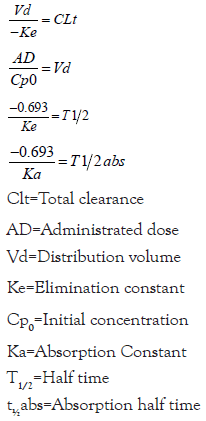
Absorption half-life time(Ta1/2): From both ka, the Absorption Half-Life (ta1/2). Using the expression t1/2=-0.693/ka it is obtained for ka1 ta11/2=0.1 h, while for ka2, ta21/2=1.9 h. These results suggest that the plasma concentration of 1,3-β-glucans remaining to be absorbed, it was halved at 0.1 h and 1.9 h after oral administration.
Elimination half-life time(T1/2): From both ke, the elimination Half-Life Time (t1/2), using the expression t1/2=-0.693/Ke. For the interval 0.5 to 1 h, t1/2=0.15 h, while, between 10 h to 16 h, t1/2=2.93 h. The Plasma concentration of 1,3-β-glucans was halved at 0.65 h and at 12.93 h of oral administration.
Volume of distribution (Vd): The apparent volume of distribution was calculated by the expression Vd=dose/Cp0. The dose administered to the animals was 120 mg of β-glucans (4 ml Maitake Pro4X). Cp0 was determined by extrapolation with the axis of the ordinates (ln Cp) in the interval 0.5 h to 1 h (Figure 1), resulting in ln Cp0=10.76, and Cp0=47098.67 pg/ml. As Vd=dose/ Cp0=120000000 pg/47098.67 pg.ml-1, resulting in Vd=2547.84 ml (2.55 liters). So, for a 20-g mouse, the Vd=2.55 l/0.02 kg, i.e. Vd=127 ml/g (127 l/kg), this is a large value of Vd.
Total clearance (Clt): Total Systemic Clearance (Clt) was determined by the expression Clt=Vd (-Ke). For Ke1, Clt=2.55 l x 4.489 h-1=11.45 l/h (190.78 ml/min), while for Ke2, its Ct=10.03 ml/min.
Area Under the Curve (AUC): The Area Under the Curve (AUC) Cp vs. time, which reflects the amount of bioavailable compound that reaches the systemic circulation and is capable of produce an effect, was determined by the trapezoidal method (Figure 1), resulting at 0.118 mg.h/ml for the time interval 0 h to 34 h.
Pharmacokinetics in intravenous administration
Plasma quantification of 1,3-β-glucans was performed after administration intravenous injection of Maitake Pro4X into the marginal tail vein of BALB/c mice. For this, two animals were used for each time (condition), performing the sacrifice of the same at 0.5 h, 1 h, 2 h, 4 h, 7 h, 10 h, 16 h, 24 h and 34 h. the determination of 1,3-β-glucans was performed by the colorimetric assay of the Glucatell kit, carried out in the Cape Cod Inc laboratory in Maryland, USA, which allowed obtaining curves of Plasma concentration (pg/ml) vs. post administration time (hours) (Figure 1). The Pharmacokinetic parameters that were determined were AUC (Area Under the Curve Cp, vs. time), T ½ (Elimination Half- Life), Cpmax (Maximum Plasma Concentration), ke (Elimination Constant), ka (Absorption Constant), Ta½ (Absorption Half-Life), Vd (apparent Volume of Distribution) and Clt (Total Systemic Clearance).
Maximum concentration (Cpmax) and Maximum time (Tmax): The results reported in Figure 1 and Table 1 suggest that plasma levels of 1,3-β-glucans showed an abrupt decrease after intravenous administration, followed by a rapid increase in concentration towards a first peak Cpmax>5000 pg/ml at the 2 hours of administration. This Cpmax gradually decreased between 2 h and 4 h, but again made a second Cpmax peak at 10 h. Subsequently, there was a third gradual decline in plasma levels of 1,3-β-glucans, which tended to remain constant for the remainder of the trial.
Elimination rate constant (Ke): Elimination of intravenously administered 1,3-β-glucans followed 1st order kinetics (monocompartmental). The semilogarithmic plot of Cp vs. Time resulted in a straight line that allowed the calculation of three elimination kinetics: Ke1 (40.4% of 1,3-β-glucans/h) for the interval 0.5 h to 1 h, Ke2 (17.6% 1,3-β-glucans/h) for the interval. 2 h to 4 h, and Ke3 (24.7% 1,3-β-glucans/h) for the interval 10 h to 16 h (Figure 1). The results obtained suggest that Ke1>Ke3>Ke2, which indicates a higher rate of elimination in the interval 0.5 h to 1 h (40.4% of 1,3-β-glucans/h), and a more gradual elimination between 2 h and 4 h (17.6% 1,3-β-glucans/h).
Absorption rate (ka): The Absorption Rate Constants (ka) were determined by the method of residuals. For this, the elimination line was drawn Log Cp vs. time was plotted over the time interval 0.68 h to 0.80 h, with its corresponding extrapolation to the ordinate axis, resulting in y=-0.2823x+3.6784, which allowed to determine ka1 (Table 1).
| Pharmacological parameters | Ways of administration | |
| Oral | Intravenous | |
| CpMax piks | 3 | 2 |
| Tmax (h) | 0.5, 10.0 and 34.0 | 2.0 and 10.0 |
| Ka (h−1) | 6.75 and 0.36 | 7.45, 7.46 and 0.60 |
| Ke (h−1) | 4.489 and 0.236 | 0.404, 0.176 and 0.247 |
| T ½ (h) | 0.65 and 12.93 | 12.81 |
| Ta ½ (h) | 0.1 and 1.9 | 0.09 and 1.20 |
| Cp0 (pg/mL) | 47098.67 | 4230.18 |
| Vd (L) | 2.65 | 28.37 |
| Cl systemic total (ml/min) | 190.78 and 10.03 | 191, 83.22 and 116.79 |
| AUC (pgx h/mL) | 118000 | 80000 |
Table 1: Comparative pharmacokinetic parameters after oral and IV administration of 5 mg/Kg β-glucans from Maitake Pro4X.
Absorption half-life time (ta1/2): From the ka values, the Absorption Half-Life (ta1/2) or time it takes to reduce by half the plasma concentration of 1,3-β-glucans remaining to be absorbed. By means of the expression t1/2=-0.693/ka, resulting in ta11/2=0.09 h for both ka1 and ka2, and 1.2 h for ka3 (Table 1).
Elimination half-life time (t1/2): From Ke, it was possible to establish the elimination half-life time (t1/2), using the expression t1/2=-0.693/Ke. I know obtained that for ke1 the t1/2=1.7 h, for ke2 it was 3.9 h, and 2.81 h for ke3, so that the plasma concentration of 1,3-β-glucans was halved at 1.7 h, at 5.9 h and at 12.81 h. It was observed that, during the first and second elimination of the compound, plasma levels of 1,3-β-glucans increased prior to reaching the half lifetime (Table 1).
Volume of distribution (Vd): The apparent volume of distribution was calculated by the expression Vd=dose/Cp0. The intravenous dose administered to the animals was 120 mg of β-glucans (4 ml of Maitake Pro4X), while the Concentration Plasma at time 0(Cp0) was determined by extrapolation with the ordinate axis (ln Cp) in the interval 0.5 h to 1 h, resulting in Ln Cp0=8.35, so that Cp0=4230.18 pg/ml. Therefore, the Vd=1.2,108 pg/4230.18 pg.ml-1=28367.59 ml (28.37 l). So, for a 20 g mouse the Vd=28.37 l/0.02 kg=1418.5 l/kg (1418.5 ml/g) (Table 1).
Total clearance (Clt): The Total Systemic Clearance (Clt) was determined through the expression Clt=Vd (-Ke). For Ke1, the Clt=28.37 l x 0.404 h-1=11.46 l/h (191 ml/min), for Ke2, the Clt=83.22 ml/min, while for Ke3, the Clt=116.79 ml/min (Table 1).
Area Under the Curve (AUC): The Area Under the Curve (AUC) of Cp vs. time in IV administration was determined by the trapezoidal method, giving 0.08 mg.h/ml in the time interval 0 h to 34 h (Table 1).
Comparison of oral vs. intravenous pharmacokinetics
Parameters Pharmacokinetics of both pathways are reported in Figure 1 and Table1. The comparison of the results obtained suggests that some elimination constants were similar in both routes (ke3IV and ke2oral). On the other hand, both routes coincided in a Tmax=10 h. The elimination t1/2 was similar in both routes (12.93 h in oral and 12.81 h in IV). I found similarity in total systemic clearance values. However, the IV route presented a higher volume of distribution, but lower AUC Cp vs. time.
Tissular Biodistribution in oral administration
Tissue quantification of 1,3-β-glucans was performed in different murine organs (liver, kidney, duodenum, colon, lung, and brain) following oral administration of Maitake Pro4X. Two BALB/c mice of both sexes were used each time. (condition), performing the sacrifice of these and removal of the organs at 0.5 h, 1 h, 2 h, 4 h, 7 h, 10 h, 16 h, 24 h and 30 h. β-Glucan determinations were performed by the colorimetric assay of the Glucatell kit, carried out in the Cape Cod Inc laboratory of Maryland, USA, allowing the obtaining of tissue uptake curves of 1,3-β-glucans vs. time post oral administration.
The determinations were made with the colorimetric method of the Glucatell kit. The Biodistribution in the gastrointestinal organs (stomach, duodenum, colon) was of the order of millions of pg.h/ ml, while in the others it was thousands of pg.h/ml. The results shown in Figure 2 regarding the uptake tissue of β-glucans after oral administration suggest that the largest concentrations occurred in the stomach (5.52 x 10⁷ pg.h/ml), duodenum (3.66 x 10⁷ pg.h/ml) and colon (3.44 x 10⁷ pg.h/ml). In addition, we have recorded a significant level of uptake in the brain (AUC of 3.77 x 10⁴ pg.h/ml) and lung, and to a lesser extent in liver (1.89 x 10⁴ pg.h/ml) and kidney (2.44 x 10⁴ pg.h/ml). The area under the curve of Tissue biodistribution of 1,3-β-glucans for the different organs is indicated in Figure 2.

Figure 2: Tissue biodistribution curves of β-glucans by IV administration. They correspond to the area low 1,3-β- glucan uptake curve from different murine organs after administration Maitake Pro4X IV. Mean values + 2 standard deviations of A: Gastric, colonic, and duodenal uptake in millions of pg/ml For B: Cerebral, pulmonary uptake, hepatic and renal in thousands of pg/ml. Significant differences were observed (*p<0.05; **p<0.01) between tissue uptakes and their times. Note: ( ) Liver; (
 ) Duodenum;(
 ) Colon; (
 ) Stomach; (
 ) Lung;(
 ) Brain; (
 ) Kidneys; (
 ) Liver; (
 ) Lung; (
 ) Brain;(
 ) Kidney.
The results reported for area under the tissue uptake curve of β-glucans, suggest that the greatest hepatic uptake (Figure 2) occurs at 7 h (*p<0.05 vs. 2 h), and the lowest at 30 h (with **p<0.01 vs. 2 h). On the other hand, the hepatic uptake at 2 h is significantly higher than that at the same time in brain (with *p<0.05). Likewise, brain uptake at 30 h is significantly lower than that which occurred at the same time in the lung (with **p<0.01).
Tissular biodistribution in intravenous administration
Tissue quantification of 1,3-β-glucans was performed in different murine organs (liver, kidney, duodenum, colon, lung and brain) after administration IV Maitake Pro4X through the marginal tail vein. were used two BALB/c mice of both sexes for each time (condition), performing at sacrifice of the same and removal of the organs at 0.5 h, 1 h, 2 h, 4 h, 7 h, 10 h, 4 p.m., 12 p.m. and 30 p.m. β-Glucan determinations were performed by the assay colorimetry of the Glucatell kit, carried out at the Cape Cod Inc laboratory in Maryland, USA, allowing the obtaining of tissue uptake curves of 1,3-β-glucans vs. time post intravenous administration. The results reported in area under the curve of tissue uptake of β-glucans after intravenous administration, suggest that the greater uptake took place at the gastrointestinal level, as in oral administration, being again the stomach, the organ with the highest accumulation (4 x 10⁷ pg.h/ml), followed by colon (1.24 x 10⁷ pg.h/ml) and duodenum (5.91 x 10⁶ pg.h/ml).
The results reported in the area under the uptake curve gastrointestinal β-glucans, suggest that the greatest duodenal uptake occurs at 4:00 p.m. (*p<0.05 vs. 7:00 a.m.). While gastric uptake at 16 h and 30 h was significantly higher than colon at the same time (*p<0.05 and **p<0.01 respectively). On the other hand, gastric uptake at 30 h was significantly greater than the duodenal at the same time (*p<0.05) (Figure 3).

Figure 3: Tissue biodistribution curves of β-glucans by oral administration. They correspond to the area low 1,3-β- glucan uptake curve from different murine organs after administration Maitake oral PRO4X. Mean values + 2 standard deviations are indicated for, A: Uptake gastric, colonic and duodenal in millions of pg/ml for B: Cerebral, pulmonary, hepatic and renal in thousands of pg/ml. Significant differences (*p<0.05; **p<0.01) were observed between the tissue uptakes and their times. Note: ( ) Liver; ( ) Duodenum;( ) Colon; ( ) Stomach; ( ) Lung;( ) Brain; ( ) Kidneys; ( ) Liver; ( ) Lung; ( ) Brain;( ) Kidney.
There was also an important level of cerebral uptake (1.5 x 10⁵ pg.h/ml) and pulmonary (1.45 x 10⁵ pg.h/ml). To a lesser extent, biodistribution was recorded in the liver (6.48 x 10⁴ pg.h/ ml) and kidney (3.29 x 10⁴ pg.h/ml). The area under the tissue biodistribution curve of β-glucans is indicated in Figure 3.
Tissue biodistribution of β-glucans in oral vs. intravenous administration
The area under the curve (pg.h/ml) of tissue biodistribution of β-glucans by both routes of administration is summarized in Figure 4. The results obtained suggest that the highest levels of uptake occurred in the gastrointestinal (stomach, duodenum, and colon) for both routes of administration. While the lowest uptake was recorded in the liver and kidney for both pathways.

Figure 4: Tissue biodistribution of 1,3-β-glucans. (ABC of the different organs is compared murine in oral (blue columns) and intravenous (red columns) administration. A: The Biodistribution in the gastrointestinal
organs (stomach, duodenum, colon) was of the order of the millions of pg.h/ml in both routes A; B) while the 1,3 β-glucan Biodistribution in brain, lungs, kidneys, and liver were in thousands of pg.h/ml. Note: ( ) Oral; (
 ) IV.
) IV.
Comparatively, gastrointestinal accumulation was greater after administration oral (Figure 4). However, cerebral, pulmonary, renal and hepatic uptake was higher after intravenous administration.
In our studies of pharmacokinetic parameters following oral administration, we observed three peaks of plasma concentration for 1,3-β-glucans from Maitake Pro4X. The first peak was recorded half an hour after administration, followed by a rapid decrease.
The second peak occurred between 2 and 10 hours later, and the last peak was observed at 34 hours’ post-oral administration of 5 mg/kg of β-glucans. Rice PJ et al., have also reported plasmatic peaks of water-soluble glucans between 0.5 and 12 hours after oral administration, with the presence of significant biological effects persisting in serum up to 24 hours after a single oral dose [8]. Our results indicate that the plasma clearance of 1,3-β-glucans follows a first-order kinetic model, with a rapid initial clearance half an hour after oral administration, followed by a gradual elimination at 10 hours. Hong F etal., have reported that orally administered β-1,3-glucans are transported by macrophages to the spleen, lymph nodes, and bone marrow, suggesting a potential plasma reduction of the compound [9].
Our observations indicate greater absorption than elimination of the compound, with a high Volume of distribution (Vd) indicating extensive tissue distribution and likely accumulation in tissues, along with poor binding to plasma proteins [10]. Therefore, it is probable that 1,3-β-glucans circulate mostly in their "free" form, enabling diffusion into extravascular compartments, interaction with their receptors, and initiation of biological responses. Miura et al., have suggested that binding of β-glucans to serum or plasma proteins may result in their inactivation and a reduction in biological activities [11].
Regarding our pharmacokinetic studies of intravenous administration, we observed two peaks in plasma concentration of 1,3-β-glucans. The first peak occurred two hours after administration, followed by a gradual decrease, and the second peak was observed at 10 hours. The clearance or plasmatic disappearance of 1,3-β-glucans following intravenous administration also followed a first-order kinetic model, with a rapid initial elimination half an hour after administration, followed by a more gradual elimination at 10 hours. The volume of distribution obtained was very high (1418.5 mL/g), higher than that observed with oral administration, indicating extensive tissue distribution, likely tissue accumulation, and poor plasma protein binding [10]. These findings suggest that intravenously administered 1,3-β-glucans circulate mostly in their "free" form, similar to oral administration, allowing for diffusion into extravascular compartments, receptor interactions, and biological responses. Our results indicate that the intravenous route has a smaller area under the Cp vs. t curve compared to the oral route, but a higher Volume of distribution (Vd). This suggests that binding to plasma proteins may be lower after intravenous administration, resulting in a higher proportion of free compound and an increased distribution volume. However, both routes of administration exhibited similar values for elimination rate, time to maximum plasma concentration (Tmax), half-life time (T1/2), and total systemic clearance.
In terms of biodistribution studies, we observed greater uptake (absorption) of the compound at the gastrointestinal level, with a predominant uptake in the stomach and significant uptake in the duodenum and colon (in the order of millions of picograms/h/ ml). These findings suggest that the increased biodistribution in the stomach for both routes of administration may be attributed to the acidic nature of the Maitake glycoprotein extract. It is likely that the acidic pH of the stomach facilitates a higher percentage of nonionized 1,3-β-glucan molecules, which are more easily absorbed and enter the circulation. Vetvicka V et al., reported greater detection of β-glucans in the stomach and duodenum within 5 minutes of administration, which decreased significantly within the first 30 minutes [12].
It has been reported by Tomita M. et al. that the intestinal transport of 1,3-β-glucans involves a specialized transporter in both absorption and secretion directions, with nonlinear absorption associated with multiple transport mechanisms. Furthermore, Rice R.J et al., reported that glucans are internalized by intestinal epithelial cells and Gut-Associated Lymphoid Tissue (GALT) cells following oral administration [13]. Vetvicka V et al., observed that most orally administered β-glucans enter the proximal small intestine and are captured by gastrointestinal macrophages, which internalize and fragment them [12]. Subsequently, macrophages transport the fragments to the bone marrow and reticulo-endothelial system. The small fragments of β-glucans are then released and taken up by other immune cells, leading to various immune responses reported by Hong F. et al. also reported the transportation of orally administered β-1,3-glucans by macrophages to the spleen, lymph nodes, and bone marrow, with large fragments of the compound being degraded into smaller soluble fragments within the bone marrow by macrophages. Additionally, Li B et al., [9]. Suggested that macrophages degrade orally administered β-glucan molecules to more bioactive fragments. Moreover, Ganda Mall JP et al., reported the co-localization of β-glucans in macrophages and dendritic cells in the intestinal level, highlighting the importance of both in β-glucan translocation [14,15].
The release of processed and fragmented particles with different properties from the original ones, which exert powerful effects on mast cells, is a possibility. It has been suggested that β-glucans could be juxtaposed to mast cells, inhibiting their degranulation, and resulting in a decrease in allergic reactions. Kimura Y et al., highlighted the presence of the Dectin-1 receptor in human and murine mast cells [16]. Activation of Dectin-1 signaling in mast cells stimulates the expression of the transcription factor NFK b and inflammatory cytokines, including monocyte chemotactic protein 1, IL-3, IL-4, IL-13, and Tumor Necrosis Factor α (TNF-α). Additionally, Pinle K.H et al., reported that mast cells can phagocytize and produce Nitric Oxide (NO) against Candida albicans through TLR2/Dectin-1 receptors [17].
Continuing with our biodistribution studies of 1,3-β-glucans, we have observed a significant level of uptake at the brain level (in the order of thousands of picograms/h/ml), indicating the ability of the Maitake glycoprotein extract to cross the Blood-Brain Barrier (BBB). Sohet F et al., explained that the BBB is a physiological structure of the Central Nervous System (CNS) that tightly regulates the exchange of molecules, ions, and cells between the blood and the brain [18]. It plays an essential role in maintaining the homeostasis of the nervous tissue. The BBB consists of endothelial cells that form a para cellular barrier with low transcytosis rates, limiting the movement of hydrophilic molecules between the blood and the brain, unlike in non-neural tissues.
The properties of the BBB are regulated by neural and immune cells. Endothelial cells interact with immune cells in the blood, as well as with cells in nervous parenchyma, such as microglia, neurons, macrophages, pericytes, and astrocytes. Although the healthy CNS has a low level of immunosurveillance with minimal presence of leukocytes, including neutrophils, T cells, and B cells, our results suggest that Maitake Pro4X β-glucans can surprisingly cross the BBB, despite their bulky structure and high molecular weight. Considering that compounds bound to plasma proteins generally cannot cross the BBB, the poor protein binding of 1,3-β-glucans would result in a high volume of distribution, partially justifying their distribution to the CNS. Shah et al., described the presence of Dectin-1 receptors on the surface of microglia cells, which are the primary phagocytic immune cells in the CNS [19,20]. Activation of microglial Dectin-1 receptors by β-glucans leads to increased tyrosine kinase phosphorylation, although it does not significantly increase cytokine or chemokine levels, unlike in macrophages and dendritic cells. Thus, the interaction between microglial Dectin-1 and β-glucans elicits a unique response and may play a crucial role in CNS antifungal immunity. Bao H et al., reported an antidepressant effect of Maitake β-glucans through the regulatory mechanism of the prefrontal Dectin-1/AMPA receptor signaling [21]. β-glucan treatment increased the levels of specific components in synapses in the prefrontal cortex, leading to antidepressant effects that persisted for several days after discontinuation of treatment. Furthermore, the effects of β-glucans were blocked by a specific inhibitor of Dectin-1 (laminarin) and an AMPA receptor antagonist.
Therefore, the presence of Dectin-1 receptors in microglial cells of the BBB suggests that 1,3-β-glucans could be internalized by BBB cells expressing Dectin-1 receptors, facilitating their transport to the CNS. Additionally, significant pulmonary uptake of Maitake β-glucans has been observed. Faro-Trindade I et al., reported that the innate recognition of fungi in the lungs is mediated by receptors such as Dectin-1, which are non-opsonic and present on the cell surface [22]. Recognition occurs in acidified phagolysosomes, a new intracellular compartment involved in the innate detection of extracellular pathogens in the lung.
In our biodistribution tests, we also found that the lowest uptake of 1,3-β-glucans occurred at the hepato-renal level, which includes organs primarily associated with drug metabolism and elimination from the body. This suggests a lower rate of inactivation and excretion of the compound, potentially explaining its prolonged presence in circulation. Rice et al., previously reported that the liver does not significantly contribute to plasma glucan removal [9]. Suda M. et al., observed slow degradation of the fraction distributed in the liver after intraperitoneal administration of 1,3-β-D-glucan [23]. This suggests that 1,3-β-glucans may not undergo significant inactivation processes in the liver's first pass effect.
Consistent with our findings, Nakao A et al., studied the presence of 1,3-β-D-glucan in rat organs and found large amounts of the compound in the small intestine and lungs, with smaller amounts in the kidneys and liver [24]. Nakao et al., reported that intravenously administered Lentinan β-glucans were predominantly incorporated in the liver and spleen, with a smaller amount detected in the kidneys, lungs, and stomach [25]. Approximately 40% of the Technetium 99 isotopic label radioactivity was excreted in urine and feces within 24 hours after dosing [26].
The results obtained from the oral and intravenous biodistribution tests suggest that β-glucans may be taken up by macrophages or mast cells, which can activate and distribute them throughout the body under normal conditions. The absorption of 1,3-β-glucans occurs in the Peyer's patches, which are lymphatic tissues in the intestine. Immune cells in the lymphatic tissue, such as macrophages and dendritic cells, recognize and bind to β-glucans using pattern recognition receptors like dectin-1. Once recognized, these cells phagocytose the β-glucans, leading to the release of cytokines and chemokines that communicate and initiate complex immune responses.
The advantageous size of Maitake's beta-glucan molecules, ranging from 2 to 4 microns, amplifies their effectiveness. Crucially, the (1,3) β-glucans from Maitake Pro4X exhibit remarkable resilience against digestive processes in the stomach and duodenum, a trait not shared by beta-glucans from commercial yeasts. This exceptional robustness ensures the preservation of the complete structure and functionality of Maitake's beta-glucans, thereby optimizing their immunomodulatory effects.
The authors would like to thank Dr. Tomas Santa Coloma, for the generous opportunity to join his Laboratory at BIOMEDUCA- Buenos Aires-Argentina. Especially thanks to Dr. Malcolm Finkelman from Associates of Cape Cod, Inc, 124 Bernard St Jean Dr, East Falmouth, MA 02536, USA, who help us with the beta glucan quantifications.
Gabriela Andrea Balogh (GAB) designed the study, reviewed the data, and drafted the manuscript. DMAB contributed to the data collection and performed all the experiments and data analysis shown in this report. All authors agree to be accountable for all aspects of the work and approved the final version of the manuscript. All authors read and approved the final manuscript.
The present study was partially fund by National Council of Research and Technology from Argentina (CONICET).
All data generated or analyzed during this study are included in this published article (Additional file 1).
This study was conducted after approval of the UCA University Institutional and Review Board from Argentina. The study did not involve human subjects.
The authors declare that they have no competing interests.
[Crossref] [Google Scholar] [PubMed]
[Crossref] [Google Scholar] [PubMed]
[Crossref] [Google Scholar] [PubMed]
[Crossref] [Google Scholar] [PubMed]
[Crossref] [Google Scholar] [PubMed]
[Crossref] [Google Scholar] [PubMed]
[Crossref] [Google Scholar] [PubMed]
[Crossref] [Google Scholar] [PubMed]
[Crossref] [Google Scholar] [PubMed]
[Crossref] [Google Scholar] [PubMed]
[Google Scholar] [PubMed]
[Crossref] [Google Scholar] [PubMed]
[Crossref] [Google Scholar] [PubMed]
[Crossref] [Google Scholar] [PubMed]
[Crossref] [Google Scholar] [PubMed]
[Crossref] [Google Scholar] [PubMed]
[Crossref] [Google Scholar] [PubMed]
[Crossref] [Google Scholar] [PubMed]
[Crossref] [Google Scholar] [PubMed]
[Crossref] [Google Scholar] [PubMed]
[Crossref] [Google Scholar] [PubMed]
[Crossref] [Google Scholar] [PubMed]
[Crossref] [Google Scholar] [PubMed]
[Crossref] [Google Scholar] [PubMed]
[Crossref] [Google Scholar] [PubMed]
Citation: Aguilera-Braico DM, Balogh G (2024)Pharmacokinetic Parameters and Tissular Biodistribution of 1,3- Glucans from Maitake Pro4x in Balbc Mice. J Pharm Care Health Syst. 10:301.
Received: 07-Dec-2023, Manuscript No. JPCHS-23-28399; Editor assigned: 11-Dec-2023, Pre QC No. JPCHS-23-28399(PQ); Reviewed: 25-Dec-2023, QC No. JPCHS-23-28399; Revised: 01-Jan-2024, Manuscript No. JPCHS-23-28399(R); Published: 08-Jan-2024 , DOI: 10.35248/2376-0419.23.10.301
Copyright: © 2024 Aguilera-Braico DM, et al. This is an open-access article distributed under the terms of the Creative Commons Attribution License, which permits unrestricted use, distribution, and reproduction in any medium, provided the original author and source are credited.