Journal of Medical & Surgical Pathology
Open Access
ISSN: 2472-4971
ISSN: 2472-4971
Image Article - (2020)Volume 5, Issue 2
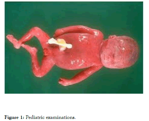
While certain adult-affecting disorders still affect children, pediatric medicine contains other diseases present primarily in patients under the age of 18. Congenital anomalies are one group of conditions which involve the infant population. When discussing congenital anomalies several terms are important to remember. A malformation caused by an underlying genetic defect is a congenital disorder. An interruption happens when another mechanism secondarily affects an ordinarily evolving organ [1]. For pediatric medicine a histological examination is mandatory. Histology is necessary to explain the quantity and nature of pre-existing infections, hypoxic changes, and other observations that suggest accidental death causes. Therefore, samples of all organs and tissues will be taken during autopsy [2-4]. A neuropathologist familiar in child mortality will examine the central nervous system whenever appropriate. In SIDS findings such as mild edema, swelling and certain gliosis foci are normal. Meningoencephalitis, aplasia, or serious dysplasia of large nuclei, or symptoms of acute or recurrent hypoxemia, may also be considered causes of death. For a limited percentage of cases, myocardial histological studies reveal results that are characteristic of myocarditis, systemic abnormalities or cardiac conductive system disruptions. In the event of an inflammation, immunohistochemistry may help to characterize the types of cells involved. Intestinal histological examination may expose malformations (congenital aganglionicmegacolon), diseases, and other abnormalities that are often not macroscopically diagnosable. Gastroenteritis may be important to the cause of death if the condition is followed by significant fatigue or if it is caused by bacteria that may excrete toxins Figure 1.

Figure 1: Pediatric examinations
Citation: Gavanji R (2020) Pediatric Pathology and Infan.t Child . J Med Surg Pathol . 5:179
Received: 03-Jul-2020 Accepted: 18-Jul-2020 Published: 25-Jul-2020 , DOI: 10.35248/2472-4971.20.5.179
Copyright: © 2020 Gavanji R. This is an open-access article distributed under the terms of the Creative Commons Attribution License, which permits unrestricted use, distribution, and reproduction in any medium, provided the original author and source are credited.