Fisheries and Aquaculture Journal
Open Access
ISSN: 2150-3508
ISSN: 2150-3508
Research Article - (2016) Volume 7, Issue 1
Introduction:Mahseers, belong to the family Cyprinidae, are well renowned for the excellent game as well as food fishes. Various aspects simultaneously considered towards the domestication of this fish species also include the study of fish health issues, preventive and remedial programmes for sustainable aquaculture. Experimental studies can be conducted in the control condition to study the pathological symptoms during bacterial infection. Considering the bacterial pathogens as a major constraint for aquaculture, the present study was carried out to standardize the bacterial concentration of A. hydrophila for the experimental challenge of Golden Mahseer for the first time, and various clinical changes were also monitored during the infection.
Materials and Methods: A total of 140 live fishes were acclimatized to the laboratory conditions for experimental study. A constant water quality was monitored during the acclimation period and throughout the experiment. The test group was injected with A. hydrophila and various physiological, biochemical and tissue level changes were observed during the experiment in comparison to phosphate buffer saline (PBS) injected control group.
Results: LD50 value of A. hydrophila for Golden Mahseer was successfully standardized. Clinical signs including weakness, slower movement, swimming closer to the surface, fin haemorrhages and red patches at the gut region were observed. Enlargement of spleen followed by tissue necrosis along with signs of haemorrhagic septicaemia was also seen in infected fishes. The necrosis of hepatocytes was observed in the histological section of liver tissue. The bacterial infection increases the superoxide dismutase activity and cortisol level in Golden Mahseer.
Conclusion: The LD50 value of A. hydrophila for experimental challenge studies on Golden Mahseer is 1.74 × 10⁵ cfu per 100 g of body weight. The symptoms of severe bacterial haemorrhage were observed. Necrosis of hepatocytes was observed in infected fishes. The superoxide dismutase activity and cortisol level also increased in infected fishes. A. hydrophila was confirmed to be the etiological agent which was re-isolated using spread plate method and confirmed by 16s rRNA sequencing.
Keywords: Bacterial infection, Experimental study, Bacterial haemorrhagic septicaemia, Disease symptoms
Mahseers, belong to the family Cyprinidae, are well known for the excellent game as well as food fishes all over the world especially the South-Asian countries [1-3]. In India, eight mahseer species are available among which Golden Mahseer, Tor putitora is considered the candidate species for hill aquaculture in the mid-Himalayan range [3-5]. The artificial breeding, seed production and scientific management of Golden Mahseer are in high priorities [6,7]. Scientific management including water quality, feed and health management are also equally important for sustainable aquaculture [8,9]. However, the data regarding the health management, mainly diseases is least available for this important fish species [2,10].
Aquaculture is primarily affected by microbial pathogens especially of bacterial origin [11], wherein, Aeromonas hydrophila and other Aeromonas are responsible for the majority of diseases [12]. A. hydrophila is reported being primary bacterial pathogen affecting freshwater fishes like Clarias gariepinus [13], Labeo rohita [14], Sparus aurata [15], Magalobrama amblycephala [16]. The severity of pathogenesis or disease outbreak can be moderate to high in fishes [17], but every type of disease results in deterioration of product and economic losses.
Aeromonad septicaemia is characterized by diverse pathological symptoms such as dermal ulceration, fin haemorrhages, fin rots, red sores, haemorrhages and necrosis of the visceral organs, etc. [17-19]. The acute form of the disease may result in fatal sepsis without any symptoms [17] while chronic infections may show the symptoms like hemorrhagic septicaemia with ulceration, inflammation, and dermal lesions [18]. Liver and kidney tissues are the primary targets of bacterial accumulation and sepsis [17,20]. The liver may become pale and show greyish to greenish coloration with foci while kidney may engorge and become friable [17,21]. Despite the knowledge of severity and symptoms of the Aeromonad septicaemia in other species, factors such as host-pathogen interaction, temperature requirement of the bacteria, course of pathogenesis, and immunity to pathogenesis may vary for fish species [12,22].
The present study was aimed to examine the effect of Aeromonas hydrophila infection on Golden Mahseer. Various macroscopic and microscopic changes in tissues and stress level was investigated to understand the fish response to bacterial infection. As the liver tissue is the primary organ affected by bacterial infection, microscopic changes in hepatic cells were also examined to ascertain the level of cell necrosis due to pathogenesis.
Ethical approval
The institutional ethical committee approved the protocols for maintenance, handling during experiments, and sacrificing of the fishes.
Experimental fish
Live fishes (size=33.32 ± 5.96 g; 146 ± 15.17 mm) were collected from Kosi River, Ramnagar (29.40°N 79.12°E) using a cast net. The fishes were initially acclimatized in ponds for three weeks and then in fibre reinforced plastic tanks (experimental tanks) for two weeks of 2000 L capacity before the experiment. Fishes were fed twice, daily with a formulated diet [23] based on 5% of the body weight. Constant aeration and water flow were maintained in experimental tanks. Water temperature and physicochemical parameters were recorded at regular intervals using multi-parameter auto-analyser (Hach®, Colorado, US) and colorimetric methods.
Bacterial strain and determination of LD50
The bacterial strain (originated from carp fishes) was obtained from Fish Pathology Laboratory (ICAR-CIFA, Bhubaneswar) and grown in our laboratory using TSB medium (Himedia, Mumbai, IN) for 24 h at 25°C. Single colony isolated on TSB agar was inoculated in TSB broth. The culture of A. hydrophila was grown overnight, and 100 μl of culture was inoculated into the fresh broth. Optical density was measured at regular intervals up to OD600=0.7 and culture was transferred to ice. The broth culture was centrifuged at 5000 × g for 15 min in a cooling (4°C) centrifuge (Eppendorf, Hamburg, DE). The pellets were washed twice with sterile phosphate buffer saline (PBS), pH 7.4 (Himedia, Mumbai, IN) and centrifuged. The final suspension was used to prepare 10 fold dilutions in 20 mL PBS having 106 cfu, 105 cfu, 104 cfu and 103 cfu mL-1. The dilutions were spread plated on TSB agar plates in duplicate for each dilution. The colony forming unit (cfu) count at optical density 600 nm was standardized with the same procedure before the experiment in three consecutive trials.
For the pathogenicity challenge test, we followed intra-peritoneal injection method using 1 ml Insulin syringe. After acclimatization in the aquarium for five days with proper aeration and feed, fish samples (33.32 ± 5.96 g) were separated into five different groups (12 fishes in each group). Four groups received 106 cfu, 105 cfu, 104 cfu and 103 cfu of bacterial culture per 100 g body weight respectively while the fifth group was injected with 100 μl of PBS. The samples were observed up to 96 h, and dead fishes were removed for routine bacteriological examination. Final LD50 values were estimated according to the method of Miller and Tainter [24]. The results were calculated as mean values from two independent experiments.
Pathogenicity experiment
A consecutive experiment was conducted in two different groups of fishes with 35 samples in each using the standardized LD50 dose to test the pathogenicity. Test group were injected with A. hydrophila intraperitoneally according to their body weight while the control group was injected with 100 μl of PBS. The fishes were observed for any physiological and pathological symptoms at various time intervals. Three samples from test group were anesthetized using tricaine methanesulfonate (200 mg L-1) (Sigma-Aldrich, St. Louis, US) and blood was drawn from a caudal vein in 3.8% sodium citrate containing vials. The blood samples were centrifuged at 7000 × g for 10 min and plasma was collected in the fresh tube. Plasma was stored at -80°C until further use. The plasma samples were used to estimate the levels of superoxide dismutase (SOD) and cortisol using ELISA and colorimetric kits (Cayman, Ann Arbor, MI), respectively. The statistical analyses were performed by the standard curve plot of known concentration of SOD and cortisol. The data was analysed using independent sample t-test (α=0.05). Liver, head kidney and spleen tissues from each sample collected at different time intervals were also plated on TSB agar plates (amp+) to detect the presence of bacteria.
Identification of A. hydrophila was also confirmed by PCR amplification and sequencing of 16s rRNA. The genomic DNA was isolated from the randomly selected clones (6 no) grown on TSB agar plates and 16s rRNA was PCR amplified using the universal primer pairs (16s rRNA F- AGAGTTTGATCCTGGCTCAG; 16s rRNA R-GGTTACCTTGTTACGACTT). The PCR reaction was performed in a total reaction volume of 50 μl containing 1 × PCR Buffer, 10 pM of each primer, 200 μM each dNTP, 50 ng template DNA and 1U Taq DNA Polymerase (Invitrogen, Waltham, USA). The reaction conditions were 94°C for 4 min; 35 Cycles of denaturation at 94°C (30 s), annealing at 58°C (30 s) and extension at 72°C (90 s) followed by a final extension at 72°C for 10 min. The PCR products (Figure 1) were sequenced using Big Dye Terminator v3.1 cycle sequencing kit in ABI3730 Genetic Analyser at Scigenom Laboratory, Kochi. The nucleotide sequences so obtained were matched with NCBI GenBank Blastn toolkit using default settings.
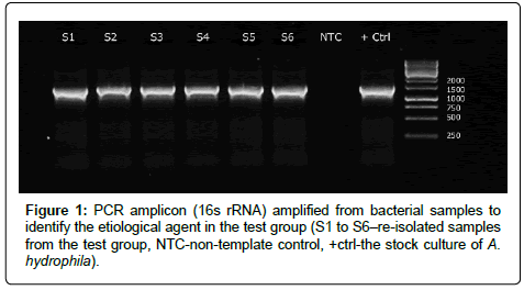
Figure 1: PCR amplicon (16s rRNA) amplified from bacterial samples to identify the etiological agent in the test group (S1 to S6–re-isolated samples from the test group, NTC-non-template control, +ctrl-the stock culture of A. hydrophila).
Histopathology
Liver tissues collected at 48 h were processed for histological examination. Tissue samples were fixed in Bouin’s Fixative and prepared for histological examination using recommended procedure [25]. Tissues were further embedded in paraffin wax and sectioned at 5 μm in a rotary microtome (Thermo Fisher Sci, Massachusetts, USA). The slides were stained with haematoxylin and eosin and observed under Leica DM2500 (Leica, Wetzlar, DE) to study any pathological changes/difference in tissues after acute bacterial infection.
Bacterial culture and experimental conditions
Aeromonas hydrophila is successfully grown at 25°C, but no growth was observed below 18°C and above 30°C. Correlation study between OD600 and cfu resulted that there are ~106 cfu mL-1 of A. hydrophila at OD600=0.7. All samples were healthy, and their movement was active. During the experiment, the ambient temperature was 22-24°C, water pH was 7.5-8.0, total hardness 80-100 mg L-1, dissolved oxygen ranged between 5.2-7.4 mg L-1 and ammonia was <0.1 mg L-1. The LD50 value determined using log-probit calculation method of Miller and Tainter [24] is 1.74 × 10⁵ cfu per 100 g of body weight of the fish (Table 1 and Figure 2).
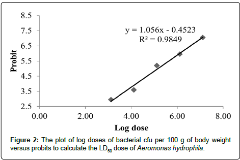
Figure 2: The plot of log doses of bacterial cfu per 100 g of body weight versus probits to calculate the LD50 dose of Aeromonas hydrophila.
| Group | Dose | Log Dose | Sample Size | Moribund fishes | Moribund fishes (%) | *Corrected % | Probits |
|---|---|---|---|---|---|---|---|
| 1 | 1.00E+07 | 7.11 | 12 | 12 | 100 | 98 | 7.05 |
| 2 | 1.00E+06 | 6.11 | 12 | 10 | 83.33 | 83 | 5.95 |
| 3 | 1.00E+05 | 5.11 | 12 | 7 | 58.33 | 58 | 5.2 |
| 4 | 1.00E+04 | 4.11 | 12 | 1 | 8.33 | 8 | 3.59 |
| 5 | 1.00E+03 | 3.11 | 12 | 0 | 0 | 2 | 2.95 |
*Corrected % Formula for 0 and 100% mortality: For 0% dead– 100(0.25/n); For 100% dead–100(n-0.25/n).
Table 1: Results of the lethal doses of Aeromonas hydrophila for the determination of LD50 after intraperitoneal injection in fishes.
Identification of Aeromonas using 16s rRNA
An amplicon of 1240 bp was successfully amplified from 6 randomly selected bacteria isolated from liver (S1-S2), head kidney (S3-S4) and spleen tissues (S5-S6) (Figure 1). The bacteria were confirmed as A. hydrophila based on nucleotide sequence blastn similarity (score-2202; Ident-99%; e-value-0.0) with 16s rRNA sequence of A. hydrophilia in NCBI GenBank database.
Macroscopic and microscopic changes on bacterial infection
Clinical signs including weakness, slower movement, swimming closer to the surface, fin haemorrhages and red patches at the gut region were observed. In the morphological examination, the hyperemia of fins was first observed at 6 h post infection (p.i.); initially on pelvic fins followed by prominent haemorrhages at caudal fin at 48 h. Darkening of the skin and red patches were observed on the ventral side of the gut region along with prominent haemorrhagic signs at the anal orifice (Figure 3). The dorsal skin and eyes did not reveal any significant symptoms of infection. Gills also did not show any haemorrhagic symptoms up to 12 h p.i.; however gills became pale after 24 h and haemorrhages appeared at 48 h p.i. The liver was seen to be yellowish brown having haemorrhagic symptoms on the surface after 6 h. The liver was pale, and greyish white foci were observed at 12 and 24 h p.i. The degenerative changes in the liver with focal necrosis of hepatocytes were observed histologically at 48 h p.i. (Figure 4). Dark red colouration and engorged spleen were observed however the spleen was friable at 48 h p.i. Haemorrhagic foci were found in internal organs while intestine was filled with yellow coloured mucoid liquid. Gall bladder was enlarged and filled with emerald-green secretion. The haemorrhage symptoms over internal organs at macroscopic and microscopic levels revealed a visceral haemorrhagic septicaemia (Figures 4A-4C).
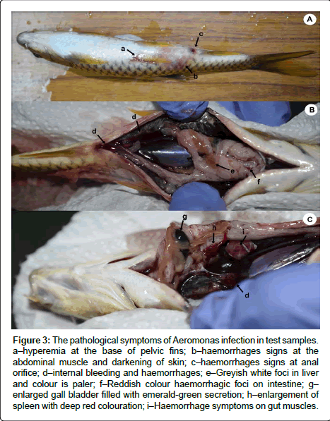
Figure 3: The pathological symptoms of Aeromonas infection in test samples. a–hyperemia at the base of pelvic fins; b–haemorrhages signs at the abdominal muscle and darkening of skin; c–haemorrhages signs at anal orifice; d–internal bleeding and haemorrhages; e–Greyish white foci in liver and colour is paler; f–Reddish colour haemorrhagic foci on intestine; g– enlarged gall bladder filled with emerald-green secretion; h–enlargement of spleen with deep red colouration; i–Haemorrhage symptoms on gut muscles.
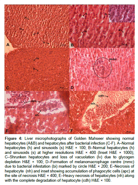
Figure 4: Liver microphotographs of Golden Mahseer showing normal hepatocytes (A&B) and hepatocytes after bacterial infection (C-F). A–Normal hepatocytes (h) and sinusoids (s) H&E × 100; B–Normal hepatocytes (h) and sinusoids (s) at higher resolutions H&E × 400 (Inset H&E × 1000); C–Shrunken hepatocytes and loss of vacuolation (lv) due to glycogen depletion H&E × 100; D–Formation of melanomacrophage centre (mmc) due to bacterial infestation (bi) marked by circle H&E × 200; E–Necrosis of hepatocyte (nh) and inset showing accumulation of phagocytic cells (apc) at the site of necrosis H&E × 400; E–Heavy necrosis of hepatocytes (nh) along with the complete degradation of hepatocyte (cdh) H&E × 100.
Stress and antioxidant activity matrices
The bacteria challenged (test group) fishes showed the stress symptoms like slow swimming and loss of appetite. The symptoms were notable after 12 h p.i. and were consistent up to 48 h p.i or morbidity. Mean superoxide dismutase activity in control samples was 2.26 U mL-1. The superoxide dismutase (SOD) activity of test samples increased significantly over control group [t (29)=3.9, p<0.05]. SOD activity increased at 3 h and was maximum at 24 h (9.81 U mL-1); followed by a decrease in activity at 48 h or 96 h p.i. (7.22 and 4.40 U mL-1 respectively) (Figure 5a). The plasma cortisol concentration of Golden Mahseer in PBS inoculated fishes was 43.24 ng mL-1. The plasma cortisol level at various time intervals showed a significant increase in test group [t (29)=2.02, p<0.05]. The level of cortisol was highest at 24 h p.i. (79.56 ng mL-1) and gradually decreases at 48 h and 96 h p.i. (62.98 and 42.71 ng mL-1 respectively) (Figure 5b).
Figure 5: (a) The superoxide dismutase activity in control and test samples, (b) Concentrations of cortisol in plasma of test and control samples.
Aeromonas hydrophila is the primary bacterial pathogen for many fish and aquatic organism, and thus, it is a great concern for the aquaculture industry [12,26]. The fish under the present study is also a valued and potential species for aquaculture in the Himalayan cold water region. Therefore, the present study was aimed to examine the effect of Aeromonas hydrophila infection on Golden Mahseer. Initially, we standardized the suitable temperature of 25°C for the growth of Aeromonas hydrophila in the laboratory condition as the ambient temperature in hill environment remains low (-2°C-25°C in different seasons). Many authors observed the growth of this bacterium within the range of 27°C-30°C [12,27] which we could not see in our conditions. The fish-pathogenic Aeromonas are also reported to be psychrophilic in nature and was isolated from diseased rainbow trout that lives at very low temperature (5°C-20°C) [27,28].
The level and dose of pathogenicity of the bacteria in several carp species were also found to be variable [26,29-31]. Therefore, the optimum dose of bacterial inoculation and pathogenicity was determined for the first time in this species and how this dose of bacteria interacts with the physiological mechanism was determined. Determination of LD50 in study organism before the experimental challenge is advantageous for successful experiment and induction of clinical signs and symptoms [32]. Where higher dose may cause mortality, the suboptimal treatment may not enrich the desired genes. In the present study, an LD50 value of 1.74 × 10⁵ cfu per 100 g of body weight was standardized for A. hydrophila in Golden Mahseer. LD50 value of A. hydrophila was previously determined for varies model and non-model fish species [26,30,33,34]. The standardized LD50 value of A. hydrophila for in vivo experiments would serve as a baseline data for future immune response studies.
The intraperitoneal inoculation of A. hydrophila produced similar signs and symptoms, reported during the disease progression in other fish species [33-35]. Clinical signs including weakness, slower movement, swimming closer to the surface, fin haemorrhages and red patches at the gut region were observed during 24-48 h p.i. Similar observations were documented in A. hydrophila infected catfish [21]. The fishes showed noticeable signs of haemorrhages and abdominal swelling. Large external ulcerative lesions also developed in the injected area and around the anal orifice. Histological lesions were observed in liver (Figure 4) and the spleen, and kidney tissues were found diffused due to acute infection. Sundus highlighted similar observations like structural fragmentation and necrosis of liver tissue and friable kidney [36]. Aeromonas hydrophila may itself initiate pathological processes and induce inflammation [32]. However, the role of extracellular products released during the infection may also play a significant role in causing degeneration and necrosis [37]. Also, histological symptoms were not performed in other tissues as the liver was the primary site of infections due to Aeromonas infection [17]. The infection was also very high in the liver in comparison to other tissues when plated on Agar plates at each time points of sample collection.
Dermal lesion with haemorrhagic foci may be correlated to bacterial haemorrhagic septicaemia mainly caused by Aeromonas sp. [21]. The study findings are similar to Cipriano and Laith & Najiah who observed the dermal ulcerative lesions, focal haemorrhages and inflammation in chronic infection of Aeromonas sp [18,21]. The histological findings of the study are similar to Laith and Najiah [21] who reported that A. hydrophila causes haemorrhagic sepsis, liver necrosis along with the accumulation of phagocytic cells at the site of necrosis. The results also agree with the finding of Afifi that various toxins and extracellular products such as hemolysin, protease and elastase released by A. hydrophila may cause severe hepatocyte necrosis [32,38]. Similarly, necrosis of hepatocytes and the presence of non-hepatocytic cells, probably the phagocytic cells (Figure 4E) in liver histology (Figure 4F) also indicate the systemic bacterial infection in the liver which is also shown previously in various pathological studies [17,20,39].
Pathogenicity of Aeromonas was also determined using the stress related SOD and cortisol parameters. Both the SOD and cortisol were elevated at a particular time and scaled down during the longer periods when the fish overcame the pathogenicity. Superoxide dismutase (SOD) acts as cyclic enzymes that catalyze the dismutation of superoxide radicals [40]. The amount of superoxide dismutase present in the cellular and extracellular environment is significant in the prevention of oxidative stress related diseases and can help to predict the defence mechanism of fish species [41,42]. Production of reactive oxygen species (ROS) is also an important mechanism of the vertebrate immune system [43]. They also possess various antioxidant enzymes to counteract the ROS and their adverse effects [41]. In the present study, a significant increase in SOD activity was observed up to 24 h p.i. Wang studied the tissue-level activity and gene expression of two superoxide dismutase (MnSOD and Cu/Zn SOD) after A. hydrophila infection and found that the activity was highest in liver and kidney at 24 h p.i [44]. However, a subsequent decrease in SOD activity suggests the failure of SOD to remove superoxide radicals. These results are similar to that of Reddy. The failure of SOD leads to accumulation of superoxides that ultimately may result in internal tissue necrosis [45]. The necrotic lesions observed in liver tissue at 48 h p.i. may be the combined effect of infection and ROS species. Although, cortisol inhibit the immune response by suppressing the inflammatory substances; the amount of cortisol may serve as a useful indicator of stress level during infection in fishes [46]. The cortisol level was significantly (p<0.05) increased in blood plasma of Golden Mahseer after A. hydrophila challenge. The highest level was detected at 24 h p.i. which could be the combined effect of stress as well as bacterial infection. Increase in plasma cortisol level was reported in many piscine systems after bacterial infection [46,47]. Small and Bilodeau also reported the similar findings on cortisol level for channel catfish (Ictalurus punctatus) after exposure to Edwardsiella ictaluri [48]. High cortisol level was observed in Atlantic cod after experimental infection with Aeromonas salmonicida [49]. Increased cortisol level is commonly associated with stressful conditions of the fish principally during disease condition [50]. Endotoxin may also increase the plasma cortisol level that may suppress the immune response of the fish [51,52]. Hence, the increased level of plasma cortisol in Golden Mahseer may be associated with the stressful condition of fish as well as bacterial endotoxins. This is the first report on plasma cortisol level of Golden Mahseer in control and stress conditions. Handling during the bacterial and PBS inoculation as well as sampling may cause stress to the fishes, and its effect may not be neglected. However in the present study the cortisol level increased almost two folds in diseased fishes compare to control which is in agreement with the findings of Olsen and Haukenes and Barton who found the increased plasma cortisol level in diseased fishes [50,53].
In the present study, we determined the LD50 concentration (1.74 × 10⁵ cfu per 100 g body weight) of A. hydrophila and examined various pathophysiological symptoms during the disease progression. The sign and symptoms of severe bacterial haemorrhage were observed, and A. hydrophila was confirmed to be the etiological agent by re-isolation of a bacterium using spread plate method followed by sequencing of 16s rRNA sequencing from infected fishes (test group). Two major stress indicators were also studied in test samples during the progression of infection. Superoxide dismutase activity and cortisol level were significantly high in test samples as compared to control. Severe liver necrosis and tissue damage were observed at 48 h p.i. Experimental conditions standardized in the present study will serve as a baseline data for further studies on Golden mahseer. Also, the knowledge of various symptoms during pathogenesis help to understand the course of pathogenesis and overall immune response of fishes which can be very beneficial to control and prevent the disease outbreaks in farmed fishes.