Journal of Osteoporosis and Physical Activity
Open Access
ISSN: 2329-9509
ISSN: 2329-9509
Research Article - (2014) Volume 2, Issue 2
This perspective appraises the pathology, etiology, clinical presentation and therapy of osteoporosis, and focuses on implications for oral healthcare. Osteoporosis is a chronic insidious, debilitating disease of aging, slowly affecting the quality of life of both genders. Very few people show no signs of OP as they age. A healthy balanced diet, rich in calcium combined with daily rigorous exercise seems to be the sole natural formula for retarding OP. Osteonecrosis of the Jaws (ONJ) is a rare complication and precise management is unresolved; management is by prevention of ONJ, is feasible mainly by completing all invasive dentistry before prescribing bisphosphonates.
<Keywords: Bisphosphonate; Bone; Fractures; Oral health; Osteonecrosis; Osteopenia; Osteoporosis; Zolendronic acid
Osteoporosis is a disease which affects the entire skeleton. Osteoporosis causes thinning of bony trabeculae and cortical bone; consequently osteoporotic bones are weakened and more prone to fracture. The prevalence of osteoporosis in North America is high, with nearly 1/3rd of people over 60 years of age suffering from osteoporosis. About 36 million Americans and 3.7 million Canadians have a low bone mass, and 44 million Americans and 4.4million Canadians have OP. Most commonly fractures of bones with osteoporosis are the spine, hip and wrist. Frequently spinal fractures are small and symptomless, but over a period of time osteoporosis leads to deformity and disability.
Prevalence and morbidity of OP
Osteoporosis (OP) is not confined to North American women. In the US, while 10 million people have OP, another 34 million have low bone mass, 40 percent of women over 50 years will suffer OP related fractures in their lifetimes, but also 14 million men have low bone mass (LBM) or OP. Osteoporosis is among the most common diseases affecting the population, with OP prevalence exceeding that of coronary thrombosis, stroke and breast cancer combined [1-3]. For example, in North America nearly 4.5 million osteoporotic fractures are recorded compared to 1.24 million cancers per year (Figure 1).
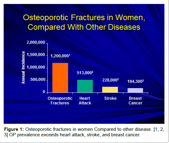
Figure 1:Osteoporotic fractures in women Compared to other disease. [1, 2, 3] OP prevalence exceeds heart attack, stroke, and breast cancer.
The risk of developing OP increases as people age. Most women start being affected from age 50, but there is a gender difference, with males becoming prone to OP ten to fifteen years later after females (Figure 2).
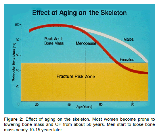
Figure 2:Effect of aging on the skeleton. Most women become prone to lowering bone mass and OP from about 50 years. Men start to loose bone mass nearly 10-15 years later.
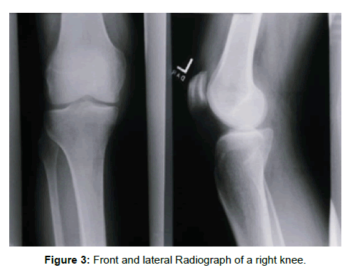
Figure 3:Front and lateral Radiograph of a right knee.
Bone mineral density (BMD mainly as Calcium hydroxyapatite and traces of other ions like Magnesium) is decreased as OP progresses. Optimal Peak Adult Bone Mass (PABM) is considered as 100 percent, but when the PABM reduces to below 45 percent, the risk from OP increases exponentially. For many decades, reduction of bone was considered to be a slow progression over decades, with small vertebral fractures. OP is deemed to be more progressively rapid than previously thought.
Once vertebral OP fractures manifest, other OP fracture sites often occur within one year. Loss of bone mass may be slow, but once fractures start, other fractures follow, especially after minor accidents or falls. Because of this, early therapy to moderate bone loss, and to slow down decreasing bone mass is indicated for women. Hip and/or head of femur fractures occur frequently when women, unwittingly suffering from OP, fall. Morbidity includes limiting perambulation, and often head of femur or total hip replacement is needed.
Symptomology of early vertebral fractures are not always clearly defined. These fractures produce slight pain, imprecise location and vague back ache. Minor collapse from body of vertebral trabecular breakage starts slowly manifesting as kyphosis. From upright posture women gradually hunch forward as their vertebrae collapse. This spinal crumple inexorably progresses to kyphosis, until walking becomes impossible. Subsequently they need walking aids (sticks, walkers etc.), or a wheel chair to get around. Their height diminishes, and the folding forward impinges on thoracic space available for chest (heart and lungs), and the upper abdominal organs (stomach, liver, spleen, pancreas) and diaphragm.
Consequent to kyphosis, organ dysfunction precipitates indigestion, esophageal reflux, respiratory difficulties… all accompanied by discomfort and pain and even incontinence from changed intraabdominal pressure. OP affects the size, mass, strength and volume of the entire skeletal system. The micro-fractures in OP are frequent in vertebrae, and over time contribute to the final collapse and overt shortening of the spine [4-6].
Bony anatomy
All bones have a thicker dense outer layer, the cortex, and an inner layer or medullary cancellous part, which is less dense. The cortex is compact bundle bone, while the medulla is traversed with thinner reticulated trabeculae, housing marrow spaces. Bones are biological calcified structures, sculpted by genetic programing to suit the functional purpose located in the body. Structural beams grow the cortex, trabeculae in the medulla, and house the bone marrow according to the needs and demands of the body. A periosteum covers the outer cortex, and has bone forming cells, osteocytes. Osteocytes are incorporated into the bone in Haversian systems, and line the thin trabeculae in endosteal sinuses. Bone and blood formation is moderated by many blood-born biochemical co-factors (such as osteocalcin), hormones and paramone molecules, and mediated through osseous stem cells, hemopoetic cells, unicellular osteocytes (bone-forming) and multinuclear osteoclasts (bone-removal). Alternate cycles of laying down bone, and bone resorption is ongoing all the time. When resorption exceeds bone formation, OP develops. Therapy is directed at these cells to mediate the rate of bone deposition and resorption (Figure 3).
3-D Micrographs of medullary bone structure shows thinning of the cortical plates, and also diminishing of trabeculae, often with medullary micro-fractures. Overall this leads to weakening of the cortex and medullary parts of the bone, and generally increases chances of fractures [5,6] (Figure 4).
Bone mass peaks when people are in their mid-twenties to midthirties. Subsequently bone mass starts decreasing over time. The World Health Organization measures of Bone Mass. Peak Bone Mass is assessed with T scores. Normal range is accepted at a positive T+ score above T =1, when T= 0, and still acceptable as healthy between 0 and -1. World Health Organization deems osteopenia to exist at T= < - 1, and OP If T= < - 2.5 (Table 1).
The healthy range is between zero loss (T= +2.5) and T= - 1 unit of Peak Bone Mass. More than one unit loss of PBM is labeled as Osteopenia. A T-score below T= -2.5 PBM is consistent with suffering from OP. So, clinical risk factors for developing OP include: being over 65 years of age; a low BMD; a T-score of T=-2.5; prior fragility fracture, most commonly at the spine, hip or forearm. All these co-factors are aggravated by a genetic calcium metabolic predisposition to OP (Table 1 and Figure 5).
Clinical diagnostic categories of osteoporosis in postmenopausal women
Osteoporosis manifests loss of bone mass due to an imbalance of plasma calcium-phosphorous levels with consequent release of calcium from bone [4]. Primary OP (senile or post-menopausal) or Secondary OP (calcium deficiency, hyperparathyroidism, osteomalacia), both result in decreased Bone Mineral Density. The normal Ca: P ratio has a range consistent with health; some healthy women (~15%, with Standard deviation T-scores at T= -1 to -2) do have a lower than normal ratio, but in general if the Ca:P ratio reaches T= -2.5 SDs, OP will become apparent. A significant number of women over 55 have a BMD below T= -2.5 and nearly all of these women have various degrees of bone loss from OP.
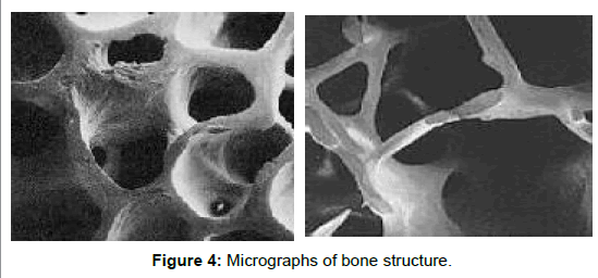
Figure 4:Micrographs of bone structure.
| Category Definition by Bone Density T-Score |
|---|
| Osteopenia Normal Range……………………………….........T-score between T= +2.5 and T= -1. |
| Osteopenia………………………………………...………....…T-score between T= -1 and T= -2.5 |
| Osteoporosis………………………………………………....………..….T-score less than T= -2.5 |
| Severe Osteoporosis……………………….............T-score less than T= -2.5 AND a fragility fracture |
Table 1: T-Scores based on WHO criteria.
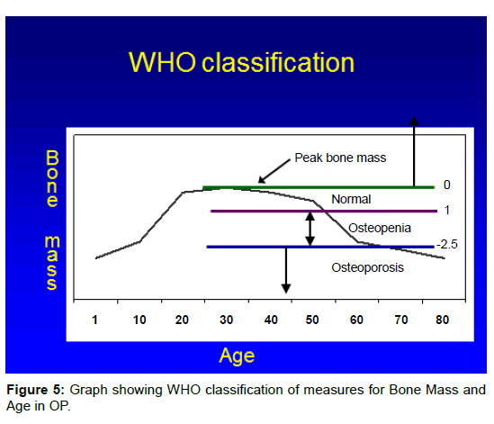
Figure 5:Graph showing WHO classification of measures for Bone Mass and Age in OP.
Red Flags and contributory co-factors include poor diets, smoking, alcohol consumption, lack of exercise all contribute to a genetically determined propensity to developing OP. With these influences, men too, when over 50 years old, will show signs of OP. Europid females are more prone to OP than Asian women. Some females may reach menopause as early as 40 years. An inactive sedentary lifestyle predisposes to develop in OP. Consequently vigorous exercise at least three times per week encourages minimizing OP development. Also people who uses steroids (like prednisone) to control allergic (chronic asthma), autoimmune diseases (Crohn’s Disease) or transplants are prone to develop OP. Women taking medications for hormone suppression are candidates for OP [6-8].
Clinical Confirmation may be done quickly with tests for BMD; Ultrasound of the foot gives a rapid but unreliable provisional diagnosis. Measures of the vertebrae using Dual X-Ray Absorptiometry (DXRA) are preferable and more consistently reliable. This DXRA measures the Calcium/Phosphate crystal density per cm2. Lower bone density may also be recognized on panoramic full mouth radiography, but it seems the skull in general is more resistant to bone resorption than other peripheral sites, like the hip, vertebrae, ribs and limbs.
Special clinical investigations needed to assess OP in females: Blood analysis laboratory investigations required include these: Calcium (albumin corrected), Complete Blood Count(CBC), Erythrocyte Sedimentation Rate (ESR), phosphate, magnesium, thyroid stimulating hormone (FSH), creatinine, Follicular Stimulating Hormone (FSH), estradiol, 25 Hydroxy Vit-D, parathyroid hormone (PTH) antigliadin anti-bodies, anti-endomysial antibodies. Also a 24 hour urine collection and analysis for calcium and creatinine assessment assists in the diagnosis.
Diets as contributing factor in OP: Deficiency of Vit-D (subtypes Vit D-2 and Vit D-3) will result in Ricketts in children and Osteomalacia in Adults. Daily doses of sunshine help in dermal synthesizing of Vit-D. An adequate dietary intake of calcium, phosphorous (and when under 25 years also fluoride) is needed to delay OP. People with lactose intolerance tend to eat less dairy products and consequently have a low calcium dietary intake.
Secondary conditions, diseases and medications implicated in causing OP
Primary or Secondary hypogonadism, primary hyperparathyroidism, thyrotoxicosis, Growth Hormone Deficiency (GH), osteomalacia, hypophosphatasia, connective tissue disorders, osteogenesis imperfecta, GIT malabsorption syndromes including coeliac disease, anorexia nervosa and excessive weight loss.
Co-factors as Medications: Glucocorticoid-steroids, excess thyroxin, anticonvulsants (like phenytoin and phenobarbital); lithium, gonadotropin-releasing hormone and medroxyprogesterone are all implicated in OP [7].
OP and Andropause in Men: Males are also at risk for developing OP. Risk factors are similar but slightly different from females. Andropause in Men is the equivalent to female menopause. Men reaching andropause, which occurs at a variable age, but in general, about ten years after women. These men are prone to decreases in bone mineral density, reduced muscle mass and strength, an increase in obesity, a declining libido, increase in erectile dysfunction, a decrease in hematopoiesis, are prone to bouts of depression with a decrease in cognitive function and general well-being.
Epidemiology of hip fractures in aging men
Both vertebral and hip fractures affect men. 20% of all vertebral fractures affect men; women show a higher percentage (>40%). Consequently women tend to be more kyphotic, earlier and more frequently, than men. Globally, there are nearly 1.7 million hip-fractures per year, and of these 30% occur in men. Hematopoiesis is reduced in elderly men, and this impacts calcium blood chemistry. For men over 65 the annual incidence is 4-5 per 1000. After six months of a hip fracture there is a 20% chance of death and a 50% chance of permanent severe disability. About 60% of elderly men with hip fractures are hypogonadal [9-11].
Major OP risk factors for men include hypogonadism, vertebral deformity, kyphosis or non-traumatic loss of vertebral height, radiographic evidence of osteopenia, age older than 65 years, hyperparathyroidism, use of systemic glucocorticosteroids, a prior fragility fracture before 40 years, a family history of osteoporotic fractures, malabsorption syndrome, and propensity to falls.
Minor OP risk factors for men influence developing OP. Any two concomitant findings of the following will predispose to developing OP in men: these include low dietary calcium intake, weight below 57 Kg (or10%of that at age 25), Cigarette smoking, excess alcohol and coffee intake, chronic use of anti-convulsants, long term use of heparin, rheumatoid arthritis, hyperparathyroidism.
Special clinical investigations needed to assess OP in males: These are similar but slightly different to that of females. The work– up includes: Complete blood count, serum calcium (total & ionized), serum creatinine, liver function and enzymes, alkaline phosphatase, TSH, PTH and serum 25-hydroxy Vit-D (to exclude PTH disease & or Vit-D deficiency). Other investigations (assist in eliminating differential causes): Protein electrophoresis over 60 year (rules out multiple myeloma), 24-hour calcium (excludes hypercalcuria in place of nephrolithiasis, hypocalcuria in place of malabsorption), 24 hour urine free cortisol, total and free testosterone, bioavailable testosterone (to exclude hypogonadism), antibodies to celiac diseases with features of calcium malabsorption.
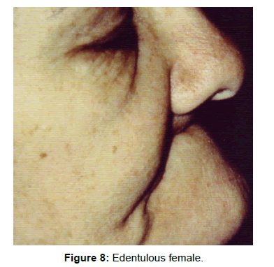
Figure 6:OP Edentulous mandible.
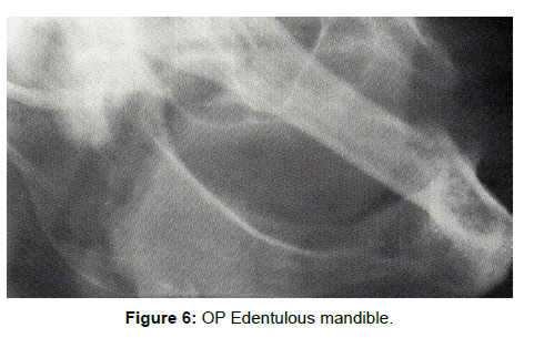
Figure 7:An intra-oral radiograph.
| Age in years | Elemental calcium | Cups (350ml) of milk |
|---|---|---|
| 9-18 | 1300mg | 4 cups |
| 19-50 | 1000mg | 3 cups |
| Over 50 | 1500mg | 5 cups |
Table 2: Daily Calcium needs and cups of whole cow’s milk needed to provide this.
Dentate patients stimulate the jaw bone metabolism on an ongoing daily regimen. Consequently osteoporosis is not always blatantly obvious on jaw radiographs [12-19]. But since bite-wing radiographs often are taken with check-ups on a regular annual basis, changes may be observed by comparison indicating marrow changes. Also by comparison, panoramic radiographs may show changes indicating OP. Diagnosis by CT is feasible [16]. More typical of OP is the development of localized OP defects, which may be confused with other bone pathology, such as simple bone cyst, giant-cell-granuloma, ossifying fibroma, osteosarcoma, aneurysmal bone cyst and others [13]. A biopsy for histopathology investigation is always necessary and will differentiate and/or confirm the diagnosis [15]. With focal OP marrow defect, nothing more than hematopoietic marrow cells are revealed. OP affects most bones, especially the spine pelvis and ribs, but less so the bones of the skull. Accordingly while focal OP marrow defects are not commonly found in both the maxilla and mandible, they do occur in these bones [14-18] With OP, edentulous jaws will show thinner cortical plates, loss of alveolar bone, a thin mandibular ridge and less dense medullary bone [19] (Figure 6).
Periodontitis
Gingivitis affects over 90 percent of the population. At most 30 percent, but more realistically only 8-12 percent of people develops periodontitis. Nearly all periodontitis is mediated through stagnated biofilms which change their invasive destructive capacity over time. While some forms of aggressive periodontitis affect young people and diabetics due to systemic predisposing dysfunctional metabolic factors, most chronic forms of periodontitis affect middle-aged and older people. Osteoporosis becomes prevalent in seniors, particularly females but also later males, and consequently when seniors suffer from osteoporosis and periodontitis, there is a tendency for periodontitis to be more aggressive, rapid and destructive of supporting alveolar bone [17-20] (Figure 7). Stagnated biofilm produces bacterial toxins enzymes, antigens and mitogens, all of which have a Zone of Influence (ZOI) and biological effect. When teeth are also distressed by occlusal trauma, there is a Zone of Traumatic Effect (ZOTE). When OP is present, the combined effect of ZOI and ZOTE is accelerated alveolar bone loss. Periodontal disease and alveolar supporting bone loss can be successfully controlled through excellent oral hygiene, and professional maintenance, monitoring and therapy. The prognosis with appropriate therapy for OP sufferers is excellent.
Therapy and management involves (1) dietary modification, (2) behavior changes (3) moderation of OP through medication (4) oral and dental implications [9,13,20-24].
Diet and Nutrition as therapy and control of OP
Because low calcium intake and diets are implicated in OP development, dairy products are promoted as high sources of calcium containing foods [25]. Dairy products include, milk (high or low fat), cream, cheeses (especially Boccancini, Cream Cheeses, Mozzarella), yoghurt, skim milk, milk shakes, ice-creams. Whole cow’s milk (3.7G total fats), is about 88 percent water, but the solids contain essential nutrients of skeletal growth. Besides calcium, phosphates and magnesium, (all nutrients essential for healthy bone growth), bovine milk also contains: proteins, potassium, sodium carbohydrates (lactose), Vit-A, Vit-D, Thiamine, Riboflavin, Niacin, Vit B-6 , Folic acid, Vit-B12, Pantothenate and Zinc.
To ensure stable calcium for bone formation the range of calcium intake is 200mg-to over 1000mg/day. The FAO/WHO minimum requirement is close to the minimum requirement, which is low for ideal health. However, the recommended calcium per day for adults is at least 800 mg, and 1200mg+ for teenagers and seniors over 55 years. People consuming less protein will remain in calcium balance with lower levels of Calcium intake. Only one quarter of calcium intake from cow’s milk is retained. Milk is a ubiquitous source of calcium, though a varied diet is easier to sustain (Table 2).
Other nutrient requirements and food sources:
Calcium: Other foods with significant calcium content include: dairy products, butter, yoghurt, almonds, prunes, and seaweed.
Vitamin D is needed for adequate calcium absorption and metabolism at 400 I.U. per day for 19-50 year olds. Over 50 the required Vit-D is 800 I.U per day, double the usual need. With a daily exposure to sunshine of about half an hour, Vit-D is synthesized in the skin. For ages 19 to 50, 4 cups of milk will provide 400 I.U. Vit-D; 8 cups of milk is needed for those over 50+ years to get the required 800 I.U Vit-D per day. Besides milk, other sources of Vit-D are in fish oils, nuts, dates, leafy greens and other fats.
Protein: First class protein contains essential amino acids which cannot be synthesized by human metabolism. Collagen is a body protein and its synthesis is a precursor to bone formation. 0.8G/kG body weight first class protein is recommended to sustain good bone density. Sources of protein are lean meat, fish, poultry, eggs, nuts and pulses (beans. lentils, peas etc.)
Sodium: A Total of 2G sodium is needed per day. Consuming excess >2Gper day will reduce bone density. High sodium intakes may disrupt stable metabolism and induce hypertension. Sources from salted foods and table salt added to food to enhance flavor.
Vitamin-C: a daily intake of 60mg a day is adequate. This is double the RDA of Vit C needed for bone synthesis. Excess intake of Vit-C may have pharmacological effects unrelated to the function of the vitamin. Sources are broccoli, parsley, guava, citrus, rose hips and fresh fruits and vegetables.
Phosphorous: The Phosphorous requirement is at least 1:1 for Ca: P, and probably needs more Phosphorous. The average Calcium intake per day is 400-1300mg/d and Phosphorous 800-1500mg/d. Sources vegetables, meats and fruits.
Vitamin K: The average mixed diet provides 300-500 ug/day Vit-K. Extra Vit-K is not recommended for improving bone density. Too much Vit-K, in combination with phytates, may decrease calcium absorption. Food sources include beans, soy products, fruit and leafy greens.
Caffeine: A large coffee (355mls) has about 180mg caffeine. More than 4 cups per day is not recommended.
Isoflavine: In post menopause women Bone mass density will be sustained with 200mg 3 X day intake of Ipriflavones, and will slow down development of OP, and maintain Bone Mass Density [12,25].
Behavior modifications
Exercise, and particularly weight bearing activities, slows development of OP. Exercise forces the body to support full weight. This stimulates bone formation and modeling. Brisk daily exercise is needed for 30 minutes. Exercise at least three times weekly is the desirable minimum. Making exercise part of a daily routine assures benefits accrue over time. Playing sports is one pleasurable form of securing benefits of exercise, such as climbing stairs, playing ball games, (like racket ball, tennis, squash) dancing, and jogging. Simply walking daily for a vigorous 30-45 minutes, contributes enormously.
A wide variety of drug therapy is available for post-menopausal OP in women. These include: hormones and mainly drugs derived from Bisphosphonates.
Hormonal therapies include calcitonin, and estrogen replacements [21,22,34].
Calcitonin and Parathyroid hormone (PTH)
A balance exists between calcitonin and PTH; calcitonin promotes calcium deposition, while PTH in physiological doses both encourages calcium deposition, but also, in excess doses PTH may promote calcium resorption. Stable calcium blood serum levels (9-11mgm/100ml) ensure healthy maintenance, but an imbalance causes calcium loss, mainly through the kidneys, and weakening of bones leading to osteoporosis. PTH will contribute to stable calcium/phosphorous blood levels, but excess PTH may cause thinning of cortical bones, giant cell granulomas in and around bones and soft tissue swelling. Excess PTH has other side effects: nausea, vomiting, fatigue, headaches are common, there is excess renal calcium secretion and bone weakening. Should a person suffer from neoplasia or be receiving radiotherapy, PTH therapy is contra-indicated. All anti-resorptive therapy should be stopped when on PTH, and anti-resorptive therapy should only start after PTH [21]. PTH works on the GIT promoting absorption of calcium, on the kidney increasing phosphorous retention, and on bone stimulating deposition or release of calcium. One commercial product for PTH available in North America is Teraparitide (Forteo®), administered Sub- Cutaneously (sc) and its cost is about 15 X times more expensive than most alternate therapies. Forteo® compares favorably to other successful medications.
Calcitonin (MiacalcinNS®) is available as a nasal spray delivering 200 I.U. sprayed into alternate nostrils daily. The annual cost of MiacalcinNS® is less than Forteo® and is well priced compared to other therapies.
Estrogen replacement therapy slows down OP, but also has side effects. Femigel® is an example, Thrombus formation is the major concern when women take hormones for OP (21,22,34). Various forms of estrogen replacement are used, but are beyond the scope of this perspective.
Bisphosphonates: These drugs inhibit bone resorption by constraining osteoclasts. Chemically they are similar to pyrophosphate which is an endogenous regulator of bone resorption [35-37]. Two phosphonate groups are linked by phospho-ether to form a bisphosphonate (P-C-P). This molecule is easily manipulated and many variants allow many varieties of drugs with the same activity but a slightly modified formula to be registered. Bisphosphonates decrease the metabolic activity and numbers of osteoclasts, and accelerates their apoptosis. It also inhibits osteoclast recruitment and concentrates in lacunae around the osteoclasts. There are 2 major groups, one which contains Nitrogen, the other a Non-Nitrogen containing Group. The Nitrogen group disrupts mevalonate pathway, the Non-Nitrogen group, enters phosphate ATP which inhibits cell functions and hastens apoptosis. In physiological doses bisphosphonates have an affinity for bone, deposit in new bone in close proximity to and in osteoclasts, and will remain in bone for many years (up to a decade) [13].
Bisphosphonate medications may be administered orally, or by intra-venous (I.V.) route.
Oral medications include Fosamax® (alendronate sodium tablets; also available as a generic), Didrocal® (etidronate and includes CaCo3); Actonel® (risedronate sodium tablets). Others include Evista® (raloxifene HCl); and Strontium renelate.
Intra Venous drugs include Aclasta® (zolendronic acid as an I.V.); Aredia® (Pamidronate disodium), and Zometa® (Zolendronic Acid).
Dental therapy and management of OP dental patients
Changing, modifying or stopping OP medication should not be done without communicating with the patients controlling physician.
For Dentate Patients with OP, most standard procedures can be successfully performed on OP patients. Smokers, especially females, are more prone to complications, like dry sockets post-extraction, and delayed healing after minor dento-alveolar or periodontal surgery. Smokers have a higher prevalence of severe periodontitis and are more resistant to periodontal treatment. Severity of alveolar bone loss increases when periodontitis presents in elderly female individuals suffering from estrogen deficiency. General alveolar ridge resorption is accelerated in women without hormone replacement therapy. Type IV bone (thin cortex and scanty medulla) is not ideal for implants; increased possibility of failure should be warned before placement, or alternate conservative therapy selected. However most OP patients sustain additive bone implants and react well to inductive (Decalcified Freeze Dried- DFDB) bone implants. Implants should be placed as early as possible, especially in females. Frequently there is a direct correlation between mandibular and lumbar spine bone mass. A finding of thin delicate mandibular bone, without a definitive diagnosis of OP, warrants immediate referral and follows-up, by a physician [14]. OP is not always obvious on skull and jaw radiographs. Diagnosis of OP is often noted in the medical anamnesis. Careful note of medications (HRT, Vit-D & Calcium supplements, PTH, Calcitonin and /or bisphosphonates) used and whether there is a history of neoplasia, and patients suffering from OP should be informed of increased probabilities of complications arising during therapy.
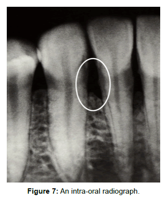
Figure 8:Edentulous female.
Edentulous Patients with OP frequently have a prognathic appearance. Other clinically noted signs are: - Thinning of the lips, a deepening of the naso-labial fold, increased deepening labial vertical lines, an increased columella/philtral angle, ptosis of peri-oral muscles, jaw-collapse with over-closure; on Radiograph there may be thinning of cortical plates and marked reduction of alveolar ridges (Figure 8).
Constructing and fitting satisfactory full prosthesis for these patients is extremely challenging. Often, placement of at least two or more stabilizing osseo-integrated implants, are needed. Bone grafts are often indicated to improve denture retention and/or allow for implant placement. Should the bone be so thin, fragile or atrophied, a permanent fixed splint, from the inferior mandibular border holding trans-osseous stabilizers, may be indicated for prostheses.
One major serious oro-dental side effect of Bisphosphonate Therapy (BPT) is the development of bisphosphonate osteo-necrosis of the Jaws (BONJ) [35-38]. BONJ is rare but serious complication, affecting less than 0.1% of cases treated with Bisphosphonate Therapy (BPT). Patients prone to developing BONJ may suffer from Paget disease, multiple bone myeloma, primary neoplasia of breast, prostate, liver, lung or kidney. BPT is also used for controlling hypercalcemia and metastatic bone lesions related to carcinoma. All these patients are candidates for developing BONJ, and require conservative non-invasive treatment.
Clinical presentation of BONJ: BONJ presents as exfoliating whitish, pale avascular bone. Pain is low level or absent. There’s peripheral gum inflammation and PSR III (probing depths >5.5 mms). Extraction sockets do not heal. Bony ridge protuberances and mylohyoid ridges are affected, particularly in edentulous jaws [35-37]. Non-vital bone (no feeling or bleeding) protrudes into the mouth and frequently loses its mucosal covering.
In cases with the aforementioned conditions, and on high doses of BPT, these patients are not ideal patients for major invasive periodontal or dento -alveolar surgery. Maxillo-facial, dento-alveolar or periodontal surgery may precipitate BONJ in these cases [35,37]. Yet people treated with BPT for osteogenesis imperfecta, with regular level BPT and can successfully receive most dental and gum treatments without BONJ complications. BPT persists in the system for years, and should extractions or major periodontal infections require treatment, these should be done and stabilized whenever possible before implementing anti-resorptive therapy by BPT. Sufferers of BONJ-prone cases require regular monitoring, anti-septic lavages and rinses (typically 0.02% Chlorhexidine is used) and antibiotic cover. Hospitalization is desirable to articulate a team of a physician, an oral-medicine specialist and a maxillo-facial surgeon. New invasive oro-dental therapies should be eschewed, but non-vital involucra must be removed to avoid developing osteomyelitis. Care of general nutrition is similar to management of patients suffering from cancer [38-40].
For diagnosis of OP, femoral neck measures and lumbar spine for low Bone Mass Density, specificity and positive predictive value are higher (over 82%) and more reliable than compared to only 73% with only Oral Panoramic radiography alone [14]. Tobacco smoking seriously aggravates OP. Smoking accelerates onset and progress of OP by its toxic effects but particularly by reducing blood flow to all organs, including bones. Smokers should quit as soon as possible. Nearly all the bisphosphonate drugs (not MiacalcinNS®, which is a calcitonin nostril spray) are taken orally (per os… po) over a long period of time. Didrocal® (Etidronate) is prescribed as 400mg po X 14days every 3months and CaCo3 as 500mg po, daily for 3 months. Costs vary for each drug, and recommended scripting, posology and delivery regimens should be followed. These drugs are effective with clinical improvements recorded over decades. Annual costs are much more affordable than hormones and consequently are widely successfully and frequently prescribed for osteoporosis [34]. Health care workers managing OP patients should have OP cases mouth fully checked out and stabilized, preferably by a dental specialist, before prescribing BPT. Dental health care workers need to be on the lookout for red-flags when treating OP patients.
Osteoporosis is a chronic insidious, debilitating disease of aging, slowly affecting the quality of life of both genders. Very few people show no signs of OP as they age. A healthy balanced diet, rich in calcium combined with daily rigorous exercise seems to be the sole natural formula for retarding OP. Traditional consumption of health liquids like water, soups or milk, is being replaced by increased drinking of soda-pop (USA teenagers up to 6 cans per day); and this popsubstitution for liquids, is contributing to increased prevalences of OP [41]. The aging process may be related to the limited number of times human cells, particularly the whole range of bone forming cells, both cartilaginous and intra-membranous, can divide before reflecting signs of break up or dysfunction.
This from the Hayflick phenomenon or limit, which is determined by the number of times a cell will multiply through mitosis, until it undergoes a programmed cell death. Telomeres loose small portions as cells multiply, till the dividing cell DNA becomes dysfunctional [42]. Age inexorably affects the entire body’s metabolism, and the skeleton is not omitted from this process. The skull seems less affected by OP than the vertebrae, hips or long bones. Possibly more bone metabolic factors will be discovered to restore healthy bone function. Falls by the elderly remains the most prevalent cause of bone morbidity. Oral health may be affected by OP; regular home and professional maintenance will ensure functional dentate status well into the last light of twilight years. Edentulous oral cripples have a wide range of successful progressive options to replace mastication with restorative dentistry. Medications help to avoid and reduce bone fractures but do not totally stop, nor totally cure, existing OP. Constant vigil, monitoring, adjusting diets, physical habits and medication, are all needed to live with the reality of OP. Osteonecrosis of the Jaws (ONJ) is a rare complication and precise management is unresolved; management is by prevention of ONJ, is feasible mainly by completing all invasive dentistry before prescribing bisphosphonates. Developed ONJ is treated by resecting dead bone and concomitant stringent oral hygiene.
Note: Cost is estimated in Can$ for one year’s supply*
Evista® (raloxifene HCI) is a registered trademark of Eli Lilly Company. 60mg, po.od…. Cost ~$706
Miacalcin® (calcitonin salmon) Nasal Spray is a registered trademark of Novartis Pharmaceuticals Corporation. 1 Spray 200 I.U., alternate nostrils daily………………………………………….. Cost$740
Fosamax® (alendronate sodium tablets) is a registered trademark of Merck & Company, Inc., 10 mg, po, od,…………………………… cost $460; or 70mg po qwk……………………………………$640
Actonel® (risedronate) 5mg po od………….Cost $622; or 35mg po qwk…………………Cost$473
Forteo® (Teraparitide) 20 mg sc od…………………………………… ……………….. Cost $9600
Didrocal® (Etidronate) 400mg x 14 days q 3months, & CaCo3 500mg X 76 q 3 mths……Cost$170
Femigel® (17-β Estradiol) 2.5G qd . Contains 1.5 mgm. Loboratoir Besins Intnl France; dispensed as dermal gel in dispenser. Each plunge = 1.25 gel. ………………………………………………Cost $400
od=every Day; po= by mouth; qwk=every week; i.u.=International Unit; q= every; sc= sub cutaneously; ; od= every day;
*Dispensing fees are not included in all costs cited here: this may be between $40 and $60 extra.