Journal of Clinical & Experimental Dermatology Research
Open Access
ISSN: 2155-9554
ISSN: 2155-9554
Case Report - (2019)Volume 10, Issue 3
79 year old lady with newly diagnosed lung adenocarcinoma which was felt to be operable. She underwent a PET/CT which showed localised disease in the lung, but also highlighted several cutaneous lesions. She was referred to dermatology for diagnosis and biopsy to exclude cutaneous deposits of tumour. There was a past history of a previous lung adenocarcinoma excised in 2015. Examination showed several wart-like, papillomatous lesions to the skin, not typical of secondary deposits of tumour. Shave biopsy of a representative lesion was taken and histology showed a benign squamous papilloma. At review in oncology, the remaining lesions had completely resolved a few weeks later. There have been case reports of benign lesions showing unexpected FDG uptake. This case highlights the phenomenon and the need to obtain timely tissue diagnosis to prevent potentially life-saving treatment such as surgery in this case.
Cloning, Stem cells, Treatment, Moral problems
The development of biology, genetics, medicine, pharmacy at the end of XX century, bring along cloning of mammals, made the map of the human genome, genetic investigation and gene therapy reach new successes, assisted reproduction to become available procedure
History
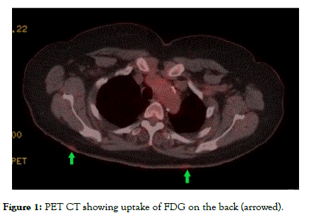
A 79 year old lady with a history of non-Hodgkin’s Lymphoma and lung adenocarcinoma that had been successfully resected three years previously was diagnosed with a new sub pleural mass during ongoing surveillance via CT scan. She underwent a CT guided biopsy. This confirmed the presence of a second lung adenocarcinoma. Due to its location, this was felt to be resectable, thereby offering a potentially curative treatment. To ensure that there were no undetected metastases, whole body PET scanning was carried out (Figure 1). The scan report stated, ‘Metabolically active enlarging lesions are concerning for broncho-alveolar carcinoma. Compared with the previous PET/ CTs, there are several new small foci of cutaneous uptake: bilaterally on the back at the level of the scapulae (T3 vertebra), in the right axilla and in the midline anterior abdomen as well as the left thigh’. It was felt that these areas of cutaneous uptake could represent cutaneous metastases which would have given a much poorer prognosis and would have meant that surgical treatment was no longer indicated.

Figure 1. PET CT showing uptake of FDG on the back (arrowed).
The lady was referred urgently to dermatology for review and biopsy.
When reviewed in the dermatology clinic, the lady reported that several of the lesions developed suddenly at the same time and that new ones were still coming. Other than their unsightly appearance, the only symptom was associated pruritus.
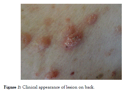
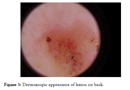
Skin examination showed exophytic, pink lesions to the upper thighs, abdomen and back, in the areas which corresponded to increased FDG uptake on the PET CT. Some lesions had superficial finger like projections with significant keratinisation, while others were flatter and had a smoother surface. The lesions each measured 5-10 mm in diameter (Figure 2). Dermoscopy showed a papillomatous surface and no signs to make us suspicious of malignancy (Figure 3). These features were in keeping with benign papillomas. A representative lesion was excised for histology.

Figure 2. Clinical appearance of lesion on back.

Figure 3. Dermoscopic appearance of lesion on back.
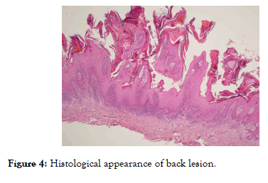
Histology was reported as a benign papillomatous squamoproliferative lesion without significant atypia. No overt features of viral aetiology were seen and there was no malignancy (Figure 4). There were no features of adenocarcinoma. It was therefore concluded that these lesions were not related to the lung tumour.

Figure 4. Histological appearance of back lesion.
Given this diagnosis, the lady was reviewed in oncology, but unfortunately she was not suitable for resection as a further nodule within the lung had been identified. Interestingly, at oncology review, many of the papillomas showed signs of regression and some of them had completely disappeared.
Benign Squamous Papillomas are relatively common lesions which arise from the stratified squamous epithelium of the epidermis these may be associated with one of the many Human Papilloma Viruses most frequently HPV-6 and HPV-11. These subtypes are not associated with cutaneous malignancy. They are diagnosed most frequently in the 4th and 5th decades of life. They may affect both skin and mucous membranes.
It is not an infrequent occurrence that in cases of multiple squamous papillomas, where one is excised, others may regress as the host immune system detects viral antigens and mounts an immune response.
Squamous papillomas may be left untreated as they have no malignant potential, but patients often seek surgical intervention for associated symptoms of itch, pain and inflammation.
Fluorodeoxyglucose Positron Emission Tomography (FDG PET) is useful in the evaluation and monitoring of several tumour types due to its high sensitivity and specificity. The procedure is minimally invasive and is therefore well tolerated by patients. Fluorodeoxyglucose is a radiopharmaceutical which emits positrons, detectable on PET scanning. The detection of FDG uptake signifies the uptake of glucose by tissue and correlates well with metabolic activity. Increased metabolic activity is seen in tumour cells, compared to background tissues. This makes PET-CT a valuable tool in staging and restaging different tumours when assessing disease progression or the efficacy of treatment. However, increased FDG uptake is not seen exclusively in malignant cells; this can also be seen in inflammatory processes [1].
False positive results due to various cutaneous aetiologies have been reported, including stomas, rhinophyma and tophaceous gout [2]. A case report in 2015 showed unexpected uptake in a seborrheic keratosis in another lung cancer case [3].
PET-FDG is also useful in the diagnosis of skin metastasis from solid tumours. Cutaneous metastases most frequently arise from the breast, large bowel and lung, with a frequency of up to 30% in patients with metastatic breast cancer [4]. The frequency of lung cancer skin metastasis has been shown to be 2.8% in a retrospective study [5].
This case highlights an uncommon uptake of FDG by benign squamous papillomas as an incidental finding which would have had significant implications had the lesions proven to be metastases from the primary lung tumour. We postulate that the increased FDG uptake as detected on the PET-CT was due to the rapidly developing lesions which were growing rapidly, thus were more metabolically active. It is essential that clinicoradiological correlation is carried out in a timely manner in cases of increased cutaneous uptake of FDG when staging tumours, so as to not delay potentially curative treatments.
Citation: Capewell N, Ford A, Diep P (2019) Multiple Benign Papillomas with Unexpected Uptake on PET Scanning. J Clin Exp Dermatol Res 10:494. doi: 10.35248/2155-9554.10.494
Received: 28-Mar-2019 Accepted: 05-Apr-2019 Published: 12-Apr-2019 , DOI: 10.35248/2155-9554.10.494
Copyright: © 2019 Capewell et al. This is an open-access article distributed under the terms of the Creative Commons Attribution License, which permits unrestricted use, distribution, and reproduction in any medium, provided the original author and source are credited.