jct
Open Access
ISSN: 2161-0495
ISSN: 2161-0495
Case Report - (2025)Volume 15, Issue 6
Introduction: We present the case of a 42-year-old male who arrived obtunded to our Emergency Department (ED) following an intentional ingestion of approximately 60 grams of extended-release Valproic Acid (VPA). Subsequent imaging and endoscopy confirmed the presence of a pharmacobezoar, a rare and underreported complication of overdose, and his course was further distinguished by the development of delayed neuromuscular toxicity. Methodology: Clinical, laboratory, imaging, endoscopic and neurophysiologic data were abstracted from the electronic medical record. Case Report: The patient was managed with early airway protection, gastrointestinal decontamination initially with single-dose and later with multiple-dose activated charcoal regimens. He received Intravenous (IV) Levocarnitine (L-carnitine), naloxone and Continuous Renal Replacement Therapy (CRRT). His hospital course was initially stable but later complicated by delayed neuromuscular toxicity with extrapyramidal features requiring prolonged inpatient rehabilitation and multidisciplinary follow-up including toxicology, intensive care, psychiatry, internal medicine and neurology teams. Discussion: This case highlights the clinical challenge of massive VPA overdose complicated by pharmacobezoar formation and delayed neuromuscular sequelae. Early risk assessment led to recognition and aggressive treatment of a potentially life-threatening ingestion in which airway protection, serial monitoring, endoscopic confirmation and extracorporeal elimination were crucial to a favourable outcome. Literature review shows that while delayed absorption and toxicity are well described in sustained-release formulations, few reports have confirmed bezoar formation endoscopically and neuromuscular toxicity after acute overdose remains rare. The combination of an endoscopically confirmed pharmacobezoar with subsequent neurological sequelae makes this case distinctive and adds to the growing literature. Conclusion: This case emphasises the importance of anticipating pharmacobezoar formation in sustainedrelease VPA overdoses and recognising that delayed neuromuscular syndromes may emerge even after initial stabilisation. Clinicians should consider early toxicology involvement, advanced imaging and endoscopy in such cases. Aggressive gastrointestinal decontamination, L-carnitine therapy and timely extracorporeal removal remain mainstays of care. Ultimately, ongoing vigilance and coordinated multidisciplinary follow-up are essential.
Valproic acid; Drug overdose; Pharmacobezoar; Neuromuscular toxicity; L-carnitine; Renal replacement therapy; Hyperammonaemia
VPA, a branched short-chain fatty acid first synthesised in 1882, was originally used as an inert organic solvent. Its anticonvulsant properties were discovered incidentally in 1963, when it was found to suppress chemically induced seizures in rodents. Since then, VPA has become a widely used antiepileptic and mood-stabilising agent, also indicated for migraine prophylaxis. Its therapeutic effects are primarily mediated through enhancement of γ-aminobutyric acid (GABA) activity, inhibition of GABA degradation, and blockade of voltage-gated sodium, calcium and potassium channels [1].
In overdose, however, VPA can cause life-threatening complications including severe Central Nervous System (CNS) depression, hepatotoxicity, hyperammonaemia and cerebral oedema. Although most cases are managed with supportive care, select patients with severe toxicity may benefit from extracorporeal elimination techniques or pharmacological interventions such L-carnitine or meropenem [2]. This report describes an unusual case complicated by endoscopically confirmed pharmacobezoar formation and delayed neuromuscular toxicity, features rarely reported in acute VPA overdose and of value to clinicians managing severe poisoning. In addition to describing this case, we provide a review of previously published reports, presented in supplementary Table 1.
A 42-year-old male with bipolar affective disorder and hypertension, presented to the ED 90 minutes after an intentional overdose of extended-release VPA totalling 60 g (120 mg x 500 mg tablets) with co-ingestion of Zolpidem 110 mg and Paracetamol 2 g. He had been discharged the previous day following a 14-day psychiatric admission without suicidal ideation. He was found drowsy on the floor surrounded by empty medication bottles; earlier that day, he had received an unspecified intramuscular injection for back pain. This was his first known overdose attempt with no history of substance misuse or prior suicidal attempts.
On arrival, he was hypotensive with a blood pressure of 85/55 mmHg and a Glasgow Coma Scale (GCS) score of 10 (Eye opening 2, verbal response 5, motor response 3). Oxygen saturation was initially normal but dropped during Computed Tomography (CT) imaging, and with snoring respirations, he was immediately intubated for depressed consciousness and persistent hypoxia. The remainder of the examination was unremarkable.
Initial laboratories showed VPA>450 μg/mL, ammonia 76 μmol/L and acetaminophen level<5.0 μg/mL. Toxicology was consulted immediately, and post-intubation gastrointestinal decontamination was initiated with 50 g of activated charcoal followed by Multiple-Dose Activated Charcoal (MDAC) every 4 hours. He received Naloxone 0.4 mg IV and L-carnitine (100 mg/kg IV loading dose over 30 minutes, then a maintenance dose of 50 mg/kg every 8 hours).
Chest radiograph revealed bilateral basal pneumonitis and pleural effusions. CT brain was unremarkable. Abdominal radiograph showed no radio-opaque foreign bodies; however, a non-contrast CT abdomen/pelvis revealed multiple small, radiolucent foreign bodies within the stomach (primarily fundus/pylorus), with similar material scattered throughout the small bowel and ascending colon without bowel obstruction or perforation (Figure 1).
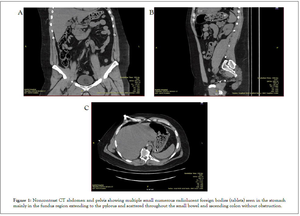
Figure 1: Noncontrast CT abdomen and pelvis showing multiple small numerous radiolucent foreign bodies (tablets) seen in the stomach mainly in the fundus region extending to the pylorus and scattered throughout the small bowel and ascending colon without obstruction.
The gastroenterology team was consulted, and upper endoscopy confirmed a large 30-cm clumped mass of VPA tablets confirming a pharmacobezoar; gastric mucosa was charcoal-stained and the duodenum appeared diffusely erythematous, with fragmented tablet material present (Figure 2).
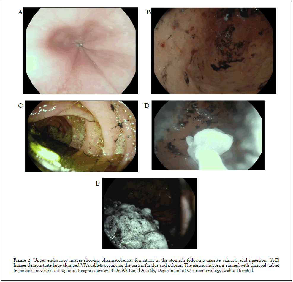
Figure 2: Upper endoscopy images showing pharmacobezoar formation in the stomach following massive valproic acid ingestion. (A-E) Images demonstrate large clumped VPA tablets occupying the gastric fundus and pylorus. The gastric mucosa is stained with charcoal; tablet fragments are visible throughout. Images courtesy of Dr. Ali Emad Alzaidy, Department of Gastroenterology, Rashid Hospital.
He was admitted to the ICU for CRRT, and serial monitoring of VPA and ammonia levels. Over 72 hours, VPA decreased to <2.8 μg/mL and ammonia gradually normalised (Figure 3). Liver Function Tests (LFT) and Urea/Electrolytes (U and E) remained unremarkable. He was extubated on Day 5 and transferred to the ward on Day 11 in stable condition.
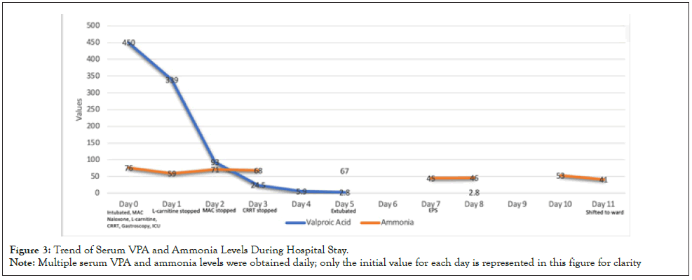
Figure 3: Trend of Serum VPA and Ammonia Levels During Hospital Stay.
Note: Multiple serum VPA and ammonia levels were obtained daily; only the initial value for each day is represented in this figure for clarity
Throughout admission, the patient was co-managed by intensive care, toxicology, psychiatry, internal medicine and neurology teams. Neurologic complications evolved during admission. He developed bilateral upper and lower limb weakness, rigidity, tremor, rotatory nystagmus and clonus. Electroencephalogram (EEG) was normal; initial nerve conduction was unremarkable, but repeat testing showed ulnar sensory neuropathy. Electromyography (EMG) of the vastus medialis and deltoid demonstrated myopathic features with profound active denervation. Magnetic Resonance Imaging (MRI) brain with contrast was normal. A diagnosis of toxic myopathy with extrapyramidal features was made, and Procyclidine was initiated. He was transferred to inpatient rehabilitation at another facility and discharged ambulatory after 54 days.
VPA is a broad-spectrum antiepileptic and mood-stabilising drug. In overdose, it disrupts mitochondrial metabolism, depleting Acetyl Coenzyme A (CoA) and carnitine stores, and shifts fatty acid metabolism from β-oxidation to ω-oxidation. This leads to the accumulation of toxic metabolites such as 2-propyl-4-pentenoic acid (4-en-VPA), which impair hepatic function and inhibit carbamoyl phosphate synthetase I (CPS-I), contributing to complications including CNS depression, hyperammonaemia, and hepatotoxicity [1,3-5].
Management strategies are guided by consensus recommendations. The 2008 American Association of Poison Control Centers (AAPCC) expert consensus guideline recommends referral from poison centers to the ED for patients with suicidal intent, symptomatic ingestions or asymptomatic ingestions >50 mg/kg; it discourages ipecac, supports activated charcoal within the preceding hour, and considers naloxone for patients with respiratory depression [6]. Our patient received airway protection, Gastrointestinal (GI) decontamination, and L-carnitine consistent with these recommendations. Although extracorporeal elimination was not addressed in the 2008 AAPCC guideline, its role has been strongly supported by the Extracorporeal Treatment (EXTRIP) Workgroup (2015), which provides structured recommendations for initiating renal replacement therapy in severe VPA poisoning. These include VPA concentration >1300 mg/L, cerebral oedema, or shock. ECTR is also suggested if the VPA concentration exceeds 900 mg/L, or if the patient has coma, respiratory depression requiring mechanical ventilation, acute hyperammonaemia, or pH ≤ 7.10 [7]. Our patient fulfilled EXTRIP criteria with CNS depression and ventilatory support and underwent 72 hours of CRRT with subsequent clinical improvement and a reduction in VPA levels.
Literature from Al Aly Z, Thanacoody RHK and Tichelbäcker T, et al., further reinforces the use of extracorporeal therapies like CRRT or haemodialysis in cases with elevated VPA levels >850 mg/L or clinical features of severe VPA poisoning such as coma, haemodynamic instability, severe hyperammonaemia and acid base disturbances [8-10]. Our patient’s presentation mirrored many of the previously published severe VPA overdose cases: CNS depression, hypotension, hyperammonaemia, and a VPA level >450 μg/mL. His treatment included early risk assessment, airway protection, MDAC, L-carnitine, and CRRT which was consistent with existing literature, including approaches described by Al Jawder S, et al. and Chan, et al. Al Jawder S, et al. similarly documented MDAC in a patient who received four 50 g doses of charcoal with good clinical response [11-12].
Our case adds two unique findings to the literature. First, while delayed clearance is often described in sustained-release VPA ingestions, limited case reports have confirmed bezoar formation endoscopically [3,13]. Representative case reports are summarised in supplementary Table 1 with full citations provided in the supplementary references. Some case reports described delayed peak levels but did not confirm presence of bezoars. In our patient, a 30-cm bezoar of fragmented tablets was directly visualized, confirming a significant source of ongoing absorption and delayed clearance. In a study by Ingels M, et al., 26 patients with initial VPA levels<100 μg/mL went on to develop significant toxicity, with some peaking more than 8 hours later [14]. This supports the for serial monitoring and imaging in suspected massive ingestion of sustained-release formulations.
Second, the patient developed delayed neuromuscular toxicity with extrapyramidal features. Bilateral upper and lower limb weakness, tremor, rigidity, clonus and nystagmus evolved during admission and EMG confirmed toxic myopathy with denervation. While neuromuscular complications such as myopathy and parkinsonism are documented in chronic VPA therapy, they are rare in the context of acute overdose and were attributed to low carnitine levels [15,16]. Mittal SH, et al. reported myopathy within days of initiating sodium valproate as a therapeutic measure for epilepsy, even in the absence of overdose, reinforcing the hypothesis that carnitine deficiency is a contributing factor [17].
The neurologic picture prompted consideration of critical illness myopathy, extrapyramidal side effects, functional neurologic disorder, and demyelination. EMG findings, clinical timing, and absence of confounding medications supported VPA-induced toxic myopathy.
VPA depletes carnitine stores through various mechanisms including excretion of valproylcarnitine, endogenous carnitine synthesis reduction, and inhibition of the membrane carnitine transporter. The resulting mitochondrial dysfunction reduces β-oxidation and impairs the urea cycle, leading to hyperammonaemia. L-carnitine is thought to decrease ammonia levels by restoring the β-oxidation process [3,18]. A Quantitative Systems Pharmacology (QSP) model by Schiavo A, et al., showed that even when started 8 hours after ingestion, a regimen of 6 g of L-carnitine followed by 1 g every four hours effectively reduced high ammonia levels [19]. A randomised controlled trial by Èincu RC, et al. similarly showed improved clinical outcomes and recovery with L-carnitine [20]. However, in a murine model, Jamshidzadeh A, et al. found that while carnitine reduced oxidative stress, it did not prevent liver injury or encephalopathy [21]. Pagali S, et al. reported hyperammonaemia rebound after discontinuation of L-Carnitine, supporting continued therapy for at least 72 hours [22]. Though high-flux HD is more efficient, CRRT can still be used as an alternative and has been used successfully in many case reports [3,7,12,23]. HD has been shown to reduce VPA half-life from approximately 30 hours to 2-3 hours in optimal settings [10].
Carbapenems such as meropenem have been used off-label to enhance VPA clearance by inhibiting acylpeptide hydrolase [24]. Al-Quteimat O, et al. reported rapid VPA reductions but also noted prolonged suppression and seizure recurrence in over 50% of patients so they advised the use of an antiepileptic drug as an adjunct with carbapenem [25]. While not used in our case, carbapenems may be considered in refractory or resource-limited settings.
Limitations of this case include the initially negative abdominal radiograph, which did not reveal any radio-opaque foreign bodies or bezoars. This reflects the radiolucency of VPA tablets and highlights the risk of delayed recognition, as plain films may be falsely reassuring in cases of massive ingestion. Given the high clinical suspicion and substantial ingested dose, early toxicology consultation prompted a CT scan of the abdomen and pelvis which confirmed multiple radiolucent foreign bodies. Additional challenges included the incomplete initial history and reliance on collateral information from family members to guide management.
This case is distinct in documenting both an endoscopically confirmed pharmacobezoar and delayed neuromuscular toxicity following acute massive VPA overdose, features that remain rarely reported in the literature. Clinicians should maintain a high index of suspicion in massive overdoses, as VPA toxicity may result in prolonged absorption, persistent toxicity and serious complications requiring aggressive interventions. Timely diagnosis and early management including airway management, activated charcoal, L-carnitine, CRRT and supportive care were essential to patient survival and contributed to a favorable outcome. Imaging and endoscopy should be considered in sustained-release overdoses or when delayed gastrointestinal clearance is suspected. Finally, this case underscores the vital role of the toxicology service for oncall consultation, risk stratification, and guidance on advanced interventions and the importance of coordinated multidisciplinary care in managing such complex toxicologic emergencies.
Informed consent was granted for the case details to be published from the patient’s legally authorised representative (spouse) during the period of incapacity. The case report contains no direct identifiers.
We thank Dr. Ali Emad Alzaidy (Department of Gastroenterology, Rashid Hospital) for provision of endoscopy images. We also acknowledge the contributions of the intensive care, toxicology, internal medicine, psychiatry, and neurology teams for coordinated multidisciplinary care.
No external funding was received for this work.
The authors declare no conflicts of interest.
[PubMed]
Citation: Alrazooqi M, Abumuaileq L, Zia S, Kazim SN (2025). Massive Valproic Acid Overdose with Confirmed Pharmacobezoar Formation and Neuromuscular Toxicity: A Case Report. J Clin Toxicol.15:593.
Received: 01-Oct-2025, Manuscript No. JCT-25-38802; Editor assigned: 03-Oct-2025, Pre QC No. JCT-25-38802 (PQ); Reviewed: 17-Oct-2025, QC No. JCT-25-38802; Revised: 24-Oct-2025, Manuscript No. JCT-25-38802 (R); Published: 31-Oct-2025 , DOI: 10.35248/2475-3181.25.15.612
Copyright: © 2025 Alrazooqi M, et al. This is an open-access article distributed under the terms of the Creative Commons Attribution License, which permits unrestricted use, distribution and reproduction in any medium, provided the original author and source are credited.