Journal of Plant Biochemistry & Physiology
Open Access
ISSN: 2329-9029
ISSN: 2329-9029
Research Article - (2017) Volume 5, Issue 4
Mimusops elengi is an ornamental plant, famous for its fragrant flowers. Its bark and fruit are astringent in nature. The different parts of the plants are frequently used in medicine. The extracts of fruits, bark and leaves were studied presently. The extract of fruit and bark confirmed the presence of both hydrolysable and condensed tannins. The crude and purified fractions of the fruit and bark was subjected to the antioxidant and antifungal activity by using simple Thiocyanate and agar diffusion method respectively. The tannins present in the fruit especially shows marvelous antioxidant activity which was not reported before.
Keywords: Ornamental plant; Tannins; Antioxidant; Antifungal
The genus Mimusops elengi belongs to the family Sapotaceae and comprises of thirty species which are distributed in the tropical parts of hemispheres. Of these Mimusops elengi , commonly known as mulsari or bakul cultivated in gardens due to its scented flowers is indigenous to the subcontinent. The plant has been studied through many years phytochemically.
The seed Kernels from Mimusops elengi have been investigated previously by Boorsma in 1902 who found 21% fatty oil and 2% saponins. The bark mainly contains saponins and tannins [1-10]. The leaves contain steroids. The pulp of the fruit contains mainly sugars and saponins. While the flowers contain volatile oil. The parts of its mostly used in medicines [11-18]. Bark is tonic and febrifuge. Unripe fruit is a useful masticator and therefore recommended to be chewed for fixing loose teeth. Pulp of ripe fruit is eaten as diet in diarrhea and is used in snake bite. Fruits and flowers are used to prepare a lotion for wounds and ulcers. The juice of the bark and unripe fruit is used by dyers to fix colors. Bark increases fertility in women [19-23].
Tannins are amorphous, rarely crystalline substance which are widely distributed in the vegetable kingdom, water soluble phenolic compounds having molecular weights between 500-3000. They have ability to form colored solutions and precipitates with iron and other metals. Hydrolysable tannins are esters of hydroxyl aromatic carboxylic acid, they are hydrolysable by the action of acids or enzymes [24-34]. They are subdivided into Gallotannins and Ellagitannins. On hydrolysis they give Gallic acid and ellagic acid. Condensed tannins are not esters. They are similar to anthocyanins and derivatives of flavonol, so these are called as Flavotannins or catechol tannins. Tannins are used in food and beverages industry in agriculture, nutrition and in herbal medicine. They are used in dyes, in the treatment of cancer and aids, used in creames and lotions as inflammatory agents [34-46]. Some tannins are strong antioxidant.
Different parts (fruit, bark, and leaves) of Mimusops elengi were collected and extracted with acetone-water (70:30) and crude extract was subjected to antioxidant activity.
Method
The fruit was collected, dried and grinded. Soaked in 2.5 L acetonewater (70:30) in dark bottle for four days. The extract filtered and evaporated. The crude aqueous extract was diluted by adding distilled water (50 ml) and petroleum ether (200 ml). The organic layer is separated and evaporated. Then extracted with ethyl acetate and n- Butanol. The organic layer was separated and evaporated. The same procedure is repeated with Bark and leaves extract. The crude aqueous extract of fruit, bark and leaves was subjected to antioxidant and antifungal activity.
Isolation and purification
Glass column is loaded with each organic layer after evaporation and run with increase in polarity pet. Ether. Pet. Ether-chloroform, chloroform, chloroform-acetone, Acetone, Acetone-methanol. Collected the different fractions, evaporated and dried. Performed different chemical test on them like FeCl3 test, NaNO2 test, and nbutanol- HCl test for Tannins, Ellagi Tannins and Condensed tannins respectively. Purification of these tannins is done by recrystallization. Confirmation of the product is done by Infra-red and UV spectroscopy.
Thiocyanate method
0.1 ml aqueous extract of fruit was taken in a conical flask. Added 0.9 ml chloroform. 10 ml absolute alcohol, 0.13 ml linoleic acid and 10 ml of acetate buffer (acetic acid and sodium acetate). The total volume was adjusted to 25 ml by adding distilled water. The mixed solution was kept in an incubator (Gallen Kamp) at 40°C. The same procedure is repeated with bark and leaves. At interval of 48 hours, the peroxide value was determined by using thiocyanate as a coloring agent. Took 1 ml of incubated material in conical flask. Added 2 ml of 0.1 M Mohr’salt. Shake it well and left for five minutes. Then added 5 ml of 20% sodium thiocyanate solution and 3 ml of 4 M HNO3 solution. Adjusted the volume up to 25 ml and then filtered. Noted the absorbance at 480 nm of the colored solution with UV spectrophotometer. The same procedure was repeated with other fractions.
Agar diffusion method
A strain of Aspergillus nigar obtained from Biochemistry department of GCU was used as a test organism to test the activity of the extract. The organism was maintained on slants having PDA (potato dextrose agar) medium (Table 1).
| Potato extract | 1 dmg |
|---|---|
| D-glucose | 20 g |
| Agar-Agar | 50 g |
Table 1: Medium preparation.
200 g sliced potato was boiled in 1 l water and filtered through fine paper. After inoculation the slants were incubated at 30°C for 72 hours. For the determination of antifungal activity, the same PDA medium was used in Petri dishes. Both the medium and plates were sterilized separately by autoclaving at 121°C for 15 minutes and by hot air sterilization at 160°C for 2 hours respectively. 20 ml of the sterilized petridishes aseptically. It was allowed to solidify then 4 ml of spore suspension of Aspergillus nigar prepared in PDA medium was poured over the first layer. After setting the second layer four pores were made in Agar plate by sterilized (red heat sterilization) stainless steel borer of 0.8 cm diameter. Suspension of the extract was made in 5% gum accatia solution two opposite pores in a plate were filled by 0.12 ml of sample and its 50% dilution. Plates were incubated at 30°C for 24 hours. Zones of inhibition were measured and reported, Antifungal activity of a sample is proportional to diameter of the zone.
The results obtained in this research showed that major amount of tannins contain gallic acid in their nucleus which was isolated by alkaline hydrolysis of fruit and bark aqueous extract. Thus, the hydrolysable tannins present in the fruit and bark are gallotannins rather than ellagitannins. Purification of these tannins is done by column chromatography (Table 2-4).
| Chemical Test | Fruit extract (water: acetone) |
Bark extract (water: acetone) |
Leaves extract (water: acetone) |
|---|---|---|---|
| FeCl3 test | Blue color | Blue color | No coloration |
| K2CO3 | Red ppt | Brown ppt | |
| Br2-water test | Yellow ppt | Brown ppt | No coloration |
| NaNO2 test | Red color | Red color | |
| n-butanol-HCl test | Red color | Red color | |
| Gelatin test | Brown ppt | Brown ppt | |
| pH test | 5.0 | 3.9 | 2.8 |
Table 2: Detection of tannins.
| Chemical Test | Fruit extract (n-butanol) |
Bark extract (n-butanol) |
Confirmation |
|---|---|---|---|
| FeCl3 test | Deep blue color | Blackish green color | Tannins present |
| n-butanol-HCl test | Red color | Red color | Condensed tannins |
| NaNO2 test | No coloration | No coloration | Ellagi Tannins absent |
Table 3: Detection of tannins.
| Part of the plant | Dry weight | Amount of Condensed Tannins | Amount of Hydrolysable Tannins | Total% |
|---|---|---|---|---|
| Fruit | 260g | 1.6g | 0.61g | |
| Bark | 204g | 0.5g | 0.2g | 0.34g |
| Leaves | 237g |
Table 4: Percentages of tannins in different parts of the plant.
Thiocyanate method was used for the determination of antioxidant activity. The graph was plotted between absorbance and time for each fraction. Tocopherol was used as a reference. The results of graphs showed that aqueous extract of fruit and bark shown significant antioxidant activity and these results are comparable with the activity of tocopherol. Chemically, phenols constitute the largest and most important class of antioxidants. The best fraction result is only given in the Table 5 and plotted in Figure 1. The results were in accordance with the results of the chemical analysis of extract of tannins. Therefore, it can be concluded that higher antioxidant activity of fruit and bark is due to the presence of tannins.
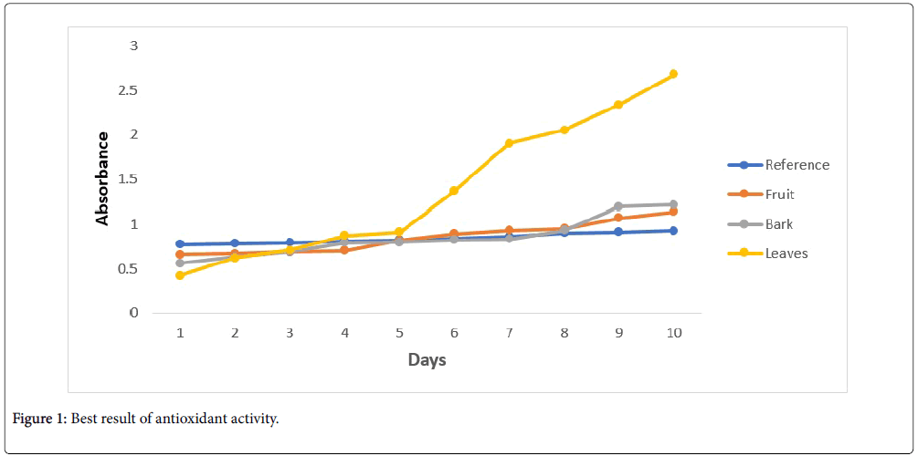
Figure 1: Best result of antioxidant activity.
| Absorbance at λ 480nm | ||||
|---|---|---|---|---|
| Days | Fruit (fraction no.3) |
Bark (fraction no.4) |
Leaves (fraction no.9) |
Tocopherol (references) |
| 2 | 0.661 | 0.563 | 0.421 | 0.771 |
| 4 | 0.671 | 0.629 | 0.621 | 0.781 |
| 6 | 0.689 | 0.690 | 0.707 | 0.790 |
| 8 | 0.705 | 0.790 | 0.870 | 0.810 |
| 10 | 0.818 | 0.798 | 0.913 | 0.820 |
| 12 | 0.889 | 0.820 | 1.362 | 0.840 |
| 14 | 0.931 | 0.831 | 1.901 | 0.861 |
| 16 | 0.95 | 0.931 | 2.051 | 0.901 |
| 18 | 1.07 | 1.191 | 2.333 | 0.910 |
| 20 | 1.135 | 1.210 | 2.673 | 0.923 |
Table 5: Results of antioxidant activity.
Agar diffusion method was used for the determination of antifungal activity. The results showed that bark and fruit shows the higher antifungal activity which is proportional to the diameter of the zone (Table 6).
| Part of the plant | 0.5% | 1% |
|---|---|---|
| Fruit | 11mm | 12mm |
| Bark | 12mm | 11mm |
| Leaves | No zone | No zone |
Table 6: Results of antifungal activity.
The research was carried out on the extract of different parts of Mimusops elengi indicated the presence of tannins in the fruit and bark. The experiments showed that the fruit and bark contain both hydrolysable and condensed tannins. Hydrolysable tannins contain gallic acid in their nucleus, ellagi tannins are absent. The analysis of fruit extract confirmed the presence of condensed tannins, which can be correlated with astringency of the fruit. The antioxidant assay was also carried out on different extracts and fractions. The results were promising and found to be comparable with the results of tocopherol. Therefore, it can be concluded that extracts of fruit and bark of the plants can be used as herbal antioxidant.