Journal of Sleep Disorders & Therapy
Open Access
ISSN: 2167-0277
ISSN: 2167-0277
Commentary - (2014) Volume 3, Issue 5
Hypnic jerks are spontaneous sudden jerking movements most often occurring just prior to falling asleep [1]. They are considered non-periodic and myoclonic in behaviour and are similar to fasciculation with polysomnography. They are more probable with fatigue and sleep deprivation [2,3]. These sudden movements are common but to date still not well understood. Approximately seventy percent of the adult population experience these movements at some time in their life. Mitchell is credited with the first cited description of these movements [4] and over the years, EEG and polysomnography have recorded the event, but even with these technological advances we arestill without a clear understanding of their motor pattern, physiological nature, and causative mechanism [5].
This reflexive phenomenon has many synonyms including: hypnic reflex, hypnagogic jerk, sleep start, sleep twitch and night start. Montagna used the term ‘physiological hypnic reflex’ and have found them to also occur during relaxed wakefulness [6] and to be increased during stage one sleep--especially in REM [7]. Others have described this phenomenon and coined the action ‘propriospinal myoclonus’ because of a suspected spinal origin [8,9]. The reflex is important to understand neurologically so we can differentiate between pathological and non-pathological origins. Many general theories have been proposed as to why this type of jerk occurs but none have made much sense. Here, I propose a simple explanation involving the quick stretch of spinal tissues relating the recovery changes of the intervertebral discs.
To give the reader a basic understanding of the spinal anatomy of interest, each vertebra (except C1-2) is separated by a hydraulic intervertebral disc. The central aspect (nucleus pulposus) consists mainly of water encased by several alternating fibrous angled sheets (anulus fibrosus). The nucleus pulposus contains hydrophilic proteoglycans which function to maintain intervertebral volumetric spacing and fundamentally work to resist compression. But variation to this volume occurs over a diurnal cycle with intervertebral discs losing approximately 25% of their volume [10] to be regained with recumbency and sleep (Figure 1).
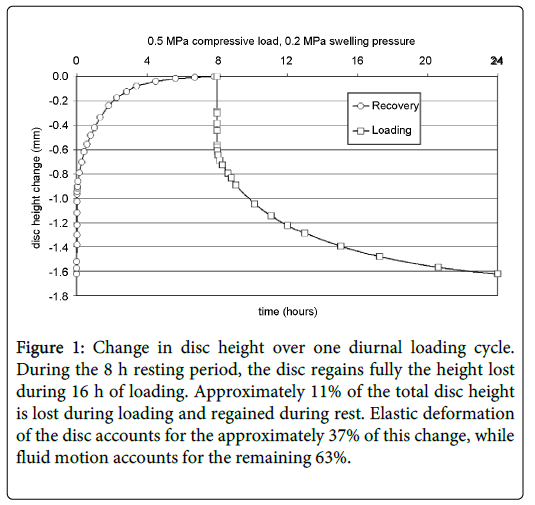
Figure 1: Change in disc height over one diurnal loading cycle. During the 8 h resting period, the disc regains fully the height lost during 16 h of loading. Approximately 11% of the total disc height is lost during loading and regained during rest. Elastic deformation of the disc accounts for the approximately 37% of this change, while fluid motion accounts for the remaining 63%.
Early in the unloading phase, the recovery rate is steepest. That is, when relaxation first occurs upon falling asleep, the intervertebral discs will begin to increase height relatively quickly. Pressure within the intervertebral discs assist with this expansion recovery by pushing outwardly against each opposing vertebra. These intradiscal pressure readings have been shown to be 0.10 MPa while lying supine and 0.5 MPa with relaxed standing. Over the course of the night, these pressures within the discs gradually increase from 0.10 to 0.24 MPa [11].
It is proposed here that as major spinal muscles relax, the rate of muscle lengthening is eventually quick enough to stimulate muscle spindles, eliciting a stretch reflex, similar in nature to the monosynaptic knee jerk reflex (Figure 2).
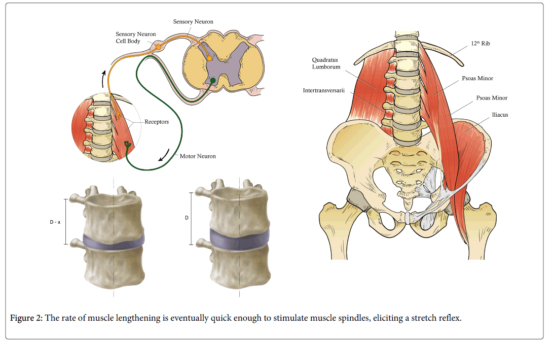
Figure 2: The rate of muscle lengthening is eventually quick enough to stimulate muscle spindles, eliciting a stretch reflex.
We know muscle spindles are abundant within the spinal tissues [12] and deep muscles have multiple attachment points to each vertebra. The psoas major, for example, has attachments not only to each vertebral body of T12 through L5 but also to the adjacent discs. When the intervertebral discs increase in length at the time of sleep onset, an associated stretch to the musculature would likely occur as origins and insertions of each muscle move away from one another in a rostral-caudal fashion. Many other muscles have attachments to vertebrae and/or related processes and therefore the anatomical location of the jerk (upper body vs. lower body) may also provide clues to the biomechanics and related neurology of the spine at that spinal level.
Furthermore, it has been observed that hypnic jerks occur when fatigue is a factor [13]. To explain this, prolonged increased physical activity and associated disc height loss is directly related to load history [14] as individuals who work in an ambulatory setting show more spinal shrinkage when compared to non-workers [15]. Intervertebral disc recovery rates are faster than compression rates due to disc fluid flow [16] so it would seem reasonable that as a person’s spine shrinks more with fatigue, the respective recovery changes will be quicker when compared to non-fatigued spines. And with this quicker rate of lengthening, the associated muscles may be more likely to experience a stretch reflex in those individuals that are fatigued when compared to those that are not.
Interestingly, there is also a common reporting of falling during or just prior to the hypnic jerk. Biomechanically, as muscles relax, the spinal discs unload and expand gaining length. Although speculative in nature, the sensation of falling may involve the cortical perception of afferent information generated from the biomechanical changes related to spinal tissue unloading. Physically, spinal tissues experience anti-gravity forces as the discs unload, perhaps generating sensory information about weightlessness that people often report with (or preceding) the hypnic jerk. Sensory information resulting from spinal unloading may explain why or how sensory pathways influence dreams.
REM sleep can occur early in a sleep cycle and is associated with lack of muscle tone. Jouvet [17] was the first to describe this relationship as he coined the term ‘paradoxical sleep’ relating to brain activity versus the atonia in his decerebrated cats. Others have described this muscle state as being similar to paralysis [18] as well as “anti-gravity” in nature [19]. Better insight into the mechanisms of the hypnic jerk may enhance our understanding of the interrelationships between spinal anatomy and neurophysiology on the one hand, and recuperative sleep on the other.
In the meantime, it is postulated that the hypnic jerk is a proximally generated monosynaptic reflex due to the quick outward expansion of the intervertebral disc(s). The resulting forces exert a quick stretch to the associated muscles (or other spindle concentrated tissues) at the axial level and elicit a subsequent efferent response, like that seen with a knee jerk reflex.