Medical & Surgical Urology
Open Access
ISSN: 2168-9857
ISSN: 2168-9857
Research Article - (2021)Volume 10, Issue 5
Background: Bladder tumor represents the second most frequent cancer of the urogenital tract after prostate cancer. The frequency of squamous cell carcinoma is higher in Africa due to the bilharzian endemic. In Western countries, urothelial carcinoma accounts for over 90% of bladder tumors. The aim of this study was to report the histological types of bladder tumors while describing the epidemiological profile of the patients.
Materials and methods: This was a retrospective study conducted in our center over the period of 4 years from January 2015 to December 2018. This study collected histopathological data of patients follow-up for bladder tumors. The samples examined were mainly biopsies and surgical specimens. The parameters studied were: age, sex, histological type, infiltrative or noninfiltrative nature of the tumor and prognosis.
Results: The mean age of the patients was 55.6 ± 14 years (18 months - 81 years). The sex ratio was 2.1. Urothelial carcinoma was the most frequent histological type (51.5%). Squamous cell carcinoma was found in 32.3% of the patients. The majority of tumors were infiltrative (66.2%). Urothelial carcinoma had a poor prognosis and was classified as high grade in 81% of the patients.
Conclusion: Urothelial carcinoma was the most frequent histological type found in our study contrary to the usual data. In the majority of cases, it was infiltrative and had a poor prognosis; hence the importance of prevention.
Bladder tumor; Urothelial carcinoma; Squamous cell carcinoma; Infiltrating
Bladder tumors are a public health problem because of their size and severity. Urothelial carcinoma is the most common histological type accounting for over 80% of bladder cancers. Squamous cell carcinoma is the most common non-urothelial tumor of the bladder. It accounts for 5-10% of bladder cancers [1]. Urinary bilharzia has been shown to predispose to squamous cell bladder cancer [2]. Squamous cell carcinoma of the bladder is rare in Western countries. In contrast, in bilharzia-endemic countries in the Middle East and West Africa, squamous cell carcinoma is the most common histological type [3]. In our context of developing countries, bladder tumors are of particular interest because of their frequency, their anatomo-pathological polymorphism, the difficulty of staging and their great prognostic uncertainty. Given the high incidence of these bladder tumors and their anatomo-pathological polymorphism, an update of their histological profile must be made, which motivated this study whose objective was to identify the histological types of bladder tumors in our center.
This was a retrospective study conducted in our center over the period of 4 years from January 2015 to December 2018. This study collected histopathological data of bladder tumors. The specimens examined consisted mainly of biopsies and surgical specimens fixed in 10% formalin and sent to the laboratory. These specimens were processed using the standard techniques of paraffin embedding, microtome cutting and haematoxylin-eosin staining. Special immunohistochemical stains (periodic acid-Schiff, Mucicarmine, Desmin, Myogenin) were used to demonstrate glandular lesions while reticulin demonstrated connective tissue. The parameters studied were: age, sex, histological type, infiltrative or noninfiltrative nature of the tumor and prognosis. Data processing was done with Excel 2016 software. The data were recorded and analysed using IBM SPSS Statistics 24 software.
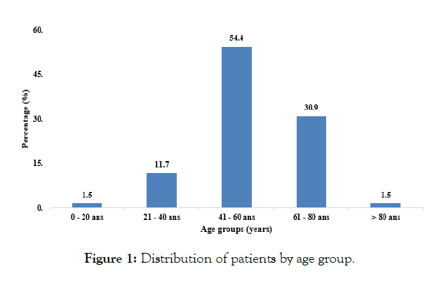
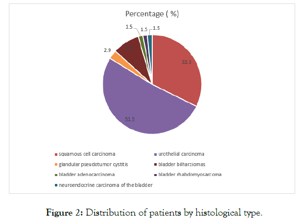
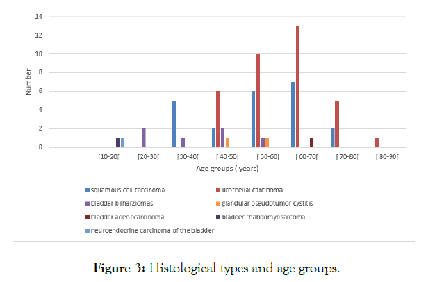
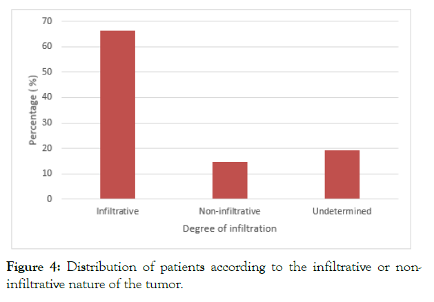
We diagnosed 68 cases of bladder tumors over a four-year period. The mean age of the patients was 55.6 ± 14 years (18 months - 81 years). The most common age range was 41 to 60 years. The majority of patients were at least 60 years old (Figure 1). The sex ratio was 2.1. Urothelial carcinoma was the most frequent histological type (51.5%). Squamous cell carcinoma affected 32.3% of patients. Bladder bilharziomas represented 8.8%. Two cases of bladder carcinoma (one bladder adenocarcinoma and one neuroendocrine carcinoma) were reported. A rhabdomyosarcoma in an 18-month-old infant was also reported as well as two cases of glandular pseudotumour cystitis (Figure 2). Urothelial carcinoma predominated in the age range of 40-70 years. For squamous cell carcinoma the age range was younger, between 30 and 60 years. Bladder bilharziomas was found in young population (83.3%) with an age range of 20-40 years (Figure 3). Urothelial carcinoma was more common in women (62.5%). The majority of tumors were infiltrative (66.2%) (Figure 4). For urothelial carcinomas, the infiltrative character was predominant (62.5%). Urothelial carcinoma had a poor prognosis and was classified as high grade in 81% of patients.

Figure 1: Distribution of patients by age group.

Figure 2: Distribution of patients by histological type.

Figure 3: Histological types and age groups.

Figure 4: Distribution of patients according to the infiltrative or noninfiltrative nature of the tumor.
The incidence of bladder tumors has been increasing in recent decades worldwide. Bladder tumors rank 7th in the incidence of cancer in men and represent 4.4% per 100,000 inhabitants of all cancers worldwide [4].
The young age of the patients in our serie is consistent with the age reported in African series. Diao et al [5] and Koffi et al [6] reported a mean age of 45.5 and 49.9 years respectively. This could be explained by exposure to bilharzia at a younger age in African countries. The role of urinary bilharzia in bladder cancer was first reported in 1878 [4]. Bilharzia eggs are thought to be responsible for the malignant transformation of the epithelium [1]. However, the young age of the patients in our serie contrasts with the age reported in western countries, such as France, where Irani [7] reports an average age of 69 years in men and 71 years in women.
The predominance of males in our serie has already been reported by other authors [5,8]. This is related to the high exposure of the male population to smoking and occupational products related to the generation of bladder cancer.
Bladder bilharziomas are inflammatory pseudotumours secondary to infection with schistosomia haematobium and result from chronic inflammation. The presence of such a tumor poses a problem of differential diagnosis. Bladder bilharziomas were found in the youngest population with an age range of 20-40 years. This result is consistent with the literature [9].
Urothelial carcinomas predominated over squamous cell carcinomas in our study. This result, in a bilharzian endemic zone, contrasts with that of other African or Middle Eastern countries where squamous cell carcinomas are more common [5,10,11]. The Senegalese health authorities had initiated a national programme to control urogenital bilharzia, which reduced the prevalence and severity of this infection. As a result, the risk of infestation has been replaced by increased exposure to industrial chemicals and smoking, which could explain the increase in urothelial carcinoma on the one hand, and on the other hand, bilharzia is also thought to be implicated in the occurrence of urothelial carcinoma. According to some authors, the eradication of bilharzia may contribute to a change in the histological type of bladder cancer, which would resemble those in Western countries [12,13]. Our results are different from those in the West where 90% of bladder tumors are of the urothelial type [1,7]. This type is mainly related to smoking, dyes and hereditary factors.
In our study, infiltrating tumors (pT2, pT3, pT4) predominated. Our results differ from those of Matalka et al. [14] and Laishram et al. [15] where the early stage cancer (pTa and pT1) accounted for 71.8% and 73%, respectively. The reason for this low rate of early presentation is due to the fact that the majority of our patients are seen at late stages but also to the preponderance of squamous cell carcinoma.
In our serie, urothelial carcinoma was of poor prognosis classified as high grade in 81% of patients. This is in contrast to the series of Laishram et al. [12] and Ahmed et al. [16] where low grade tumors represented the majority of cases. Carcinomas, sarcomas and glandular cystitis remain rare as in other studies [11,12,17].
This study has proved that bladder tumors are more frequently encountered in men, often from the sixth decade onwards. Urothelial carcinoma was the most frequent histological type found in our serie, contrary to the usual data. In the majority of cases, urothelial carcinoma was infiltrative and had a poor prognosis; hence the importance of prevention. However, squamous cell carcinoma remains the most common type in our country.
All authors have read and approved the final version of the manuscript.
The authors declare no competing interests.
Citation: Sow O, Ndiaye M, Ndiath A, Sarr A, Sine B, Ondo CZ, et al. (2021) Histopathological Profile of Bladder Tumors at Aristide Le Dantec University Teaching Hospital. Med Surg Urol 10:247.
Received: 05-May-2021 Accepted: 18-May-2021 Published: 25-May-2021 , DOI: 10.35248/2168-9857.21.10.247
Copyright: © 2021 Sow O, et al. This is an open-access article distributed under the terms of the Creative Commons Attribution License, which permits unrestricted use, distribution, and reproduction in any medium, provided the original author and source are credited.