Fisheries and Aquaculture Journal
Open Access
ISSN: 2150-3508
ISSN: 2150-3508
Research Article - (2018) Volume 9, Issue 2
Microbiological and helminthiasis examination of fish from Tinike and Adelle Lakes were conducted at Haramaya District, Ethiopia. The types of fish available in the lakes were also assessed. Adelle Lake has only Clarias gariepinus while the Tinike Lake has only Oreochromis niloticus fish species. Ten fish of each species, 20 in total, were collected from the lakes and post-mortem examined for the presence of adult helminthiasis in the body parts. Twelve samples from different body parts of each fish, 240 samples in total, and 11 samples of lakes water were aseptically collected and examined for gram-negative enteric bacteria. No adult parasites were observed in fish from Adelle Lake. However, 15% and 20% of the fish from Tinike Lake had cestodes in their intestine, and nematodes in their heart respectively. Of 251 total samples from fish and the lake water, 178 (70.9%) were positive for pathogenic microbial. Except for muscle tissue to which only 5% are positive, 50% and above samples were found positive for some Gram’s-negative bacteria. From total of fish and water sample, 43%, 36.3%, 15.1%, 12.8%, and 1.2% are positive for Proteus, E. coli, Salmonella, Yersinia and Klebsiella respectively in the descending order. Two bacterial genera, Proteus and E. coli (13.6%), Proteus and Yersinia (2%), Proteus and Salmonella (4%), E. coli and Yersinia (2%), E. coli and Salmonella (1.2%), and E .coli and Klebsiella (0.4%) while triplet of genera Proteus, E. coli and Yersinia (6%), Proteus, E. coli and Salmonella (0.8%), and Proteus, E. coli and salmonella (0.4%) were observed. Salmonella spp. was not detected from fish from Lake Adelle compared to the 30% in fish from Lake Tinike (P<0.0001). Proper cooking of fish could destroy those spoilage and public health risk. Observing similar levels of gram-negative enteric bacteria in the fish and water suggesting contaminated water as a source for the fish contamination.
Keywords: Gram Negative Bacteria; Freshwater; Fish parasites; Lakes; Haramaya
Ethiopia depends on its inland lakes and rivers for its fish production [1,2] with an estimate of more than 200 edible fish species [3]. The annual fish production potential in Ethiopia is estimated between 30 and 51 thousand tons [3] with significant contribution to the country's economy and food supply. Microbial activities create undesirable changes in flavour, texture and appearance [4,5] results in cross contamination and multiplication of microorganisms [6,7]. One of the factors that hinders fish production is disease mainly interaction with pathogens (parasites and bacteria) and environmental stress causing mortality both in aquaculture and inland fisheries industry. Some of these fish pathogens are also zoonotic with the potential to infect humans [8-10]. Multiple fish parasitic diseases were also reported [4,11]. Some of them are Anisakis simplex, Pseudoterranova decipiens , Diphyllobothrium spp., Heterophyidae spp. , Opisthorchiidae spp. and Nanophyetidae spp. and known to infect humans [11,12]. On the other hand, the potential biological contaminants of aquaculture also bacteria [13,14] including enteric bacteria such as Salmonella, E. coli, Yersinia, Proteus, Klebsiella and others [15-17]. People get infected through consumption of undercooked fish, and contact with infected fish, fish products, fish processing industry and aquaculture water [18,19]. With an increase in bacterial flora and load, fish decomposition is rapid. Isolation and identification of bacterial community play a considerable role as potential indicator of fish health and product quality, and potential public health risk from bacterial pathogens [20]. The objectives of this study were to assess available fish species, its infection with adult helminthes and contamination by gram negative enteric bacteria at selected lakes of Haramaya District, Ethiopia.
Location of study lakes
This study was conducted on fish population at two public lakes (Lake Tinike and Lake Adelle) in Haramaya District located at 41° 59’ 58’’ North and 90° 24’ 10’’ South in Ethiopia. The fish are caught by local fishermen for local consumption.
Fish sample collection
From each lake 10 fish were directly purchased and separately packed into sterile plastic bags. In addition 11 water samples (five from Adelle Lake and six from Tinike Lake) from the two lakes were collected into sterile glass bottles. All samples were transported on ice to the Microbiology Laboratory, College of Veterinary Medicine in Haramaya University for microbiological and parasitological examinations. The species of the fish was determined based on their morphological appearance [21].
Parasitological examinations
The presence of adult parasites in various tissues and organs of the fish were examined by gross post mortem examination. Hence, pre sterile surgical scissors and scalpel bled were used during any postmortem incision. Any observed parasitic worms were collected and examined under stereomicroscope. During post-mortem gross anatomical examinations, specimens were aseptically collected from each tissue and organ of the fish for microbiological analysis.
Isolation and identification of Gram negative enteric bacteria
Specimens for bacteriological culture consisted of external body surface, gills, heart, liver, stomach content, intestinal content, kidneys, pyloric caeca and vents were taken during post-mortem parasitological examination. Sterile cotton swabs were used for the swab samples and surgical were used for tissue sampling from the body organs. Fish and water samples were prepared in buffered peptone water (BPW) (Merck, Germany) at 1:9 ratios and incubated at 37°C for 24 hours. A loop-full of culture from the BPW enrichments were streaked on to blood agar supplied with 5% sheep blood (Oxoid, UK) and incubated at 37°C for 24 hours. Representative colonies with different morphologies on the blood agar were further streaked on to nutrient agar (Oxoid, UK) and incubated at 37°C for 24 hours for the isolation and identification of Gram negative enteric bacteria.
Isolated colonies from the nutrient agar were presumptively identified by morphological appearance, Gram staining, biochemical testing, and by culturing further on differential media according to Jay et al. [22]. Hence, Violet red glucose bile (VRGB) agar (Oxoid, UK) was used to identify the isolates as Enterobacteriaceae, followed by differential media specific for each bacterial genus (E. coli , Salmonella , Shigella , Proteus , Yersinia and Klebsiella ).
Salmonella-Shigella agar (Oxoid, UK) was used for the isolation and differentiation of Salmonella and Shigella followed by biochemical testing using triple sugar iron (TSI) agar (Oxoid, UK). E. coli isolates were presumptively identified by their metallic shinny appearance on EMB agar (Oxoid) and indole, methyl red, Voges-Proskauer, and citrate utilization (IMViC) test according to Jay et al. [22]. Proteus species were presumptively identified by their swarming growth on nutrient agar and blood agar media. Yersinia was presumptively identified by their readily growth, phenotypic characteristics and growth at low temperature (25°C) on nutrient agar or blood agar media. The suspected isolates were further differentiated based on their biochemical reactions (lactose fermentation and TSI tests) and motility test. Klebsiella species were identified by their characteristic growth (lactose positive, dome-shaped, mucoid and sticky colonies on MacConkey agar (Oxoid) and by biochemical tests (IMViC test). The colonies were further characterized by their voluminous, rounded, mucoid, translucent and confluent growth after overnight incubation at 30°C.
Data analysis
Data was entered in Excel Microsoft word 2007© (Microsoft Corp., Redmond, WA) and analyzed using STATA version 11 (Stata Corp., College Station, TX). Binary outcomes for the prevalence of bacterial species detected from various parts of the fish were expressed as percentages with their 95% exact binomial confidence intervals (CI), and P<0.05 was used to compare the prevalence of the bacterial species between the two lakes.
Fish species and parasites
The fish samples (n=20) obtained from Lake Adelle were identified as C. gariepinus and all the fish (n=20) obtained from Lake Tinike were identified as O. niloticus. Among the O. niloticus fish (n=20) obtained from Lake Tinike, cestodes were observed in the small intestine of 15% of the fish. Nematodes were observed in the heart of 20% of the O. niloticus fish (n=20) at a rate of 2-3 nematodes per positive fish (Figure 1). All the C. gariepinus fish (n=20) that were obtained from Lake Adelle were negative both for the cestodes and nematodes.
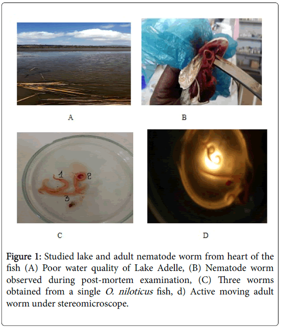
Figure 1: Studied lake and adult nematode worm from heart of the fish (A) Poor water quality of Lake Adelle, (B) Nematode worm observed during post-mortem examination, (C) Three worms obtained from a single O. niloticus fish, d) Active moving adult worm under stereomicroscope.
Gram negative enteric bacteria from various body parts of fish and water samples
Across the total samples (n=240) collected from the various body parts of the fish, 71% of them were positive for the Gram negative enteric bacteria examined (Table 1). Only a single muscle sample was positive for Proteus spp . while 50% to 100% of the samples from the other body parts were positive for at least one bacterial spp. The lowest prevalence was observed in the muscle tissue samples (5%; 95% CI: 0.13%-25%). The prevalence (50%; 95% CI: 27.2%-72.8%) of the Gram negative enteric bacteria observed in the stomach was lower compared to 95% (95% CI: 75.1%-98.9%) prevalence observed both in the small intestine and pyloric caeca. In the other body parts similar prevalence’s were observed ranging from 65.0% to 90.0% of the body parts examined. Seventy three percent (n=11) of all the water samples tested were positive for one or more of the enteric bacterial spp. (Table 1).
| Fish organ and tissue | Total no. of samples examined | No. (%) positive | Exact 95% confidence interval for %positive |
|---|---|---|---|
| External surface | 20 | 15 (75.0) | 50.9-91.3 |
| Heart | 20 | 14 (70.0) | 45.7-88.1 |
| Intestine | 20 | 19 (95.0) | 75.1-98.9 |
| Kidney | 20 | 13 (65.0) | 40.8-84.6 |
| Left gill | 20 | 15 (75.0) | 50.9-91.3 |
| Liver | 20 | 13 (65.0) | 40.8-84.6 |
| Oral cavity | 20 | 15 (75.0) | 50.9-91.3 |
| Muscle | 20 | 1 (5.0) | 0.13-24.9 |
| Pyloric caeca | 20 | 19 (95.0) | 75.1-98.9 |
| Right gill | 20 | 18 (90.0) | 68.3-98.8 |
| Stomach | 20 | 10 (50.0) | 27.2-72.8 |
| Vent | 20 | 18 (90.0) | 68.3-98.8 |
| Total positive fish organs | 240 | 170 (70.8) | 64.6-76.5 |
| Water samples from lakes | 11 | 8 (72.7) | 39.0-94.0 |
Table 1: Overall prevalence of Gram negative enteric bacteria detected from various parts of fish obtained from two public lakes in Haramaya, Ethiopia.
The prevalence of Gram negative enteric bacteria detected from various body parts and water samples is compared by fish species/lakes (Table 2). The overall prevalence for the detection of enteric Gram negative bacteria from various body parts of fish was significantly (P=0.0018) higher in the Lake Adelle (80%) compared to that of Lake Tinike (62%). The prevalence of enteric Gram negative bacteria detected from the external surface (100%), left gill (100%) and stomach (100%) were significantly higher in the fish obtained from Lake Adelle compared to their corresponding prevalence of 50%, 50% and 0% respectively in fish obtained from Lake Tinike. The prevalence of the Gram negative enteric bacteria detected from the remaining body parts of the fish and water samples were not significantly different.
| Body parts | Lake Adelle (C. gariepinus) | Lake Tinike (O. niloticus) | P-value | ||
|---|---|---|---|---|---|
| No. examined | No. (%) positive | No. examined | No. (%) positive | ||
| External surface | 10 | 10 (100) | 10 | 5 (50.0) | 0.0098 |
| Heart | 10 | 6 (60.0) | 10 | 8 (80.0) | 0.3291 |
| Intestine | 10 | 10 (100) | 10 | 9 (90.0) | 0.3049 |
| Kidney | 10 | 5 (50.0) | 10 | 8 (80.0) | 0.1596 |
| Left gill | 10 | 10 (100) | 10 | 5 (50.0) | 0.0098 |
| Liver | 10 | 5 (50.0) | 10 | 8 (80.0) | 0.1596 |
| Oral cavity | 10 | 9 (90.0) | 10 | 6 (60.0) | 0.1213 |
| Muscle | 10 | 1 (10.0) | 10 | 0 | 0.3049 |
| Pyloric caeca | 10 | 10 (100) | 10 | 9 (90.0) | 0.3049 |
| Right gill | 10 | 10 (100) | 10 | 8 (80.0) | 0.136 |
| Stomach | 10 | 10 (100) | 10 | 0 | <0.0001 |
| Vent | 10 | 10 (100) | 10 | 8 (80.0) | 0.136 |
| Total parts | 120 | 96 (80) | 120 | 74 (61.7) | 0.0018 |
| Water | 5 | 5 (100) | 6 | 3 (50.0) | 0.0637 |
Table 2: Comparison of the prevalence of Gram negative enteric bacteria between Lake Adelle and Lake Tinike.
The prevalence of specific Gram negative enteric bacterial genera isolated from various body parts of the fish samples obtained from both lakes were 43% (Proteus spp .), 37% (E. coli ), 15% (Salmonella spp. ), 13% (Yersinia spp. ) and 1% (Klebsiella spp. ) in descending order (Table 3). Similarly more than half (55%) of the water samples were positive for Proteus spp. With the exception of a single muscle sample which was positive for Proteus spp ., the remaining muscle samples were negative for any of the enteric Gram negative bacterial spp.
| Fish tissue and organs | Total no. of samples examined | No. (%) positive for specific bacterial genera | ||||
|---|---|---|---|---|---|---|
| Proteus spp. | E. coli | Salmonella spp. | Yersinia spp. | Klebsiella spp. | ||
| External surface | 20 | 6 (30) | 10 (50) | 4 (20) | 2 (10) | 0 |
| Heart | 20 | 11 (55) | 5 (25) | 4 (20) | 0 | 1 (5) |
| Intestine | 20 | 13 (65) | 11(55) | 6 (30) | 4 (20) | 0 |
| Kidney | 20 | 9 (45) | 4 (20) | 2 (10) | 0 | 0 |
| Left gill | 20 | 12 (60) | 9 (45) | 1 (5) | 1 (5) | 1 (5) |
| Liver | 20 | 5 (25) | 6 (30) | 5 (25) | 3 (15) | 0 |
| Oral cavity | 20 | 6 (30) | 7 (35) | 2 (10) | 4 (20) | 1 (5) |
| Muscle | 20 | 1 (5) | 0 | 0 | 0 | 0 |
| Pyloric caeca | 20 | 10 (50) | 12 (60) | 3 (15) | 5 (25) | 0 |
| Right gill | 20 | 10 (50) | 7 (35) | 4 (20) | 5 (25) | 0 |
| Stomach | 20 | 8 (40) | 5 (25) | 0 | 0 | 0 |
| Vent | 20 | 11 (55) | 12 (60) | 5 (25) | 6 (30) | 0 |
| Total fish parts positive | 240 | 102 (42.5) | 88 (36.7) | 36 (15) | 30 (12.5) | 3 (1.2) |
| Water from lakes | 11 | 6 (54.5) | 3 (27.3) | 2 (18.2) | 2 (18.2) | 0 |
Table 3: Prevalence of specific Gram negative enteric bacterial species in various body parts of fish obtained from public lakes in Haramaya District, Ethiopia.
The prevalence of Gram negative enteric bacteria was compared between the two lakes both for the body parts of fish and water samples (Table 4). The prevalence of Proteus spp. , E. coli and Yersinia spp. from the various body parts of fish were significantly higher in Lake Adelle than that of Lake Tinike. Salmonella spp. was not detected from any of the various body parts of fish obtained from Lake Adelle compared to the 30% prevalence observed from the various body parts of fish examined from Lake Tinike (P<0.0001). With the exception of Proteus spp ., which was significantly higher in Lake Adelle, no significant differences were observed in the water samples obtained from the two lakes.
| Organism | Various body parts of fish | Water samples | ||||
|---|---|---|---|---|---|---|
| Lake Adelle (n=120)% (95%CI) | Lake Tinike (n=120)% (95%CI) | P-value | Lake Adelle (n=5)% (95%CI) | Lake Tinike (n=6)% (95%CI) | P-value | |
| Proteus spp. | 54.2 (44.8-63.3) | 30.8 (22.7-39.9) | 0.0002 | 100 (47.8-100) | 16.7 (0.4-64.1) | 0.0057 |
| E. coli | 53.3 (44.0-62.5) | 20 (13.3-28.3) | <0.0001 | 40 (5.3-85.3) | 0 (0.0-45.9) | 0.0868 |
| Yersinia spp. | 25 (17.5-33.7) | 0 (0-3) | <0.0001 | 40 (5.3-85.3) | 0 (0.0-45.9) | 0.0868 |
| Salmonella spp. | 0 (0-3) | 30 (22-39) | <0.0001 | 20 (0.05-71.6) | 33.3 (4.3-77.7) | 0.6218 |
| Klebsiella spp. | 0 (0-3) | 1.7 (0.2-5.9) | 0.1515 | 0 (0.0-52.2) | 0 (0-45.9) | - |
Table 4: Comparison of the prevalence of Gram negative enteric bacteria from various body parts and water samples between Adelle and Tinike lakes in Haramaya District, Ethiopia.
As shown in Table 5, the various fish organs and tissues examined as well as water samples from the lakes were positive for up to three bacterial species. Out of the total samples from fish (n=240) collected from the various body parts of fish 17%, 10%, 10%, 3% and 0.4% were positive for Proteus spp. , E.coli , Salmonella spp. , Yersinia spp. and Klebsiella spp. respectively were detected singly. The highest codetection of two genera was Proteus and E. coli (13.8%) followed by Proteus and Salmonella (4%), Proteus and Yersinia (2%), E. coli and Yersinia (2%), E. coli and Salmonella (1.2%), and E. coli and Klebsiella (0.4%) while triple genera Proteus, E. coli and Yersinia (6%), Proteus, E. coli and Salmonella (0.8%), and Proteus, E. coli and Salmonella (0.4%) were observed as mixed bacterial contamination.
| Body parts | No. examine | No. (%) positive for single genus | No. (%) positive for mixed genera | ||||||||||||
|---|---|---|---|---|---|---|---|---|---|---|---|---|---|---|---|
| P | E | Y | S | K | P,E | P,Y | P,S | E,Y | E,S | E,K | P,E,Y | P,E,S | P,E,S | ||
| External surface | 20 | 2 (10) | 4 (20) | 1(5) | 1 (5) | 0 | 3 (15) | 1(5) | 1(5) | 0 | 2(10) | 0 | 0 | 0 | 0 |
| Heart | 20 | 5 (25) | 0 | 0 | 1 (5) | 1 (5) | 4 (20) | 0 | 2(10) | 0 | 1(5) | 0 | 0 | 0 | 0 |
| Intestine | 20 | 2 (10) | 1(5) | 0 | 5 (25) | 0 | 6 (30) | 0 | 1(5) | 0 | 0 | 0 | 4(20) | 0 | 0 |
| Kidney | 20 | 7 (35) | 3 (15) | 0 | 1 (5) | 0 | 1 (5) | 0 | 1(5) | 0 | 0 | 0 | 0 | 0 | 0 |
| Left gill | 20 | 4 (20) | 1(5) | 0 | 1 (5) | 0 | 7 (35) | 1(5) | 0 | 0 | 0 | 1(5) | 0 | 0 | 0 |
| Liver | 20 | 1(5) | 2 (10) | 2 (10) | 4 (20) | 0 | 2 (10) | 0 | 0 | 0 | 0 | 0 | 1(5) | 1(5) | 0 |
| Oral cavity | 20 | 4 (20) | 3 (15) | 2 (10) | 2 (10) | 0 | 1(5) | 0 | 0 | 2 (10) | 0 | 0 | 0 | 0 | 1(5) |
| Muscle | 20 | 1(5) | 0 | 0 | 0 | 0 | 0 | 0 | 1(5) | 0 | 0 | 0 | 0 | 0 | 0 |
| Pyloric caeca | 20 | 4 (20) | 6 (30) | 0 | 2 (10) | 0 | 1 (5) | 0 | 1(5) | 0 | 0 | 0 | 0 | 0 | 0 |
| Right gill | 20 | 3 (15) | 3 (15) | 2 (10) | 3 (15) | 0 | 3 (15) | 2(10) | 1(5) | 0 | 0 | 0 | 0 | 0 | 0 |
| Stomach | 20 | 5 (25) | 2 (10) | 0 | 0 | 0 | 3 (15) | 0 | 0 | 0 | 0 | 0 | 0 | 0 | 0 |
| Vent | 20 | 2 (10) | 2 (10) | 0 | 2 (10) | 0 | 3 (15) | 1(5) | 2(10) | 2(10) | 0 | 0 | 3(15) | 1(5) | 0 |
| Total parts | 240 | 40 (16.7) | 24 (10) | 7(9.6) | 24(2.9) | 1(0.4) | 33(13.8) | 5(2.1) | 9(3.8) | 4(1.7) | 3(1.3) | 1(0.4) | 8(3.3) | 2(0.8) | 1(0.4) |
| Water | 11 | 3 (27.3) | 3 (27.3) | 0 | 2(18.2) | 0 | 1(9.1) | 0 | 1(5) | 0 | 0 | 0 | 2(18.2) | 0 | 0 |
Note: *P=Proteus spp.; E=E. coli; Y=Yersinia spp.; S=Salmonella spp.; K=Klebsiella spp.
Table 5: Singly and multiply co-isolated Gram negative enteric bacterial species* from various body parts of fish obtained at two lakes in Haramaya, Ethiopia.
Fish species at Lakes of Haramaya District, Ethiopia
Under current investigation, one fish species was identified in each of the lakes where C. gariepinus in Lake Adelle and O. niloticus in Lake Tinike. The ecology and population dynamics of fish species in these two lakes was beyond the scope of our study and requires further study. Both C. gariepinus and O. niloticus were reported from various Ethiopian Rift Valley lakes including Lake Ashenge, Lake Koka, Lake Langano, Lake Awassa and Lake Ziway as well as in highland Lake Tana (altitude of 1830 m) [23]. This finding also indicated presence of these fish spices in Haramaya district Lakes indicating the diversity and endemics of these fish species. Despite favourable conditions, aquaculture has not taken off in Ethiopia where the population frequently faces a deficit in animal proteins. Aquaculture is promising alternative protein sources needed to be improved sector in the country. Water bodies located in the Rift Valley show signs of overexploitation [24] whilst those located in remote areas like Tinke and Adele lakes were with poor infrastructure and affected with recurrent drought which make them remain underutilized.
Fish helminthiasis
The safety and health of the available fresh fish sold by local fishermen at two local lakes were assessed. Thus, cestodes and nematodes in 15% and 20% of the total samples collected from the various body parts of fish (O. niloticus ) obtained from Lake Tinike. Bekele et al. [25] reported a similar prevalence of 17% of the O. niloticus fish harboring helminth parasites. Higher prevalence levels of cestodes (53%), Capillaria spp. (40%) and Contraceacum spp. (27.6%) helminths were also reported in C. gariepinus fish examined at Lake Hawassa [26]. Detection of these helminths parasites in the fish from this lake indicates potential public health risk associated with consumption of fish. One study [27] reported an overall 92.7% prevalence of helminth infection among children at Lake Awassa area, in South Ethiopia with A. lumbricoides (76%), hook worm spp . (62.5%), T. trichuria (60%) and S. mansoni (33%). No helminth parasites were observed from any of the fish (C. gariepinus ) that were obtained from Lake Adelle which may suggest that fresh fish from this lake has no human risk with respect to zoonotic parasites. However, further studies are needed to collaborate this finding since our study was limited by sample size.
Grams negative bacteria affecting fish quality
Enteric bacteria (Proteus, E. coli , Yersinia, Salmonella and Klebsiella) were detected in 71% of the various body parts of fish and with high level contamination (73%) of water in the corresponding lakes. This high level of infection in the fish and high level contamination of the lakes is alarming. Contamination of the lakes can occur from the faeces of animals and aquatic birds, and human excreta (and sewage), contaminated with Enterobacteriaceae, that is washed into the lakes from the surrounding areas. This will lead to the infection of the fish in the lakes [28]. The level of microbial isolation from C. gariepinus (80%) fish examined from Lake Adelle was higher than that of O. niloticus (61.7%) fish obtained from Lake Tinike. There was a concomitantly higher water contamination at the Lake Adelle (100%) than that of Lake Tinike (50%). This suggests high chance of infection of fish reared in contaminated water. This infectioncontamination cycle in the lakes will increase the level of enteric bacteria with subsequent infection of fish destined for human consumption [29]. Moreover, the level of contamination of fish tissues and organs with specific bacterial genera were in parallel with the levels of water contamination in the respective lakes. Sujatha et al. [30] reported the presence Enterobacteriaceae which attributed to the infection of the fish by the faecal material fed to the fish in India.
The presence of Proteus (43%), E. coli (36.3%), Salmonella (15.1%), Yersinia (12.8%) and Klebsiella (1.2%) in the fish tissues and organs, as well as the water samples taken from the Adelle and Tinike lakes suggests the potential public health risk associated with pathogenic bacteria. On the other hand the absence of Klebsiella from all samples taken from the Lake Adelle and Yersinia from all samples taken from the Lake Tinike but the presence of Proteus, E. coli and Salmonella in samples from both lakes indicates the similarities and differences in the microbial community biodiversity of these lakes. The presence of E. coli (36.3%) and Salmonella (15.1%) was higher than 15.5% and 3% of the respective pathogens reports of Velmurugan et al. [31] from fish at Bale Zone Rivers, Ethiopia could be due to microbial contamination and the stability of lake water acting as a source of infection for fish than a flowing river. The level of Klebsiella (1.2%) observed in this study is lower than the 8.5% reported by Velmurugan et al. [31] which may be explained by differences in the prevalence of this bacteria associated with geographic ecology. The present finding and that of Velmurugan et al. [31] indicated that these pathogenic bacteria are diversified in aquatic environment associated with faecal oral contamination of fish by gastro intestinal tract bacteria commonly found in warm blooded animals [32].
The mixed isolation of Proteus and E. coli (13.6%), Proteus and Yersinia (2%), Proteus and Salmonella (4%), E. coli and Yersinia (2%), E. coli and Salmonella (1.2%), E. coli and Klebsiella (0.4%), Proteus, E. coli and Yesinia (6%), Proteus, E. coli and salmonella (0.8%), Proteus, E. coli and Salmonella (0.4%) from different fish organs and tissues with one or more microbial community of different genera indicated the chance of mixed infection which could act as source of infection for the public. Mixed pathogenic bacteria were also isolated from tissues and organs of fish in Egypt [26].
The current 43% Proteus prevalence was higher than 2.7% prevalence while the 15.1% Salmonella prevalence was lower than the 21.6% prevalence reported from Sudan [33]. The present study indicates a relatively low prevalence of 12.2% Salmonella in O. niloticus was similar with 14.3% reported from South India [7] but higher than reports of Heinitz et al. [34]. Contamination of the water body where fish live from the surrounding run off (flooding) and aquatic birds coupled with poor hygienic practices at all stages of fish handling contribute significantly for spoilage and zoonotic microbial community including Salmonella to prevail in fish, as Zewdu et al. [35] reported 2.3% Salmonella prevalence from fish meat in Addis Ababa. The bacteria isolated from fish such as Proteus, E. coli , Klebsiella, Yersinia and Salmonella, there is also tendency for humans to be infected by these pathogenic zoonotic bacteria [20,36,37] and also spoil postharvest product at any point in time.
Fish production has a contribution in food security. Its availability by types, health and safety are the issues of concern. The presence and distribution of parasitic helminths with similar spoilage and pathogenic enteric microbial community in the studied fish and the water indicates potential fish health issue and public health risk in the studied lakes. Proper lake management with cooking of fish prior to human consumption is recommended to destroy parasitic and pathogenic bacteria to reduce human infections.
The cooperation of small scale fish producers at Lake Adelle and Lake Tinike is highly appreciated.