International Journal of Physical Medicine & Rehabilitation
Open Access
ISSN: 2329-9096
ISSN: 2329-9096
Case Report - (2023)Volume 11, Issue 4
Soft tissue sarcomas represent rare, heterogeneous and complex malignant neoplasm, which require multidisciplinary treatment to improve clinical outcome, survival and quality of life of affected subjects. Whenever possible, limb saving is considered the preferable choice over amputation.
This clinical case describes the surgical intervention and rehabilitative treatment in the case of a young patient suffering of fibromyxoid sarcoma of the popliteal fossa, with the aim of obtaining the best clinical outcome in terms of survival, limb function recovery, walking and the performance in daily living activities.
Design: A single clinical case.
Participant: An active 38-year-old woman affected by fibromyxoid sarcoma of the right popliteal fossa underwent neoadjuvant radio-chemotherapy before surgical excision of the lesion with sacrifice of sciatic nerve and hamstrings, endto- end vascular bypass of the femoral artery and vein with contralateral saphenous graft. Subsequent motor rehabilitation treatment in a sub-intensive regime and evaluation of aids were done.
Rehabilitation exercise program: exercises to improve the range of motion of the hip and knee and strengthen the muscles involved in flexion and abduction of the hip and in flexion and extension of the knee, trying to discourage compensation at the level of the pelvis and spine; multidirectional massage at the level of surgical scars to improve trophism, proprioceptive exercises to facilitate right knee perception control, walking training, initially with the aid of two crutches, then with only one crutch on the right side, stairs training, strategies aimed at recovering autonomy in daily activities.
Conclusion: Demolitive surgery, followed by a well-structured rehabilitation program combined with the use of appropriate aids, was useful to maximize the patient's functional results and provide a satisfactory return to the activities of daily living. Pre-surgical management is desirable to avoid the establishment of abnormal body pattern.
Fibromyxoid sarcoma popliteal cavity; Rehabilitation; Orthopedic surgery
Mrs. D.M. underwent outpatient physiatric consultation after surgery. She sat on a wheelchair. The left lower limb was in a neutral position without sensory-motor deficits [1-4]. The right lower limb was extra-rotated with foot drop due to a complete deficit of the Standardized Patient Instructor (SPI) and Serum Protein Electrophoresis (SPE). Passive mobilization of the right hip was limited to 55° flexion, 22° abduction, 20° extension; passive mobilization of the right knee was limited to 60° flexion, -20° extension. Stiffness of the scars was present. No muscletendon retractions were found at the tibio-tarsic joint. Modified Medical Research Council scale (mMRC) was used to evaluate muscle strength: antigravity movements in hip flexion (MRC 3/5), movements against minimal resistance in hip abduction and adduction (mMRC 4-/5), partially antigravity in knee extension (mMRC 3-/5), in favor of gravity in knee flexion (mMRC 2/5), absence of flexion-extension movements of TT and of the toes and pronation-supination of TT (mMRC 0/5)[5]. There was anesthesia at the level of the innervation territories of the tibial nerve and common peroneal, i.e., in correspondence of the lateral and posterior area of the leg, dorsal and plantar surface and of the toes; epicritic tactile sensitivity preserved in the remaining districts of the leg. Tingling-like and occasionally shock-like paraesthesia in the medial region of the leg and foot were reported. Deficit of proprioception of the right knee was present [6,7]. Deambulation was possible with the aid of two crutches and the use of a Codivilla spring for the right foot. Regarding the degree of disability Barthel Index was 65/100, ADL=2/6, IADL=3/8.
Rehabilitation program and path
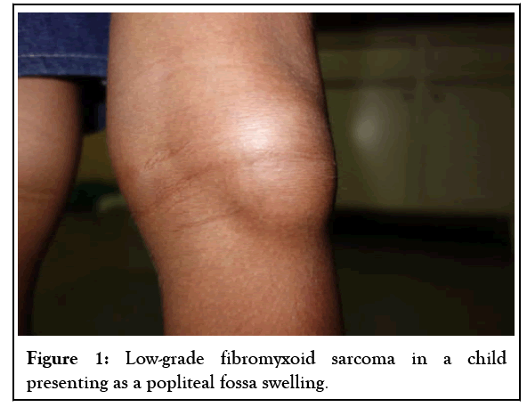
Six weeks after surgery the patient began sub-intensive rehabilitation treatment aimed to functional recovery of the right lower limb along with proprioceptive stimulatio, reabsorption of edema/hematoma with application of kinesio taping at the level of the right popliteal fossa to avoid muscle-tendon retractions, massage of surgical scars with elasticizing cream, improvement of autonomy in ADL/IADL, assisted deambulatory training, climbing the stairs, prescription of aids [2]. The rehabilitation program consisted in 10 sessions of 60 minutes three times per week, followed by 20 sessions of 40 minutes twice per week [8] (Figure 1).

Figure 1: Low-grade fibromyxoid sarcoma in a child presenting as a popliteal fossa swelling.
Treatment involved passive and actively assisted physiotherapy. Exercises in standing position for weight distribution between legs were initially performed [5]. Massage of the scar was started in the prone position. Progressively, a home-based posture maintenance program was conducted to increase flexion of the hip in sitting position, passive extension of the knee in the prone position and to facilitate walking with two crutches. Exercises for recovery and maintenance of lower limbs muscle tone were proposed as well as proprioceptive exercises. After the first 10 sessions of physiotherapy articular and strength measures were taken by the same physician of the first evaluation: passive movements of the knee were limited at 80° in flexion, n-10° in extension and passive movements of the hip reached 70° in flexion and 25° in abduction. Sitting position was better maintained and deambulation was possible with only one crutch [9]. The proprioceptive deficit of the right knee remained if vision was not used. Trophism of surgical scars was improved; popliteal cavity and right foot appeared less edematous; hypotrophism of the right lower limb remained.
Stenic deficit was still present in hip flexion (mMRC 3+/5), knee extension (mMRC 4-/5), and knee flexion (mMRC 2/5); there was no active movements of the ankle and toes (mMRC 0/5). The asymmetric pattern of sitting position was improved [10].
The patient had acquired good motor skills; she was able to perform the main postural passages and postural transfers with supervision. She maintained the standing position and was able to walk wearing a Codivilla spring on the right foot and the aid of a crutch with the right arm; deambulation was prudent but she could take the stairs with greater difficulty in the descent due to the absence of motor and propioceptive control of the right lower limb. Barthel Index=90/100, ADL=6/6, IADL=6/8 [6]. The aids were then re-evaluated, with the replacement of the codivilla plastic spring with a “toe- off” carbon brace, associated with custom-made insoles and shoes. After the successive 20 sessions, in the interval 10-30 weeks after surgery, the final evaluation showed further recovery of passive range of motion of the right lower limb joints, in particular hip flexion was possible up to 95°, knee flexion was complete, knee extension reached -10°. There was a complete recovery of the strength in hip flexion (mMRC 5/5), knee extension and flexion (mMRC 5/5); there was no motor recruitment at the ankle and toe level (mMRC 0/5). The remaining findings were unchanged. The patient was autonomous in performing postural passages and transfers, maintaining the standing position and walking, wearing the before mentioned carbon brace on the right. The patient was able to go up and down the stairs with support on the handrail. Barthel index=100/100, ADL=6/6, IADL=8/8 [7].
Complete autonomy in the activities of daily living at 30 weeks from the surgery was obtained. Despite the sacrifice of the sciatic nerve and right hamstring muscles, active right knee flexion was restored and joint range recovery was observed, most likely thanks to the secondary function of sartorius, gracilis and tensor fascia lata muscles. As for posture, the patient was able to change the asymmetrical body pattern acquired after the surgery. Therefore, an adequate and well-structured rehabilitation intervention may influence the recovery after a demolitive surgery for cancer of the popliteal fossa.
[Crossref] [Google Scholar] [PubMed]
[Crossref] [Google Scholar] [PubMed]
[Crossref] [Google Scholar] [PubMed]
Citation: Vedovi E (2023) Functional Recovery and Rehabilitation after Popliteal Sarcoma Excision in a Young Woman. Int J Phys Med Rehabil. 11:671.
Received: 17-Feb-2023, Manuscript No. JPMR-23-21827; Editor assigned: 22-Feb-2023, Pre QC No. JPMR-23-21827 (PQ); Reviewed: 13-Mar-2023, QC No. JPMR-23-21827; Revised: 20-Mar-2023, Manuscript No. JPMR-23-21827 (R); Published: 27-Mar-2023 , DOI: 10.35248/2329-9096.23.11.671
Copyright: © 2023 Vedovi E. This is an open-access article distributed under the terms of the Creative Commons Attribution License, which permits unrestricted use, distribution, and reproduction in any medium, provided the original author and source are credited.