
International Journal of Physical Medicine & Rehabilitation
Open Access
ISSN: 2329-9096
ISSN: 2329-9096
Review Article - (2026)
Fat Embolism Syndrome (FES) is a complication arising from skeletal trauma. It is characterized by a combination of delayed onset respiratory insufficiency, neurologic dysfunction, and petechial rash. These manifestations typically arise 24-72 hours after injury. Current consensus supports a model involving the initial fat embolization being followed by secondary inflammation and an amplification cascade that leads to organ injury and dysfunction. Fat embolization is common, but clinically significant FES only occurs in a small proportion of patients. Risk increases with multiple injuries and fractures and when stabilization is delayed. Morbidity and mortality risk increases when there is cerebral involvement. FES management focuses on supportive care, and most of those who survive FES end up achieving significant neurologic recovery.
Fat Embolism Syndrome (FES); Long-bone fractures; Cerebral fat embolism; Intramedullary reaming; Two- hit injury mechanismmanagement
Fat Embolism Syndrome (FES) occurs when fat particles enter the bloodstream and obstruct small vessels, leading to multiorgan dysfunction that typically manifests 24 to 72 hours after skeletal trauma [1]. The syndrome presents with a classic triad of symptoms that includes respiratory insufficiency, neurological dysfunction, and petechial rash [2]. Cerebral involvement occurs in the majority of FES cases and represents the most severe manifestation, with mortality higher when neurological involvement occurs [3,4].
Here we review FES, including its epidemiology and clinical presentation, as well as how it is diagnosed and managed.
Fat embolism syndrome arises from combined mechanical and biochemical injury
Research suggests that FES occurs as a “two-hit” process in which an initial mechanical embolization is followed by secondary inflammatory and biochemical injury that amplifies end-organ dysfunction [5]. Increased intramedullary pressure following fracture or surgical manipulation forces fat globules from bone marrow into disrupted venous sinusoids, resulting in systemic embolization [6]. Fat particles measuring 10 to 40 μm may lodge within pulmonary capillaries or traverse the pulmonary circulation or a patent foramen ovale to reach cerebral arterioles [7].
In addition to these mechanical changes, biochemical toxicity occurs due to the stimulation of cytokine and reactive oxygen species [8,9]. The interaction of mechanical obstruction and biochemical toxicity explains the delayed onset of symptoms, as well as the predominance of pulmonary and cerebral involvement. It also highlights the potential to impede damage with prompt recognition and supportive care.
The most significant cases arise due to long-bone fractures It is estimated that 90% of FES cases are due to long-bone fractures, such as fractures of the femur or tibia [10]. Nonetheless, FES that is considered clinically significant occurs in only 0.9% to 2.2% of those who suffer long-bone fractures [11,12]. Risk is higher in those who endure more fractures and more severe fractures, reaching 5% to 10% in people who experience bilateral femoral fractures. In contrast, those with isolated fractures develop FES less than 1% of the time [13].
Younger patients are more susceptible to FES, though the mechanism underlying this differential vulnerability is not known [14]. When stabilization does not occur in the first 24 hours, FES is more likely to occur, likely due to inflammation exacerbating injuries [15].
FES risk is higher in the context of intramedullary reaming
It is common for fat globules to be released into the bloodstream during surgeries that involve intramedullary reaming. In nearly 90% of patients undergoing intramedullary nailing or reaming of fractures, fat passing through the right atrium has been detected [16,17]. This effect is thought to occur due to intramedullary reaming putting pressure on the bone canal, pushing marrow fat into venous sinusoids [16].
Strategies to reduce the volume of fat that is embolized during reaming have been developed. For instance, slower reamer advancement combined with quick reamer rotation may reduce intramedullary pressure and minimize the amount of fat entering circulation [16-20]. Sharp reamers that have a smaller shaft diameter are associated with a lower embolic load compared to reamers that are dull with larger shift diameters [17-20].
Venting the medullary cavity by drilling a small cortical hole (typically between the greater and lesser trochanter) provides a pressure relief valve during reaming and nail insertion. One randomized trial demonstrated that venting reduced major embolic events from 85% to 20%, representing a substantial risk reduction [21,22]. It has also been suggested that using suction to clear marrow could provide complementary support [20].
The Reamer-Irrigator-Aspirator (RIA) system represents the most technologically advanced approach to mitigating reaming-related embolization (Figures 1-4). By simultaneously irrigating and aspirating intramedullary contents during reaming, the RIA system has been demonstrated to significantly reduce the volume of embolized fat compared to conventional reaming [16,17,22].
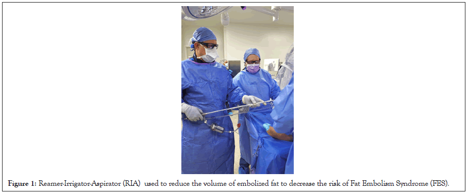
Figure 1: Reamer-Irrigator-Aspirator (RIA) used to reduce the volume of embolized fat to decrease the risk of Fat Embolism Syndrome (FES).
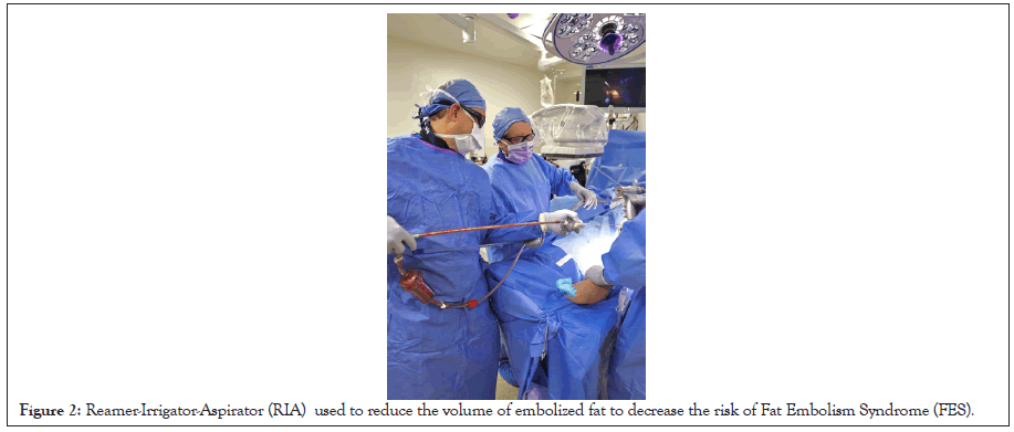
Figure 2: Reamer-Irrigator-Aspirator (RIA) used to reduce the volume of embolized fat to decrease the risk of Fat Embolism Syndrome (FES).
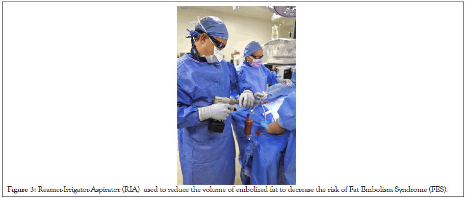
Figure 3: Reamer-Irrigator-Aspirator (RIA) used to reduce the volume of embolized fat to decrease the risk of Fat Embolism Syndrome (FES).
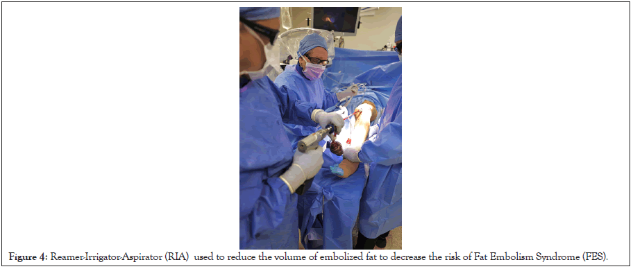
Figure 4: Reamer-Irrigator-Aspirator (RIA) used to reduce the volume of embolized fat to decrease the risk of Fat Embolism Syndrome (FES).
While its direct impact on preventing clinical FES remains under investigation, the RIA system is particularly favored in high-risk patients, including those with polytrauma, compromised pulmonary function, or bilateral long-bone fractures.
Fat embolism syndrome presents with delayed respiratory and neurological manifestations
FES typically manifests within 24 to 72 hours of injury and follows a characteristic clinical pattern [2]. Respiratory manifestations are usually the earliest and most consistent findings, presenting as progressive hypoxemia (PaO2 <60 mmHg on room air), tachypnea and bilateral pulmonary infiltrates resembling acute respiratory distress syndrome [21].
Neurological involvement ranges from subtle confusion and agitation to seizures, focal neurological deficits, and coma, typically developing 12 to 72 hours after injury [3]. Neurologic dysfunction is the principal determinant of morbidity and mortality.
A petechial rash occurs in 20% to 50% of cases but is highly specific when present; it may involve the anterior chest, axillae, neck, conjunctivae, and oral mucosa [23]. Thrombocytopenia develops in up to 50% of patients within 24 to 48 hours due to platelet consumption within microvascular thrombi [24].
Diagnosis depends on clinical criteria and exclusion of competing etiologies
Diagnosing FES is challenging because its presentation is nonspecific, and there are no definitive laboratory or imaging tests. The gold standard for FES diagnosis is the use of the Gurd and Wilson criteria. The Gurd and Wilson criteria requires two major criteria, which may include petechial rash, respiratory insufficiency with bilateral infiltrates, and cerebral involvement [2]. It also requires four minor criteria, which may include tachycardia, fever, retinal changes, jaundice, renal dysfunction, thrombocytopenia, anemia and elevated erythrocyte sedimentation rate. The Schonfeld Index and the Lindeque criteria are other indices that may support diagnosis. Nonetheless, clinical judgment cannot be replaced.
Brain MRI provides the highest diagnostic sensitivity for cerebral involvement
Imaging can help with the recognition of cerebral involvement. Unfortunately, Computer Tomography (CT) often fails to detect such involvement due to the nonspecific findings it produces [25]. On the other hand cerebral involvement is regularly identified with brain Magnetic Resonance Imaging (MRI) [26]. A starfield pattern is a characteristic imaging phenomenon in FES and involves multiple parts of the brain including the basal ganglia, corpus collasum, internal capsule, and centrum semiovale. (Figure 5). As the clinical course of FES progresses, so too do the lesions, and prompt intervention may help to reverse any early cytotoxic effects [27].
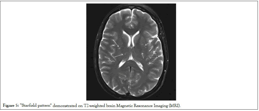
Figure 5: “Starfield pattern” demonstrated on T2-weighted brain Magnetic Resonance Imaging (MRI).
Management emphasizes supportive care and early fracture stabilization
There is no established therapy for FES, but early identification and intervention are critical for successful management [21]. Up to half of all patients require mechanical ventilation [13]. Supportive care typically involves oxygenation maintenance, stabilization hemodynamics, managing fluids, Deep Vein Thrombosis (DVT) prophylaxis, stress ulcer prevention, nutritional support and frequent assessment of neurologic status [28].
Early fracture stabilization within 24 hours represents the most effective preventative intervention [15]. Bone’s randomized trial demonstrated reductions in Acute Respiratory Distress Syndrome (ARDS) incidence, ICU length of stay, and pulmonary complications with early fixation. It has been suggested that, in unstable patients, temporary external fixation followed by definitive repair once stability is achieved could minimize injury caused by a surgical “second-hit” [29].
Pharmacologic therapies have not demonstrated definitive efficacy for treatment. Prophylactic corticosteroids have been associated with reduced incidence in high-risk patients [14,23,30]. However, concerns regarding infection risk, fracture healing, and avascular necrosis have limited routine adoption [31]. Neuroprotective strategies beyond maintenance of cerebral perfusion remain investigational and decompressive hemicraniectomy has been described only in rare catastrophic cases [16,32].
Most survivors experience substantial neurological recovery
The majority of survivors achieve complete or near-complete neurologic recovery [33]. Serial MRI demonstrates gradual lesion resolution that correlates with clinical improvement [27]. Importantly, even profoundly comatose patients may achieve meaningful recovery with aggressive care [34].
FES is a potentially devastating complication of skeletal trauma that requires heightened clinical awareness and prompt intervention. Early fracture stabilization, vigilant supportive care and timely recognition of neurologic involvement form the foundation of modern management. Advances in neuroimaging and trauma systems have improved diagnostic accuracy and outcomes. Despite severe initial presentations, most survivors achieve functional independence, underscoring the importance of aggressive early care and sustained clinical vigilance.
[Crossref] [Google Scholar] [PubMed]
[Google Scholar] [PubMed]
[Crossref] [Google Scholar] [PubMed]
[Crossref] [Google Scholar] [PubMed]
[Crossref] [Google Scholar] [PubMed]
[Google Scholar] [PubMed]
[Google Scholar] [PubMed]
[Crossref] [Google Scholar] [PubMed]
[Crossref] [Google Scholar] [PubMed]
[Crossref] [Google Scholar] [PubMed]
[Crossref] [Google Scholar] [PubMed]
[Crossref] [Google Scholar] [PubMed]
[Google Scholar] [PubMed]
[Crossref] [Google Scholar] [PubMed]
[Crossref] [Google Scholar] [PubMed]
[Crossref] [Google Scholar] [PubMed]
[Crossref] [Google Scholar] [PubMed]
[Crossref] [Google Scholar] [PubMed]
[Crossref] [Google Scholar] [PubMed]
[Crossref] [Google Scholar] [PubMed]
[Crossref] [Google Scholar] [PubMed]
[Crossref] [Google Scholar] [PubMed]
[Crossref] [Google Scholar] [PubMed]
[Crossref] [Google Scholar] [PubMed]
[Crossref] [Google Scholar] [PubMed]
[Crossref] [Google Scholar] [PubMed]
[Crossref] [Google Scholar] [PubMed]
[Crossref] [Google Scholar] [PubMed]
[Crossref] [Google Scholar] [PubMed]
[Crossref] [Google Scholar] [PubMed]
[Crossref] [Google Scholar] [PubMed]
[Crossref] [Google Scholar] [PubMed]
[Crossref] [Google Scholar] [PubMed]
Citation: Lichtblau C, Quinnan S, Deszczynski J, Meli G, Walachowska M, Prowotorow M (2026). Fat Embolism Syndrome: A Two-Hit Mechanism of Mechanical and Biochemical Injury. Int J Phys Med Rehabil. 14:001.
Received: 20-Feb-2026, Manuscript No. JPMR-26-40955; Editor assigned: 23-Feb-2026, Pre QC No. JPMR-26-40955 (PQ); Reviewed: 09-Mar-2026, QC No. JPMR-26-40955; Revised: 16-Mar-2026, Manuscript No. JPMR-26-40955 (R); Published: 23-Mar-2026 , DOI: 10.35248/2329-9096.26.14.001
Copyright: © 2026 Lichtblau C, et al. This is an open-access article distributed under the terms of the Creative Commons Attribution License, which permits unrestricted use, distribution and reproduction in any medium, provided the original author and source are credited.