Internal Medicine
Open Access
ISSN: 2165-8048
ISSN: 2165-8048
Original Research Article - (2022)Volume 12, Issue 1
Pigmented rice such as black rice is known for its health-promoting effects and functional activities, owing to the high contents of flavonoids, phenolic compounds, anthocyanins and dietary fiber. Being as a functional food source, black rice provides potential benefit in the prevention of cancer. In this study the modification techniques applied for the preparation of sample were enzymatic modification and heat moisture treatment, with the objective to evaluate cytotoxicity of native and dual modified black rice flour against Human Colorectal Adenocarcinoma cell line (HCT116) and Mouse embryo fibroblast cell line (3T3-L1) by using the MTT assay. In this study, the IC50 of NBRF and DMBRF were 255.78 μg/mL and 340.85 μg/mL, respectively. The result confirms that the NBRF having significant cytotoxic and anti-cancer potential against Human colon cancer cells. Also the IC50 Value of NBRF and DMBRF on 3T3-L1 cell line were found to be 345.96 μg/mL and 1106.94 μg/mL respectively. Therefore, it was proved on normal cell line, the NBRF had weak cytotoxicity and DMBRF is a non-toxic. Black rice contains high levels of phytochemicals and has potential role in the prevention of colon cancer. Black rice as a cancer chemo preventive dietary agent represents a unique approach to evaluate effective whole food compared to the individual phytochemical.
Functional food; Potential; Cytotoxicity; Phytochemical; Prevention
Cancer is a significant health concern. One in eight deaths worldwide is due to cancer. It represents the first or second cause of death in advanced countries. Therefore, urgent action is warranted to reduce the threats of this disease, particularly in developing countries in which the prevalence and incidence of this disease are expected to increase. Genetic defects only account for nearly 5%-10% of all cancer cases, whereas more than 90% are due to the environmental and lifestyle factors. Therefore, most of the cancer cases and deaths worldwide are actually preventable. It has also been reported that up to 30% of human cancers could be prevented via an appropriate dietary modification. Carcinogenesis is a complex multistage process comprised of initiation, promotion, and progression stage. In chemoprevention, a vital goal is to block tumor progression [1,2].
Oxidative stress, which results from an imbalance between prooxidants and antioxidants and leads to the constant and unregulated generation of reactive oxygen species which is involved in several phases of carcinogenesis. Diet is recognised as a major modulator in the development of colorectal cancer; a high consumption of plant foods containing multiple bioactive phytochemicals, such as vegetables, fruits, and whole grains, has been linked to a decreased risk of colorectal cancer. As a result, identifying plant food sources and their bioactive components that target oxidative stress as well as important events in the multi-step carcinogenesis is vital for developing colon cancer prevention and treatment strategies [3]. Especially, rice is one of the cereal grains used as a staple food by around half of the world's population and by the majority of the population in India. When contrasted to polished white rice, coloured rice variants are regarded valued due to their health advantages. Unpolished rice with bran has more nutrients than milled or polished white rice. It has been shown that pigmented rice types have large quantities of antioxidant chemicals and have good antioxidant capabilities. Due to high levels of dietary fiber,flavonoids, phenolic compounds, and anthocyanins, pigmented rice varieties, particularly those with dark coloured pericarps such as black or purple rice, are recognised for their healthpromoting benefits and functional activities [4].
Black rice is high in dietary fiber and has been linked to a decreased risk of cancer. Whole grains such as black rice, have also been linked to a decreased risk of colon cancer in studies. This may be due to its high fiber content, as fiber attaches to carcinogenic compounds and poisons, aiding in their elimination from the body and preventing them from sticking to cells in the colon. Based on epidemiological and in vivo animal and human research, anthocyanins, which are rich in black rice, exhibit anti-carcinogenic characteristics. Previous studies have reported that black rice has a higher content of anthocyanin than other rice varieties.
Chemo preventive drugs could inhibit the growth of cancer cells, but they exert many adverse side effects. So dietary factor plays a crucial role in the prevention and management of cancers. Being an antioxidant and dietary fibre rich source, black rice display encouraging results in the prevention of the cancer. This study was carried out with the objective to evaluate the toxicity of native and dual modified black rice flour extract on 3T3 normal cells and to know the anticancer effects of native and modified black rice flour extract on colon cancer cell line HCT-116 [3].
Raw materials and sample preparation
Black rice was purchased from local organic shop at Tiruchirapalli district, Tamil Nadu, India. The selected rice was ground separately in an analytical mill and then passed through sieve having 100 meshes (150 μm). The rice flours were collected and stored in an air tight container at 4˚C.This sample was termed as native black rice flour. For preparing dual modified rice flour, the native rice flour was wet-milled to produce 10% (w/v) rice flour slurry, adjusted to pH 4.5 with a 0.1 M sodium acetate buffer. The enzyme alpha-amylase (0.2 g) was added into the flour. The solution was incubated in a shaking water bath at 55 °C for 24 hours. The solution was then centrifuged (3000 g) for 10 min, the precipitate was washed twice with distilled water and collected by centrifugation [5,6]. The precipitate was oven dried at 40°C upto 25% moisture content to be obtained. The samples were sealed in a screw-cap container and equilibrated at room temperature for 24 hours. Then the equilibrated containers were placed in a hot air oven (100°C) for 1 hour. Then the treated flour was taken out and dried in a hot air oven at 40°C to obtain 12% moisture content. The sample was milled and sieved to a particle size of 100 meshes (150 μm), sealed in an air tight container and kept at 4°C. The dual modification methods applied were enzymatic modification and heat moisture treatment. This sample termed as dual modified black rice flour.
Extraction of sample
One gram of each sample was dissolved in 25 ml of methanol solvent, kept in shaking incubator for 24h. The extracts were filtered with Whitman filter paper No.1 and stored at 4°C until analysis.
Cell Lines and Culture Medium
Human colorectal adenocarcinoma cell line (HCT116) and Mouse embryo fibroblast cell line (3T3-L1) were obtained from National Centre for Cell Science (NCCS), Pune, Maharashtra, India. The cells were maintained in a CO2 incubator at a pH of 7, at a temperature of 37 ± 0.5°C and a relative humidity of 80%. The culture medium DMEM-High Glucose was also incubated for about 24-72 h.
MTT assay
MTT assay is a colorimetric assay that measures the reduction of yellow 3-(4,5-dimethythiazol-2-yl)-2,5-diphenyl tetrazolium bromide (MTT) by mitochondrial succinate dehydrogenase. The assay depends both on the number of cells present and on the assumption, that dead cells or their products do not reduce tetrazolium. The MTT enters the cells and passes into the mitochondria where it is reduced to an insoluble, dark purple coloured formazan crystals. The cells were solubilized with a Dimethyl Sulfoxide (DMSO) and the released, solubilized formazan reagent was measured spectrophotometrically at 570 nm. Cell viability was evaluated by the MTT assay with Human colorectal adenocarcinoma cell line (HCT116) and Mouse embryo fibroblast cell line (3T3-L1) with Sample. A Sample (25, 50, 100, 200 and 400 μg/mL) comparing with cell control and standard control [4]. Cells were counted by haemocytometer and seeded at density of 5.0 × 10³ cells/well in 100 μL media in 96 well plate culture medium and incubated overnight at 37°C. After incubation, take off the old media and add fresh media 100 μL with different concentrations of test compound in representative wells in 96 plates. After 48 h, discarded the drug solution and add the fresh media with MTT solution (0.5 mg/ mL-1) was added to each well and plates were incubated at 37°C for 3 h. At the end of incubation time, precipitates were formed because of the reduction of the MTT salt to chromosphere formazan crystals by the cells with metabolically active mitochondria. The optical density of solubilized crystals in DMSO was measured at 570 nm on a micro plate reader. The percentage growth inhibition was calculated using the following formula and concentration of test drug needed to inhibit cell growth by 50% values is generated from the dose-response curves for each cell line using with origin software [7].
Cell morphology observation
The effects of rice extracts on the cellular morphological changes were determined using the method. In this method, the effective dosage concentration of the extract is based on the Inhibition Concentration (IC50) value determined using the MTT assay. The morphological observation was performed at 37°C for 24 h (25, 50, 100, 200 and 400 μg/mL) using a light inverted microscope (Nikon, Japan) at magnification 40X.
Cytotoxicity assay for HCT-116 cell line
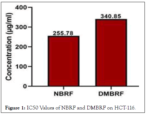
The 3-(4,5-dimethylthiazol-2-yl)-2,5-diphenyltetrazolium bromide (MTT) assay is a simple and reliable assay that measures the viability of cells, and it can be used to screen antiproliferative agents. Different doses of NBRF and DMBRF extracts (methanol), ranging from 25-400 μg/mL, were applied against Human colorectal adenocarcinoma cell line (HCT116). The results showed that the extracts had cytotoxicity activity against colon cancer cell line. The concentration of NBRF and DMBRF were increases the cell viability was decreases range from 96.69% to 32.72%. Similarly reported in HeLa untreated cells showed 100% viability and the cells treated with Methanolic extract of black rice from 5 μg/mL to 100 μg/mL showed gradual decrease in the viability of HeLa cancer cells. This clearly indicates that when the concentration of black rice increased, the viability of cancer cells decreased [8]. Overall, in all the concentrations the NBRF had highest anticancer activity, when compare to DMBRF. Because, due to enzyme digestion and HMT results in loss of some phytochemicals and antioxidants in DMBRF similarly reported that dual modified black waxy and red jasmine rice flour resulted in decreased antioxidants, when compared with native varieties. The anticancer activity of the NBRF and DMBRF extracts is expressed as IC50, which is the concentration causing a 50% inhibition of cell proliferation. In this study, the IC50 of NBRF and DMBRF extracts were 255.78 μg/mL and 340.85 μg/mL, respectively. The result confirms that the NBRF had significant cytotoxic and anti-cancer potential against Human colon cancer cells (Figure 1).

Figure 1: IC50 Values of NBRF and DMBRF on HCT-116.
Originally, the phase contrast microscopic observation displays the morphological changes in the NBRF and DMBRF treated cancer cells. Apoptotic induced cells seen common features such as cell shrinkage, nuclear condensation, membrane blebbing, chromatin breakage, and the production of pyknotic aggregates of condensed chromatin. These unique morphological alterations in apoptotic cells were commonly exploited for apoptosis detection and quantification. Thus, the inverted phase contrast microscope was used to visualise the morphological alterations that identify apoptosis. In this study, contrast to control cells, cytomorphological changes in HCT-116 cells was identified after 24 h of treatment with tested NBRF and DMBRF. The untreated cells maintained their original morphology, which included many nucleoli. The majority of the control cells remained attached to the tissue culture dishes [9,10].
The most noticeable morphological changes after revealing cells to 200 μg/mL and 400 μg/mL treated concentrations of NBRF and DMBRF counting cell contraction, cytoplasmic compression and nuclear chromatin condensation.
Cells enduring apoptosis resulted in different sorts of morphological alterations, such as echinoid spikes on the surface of apoptotic cells, apoptotic bodies, and cell number decrease, when the dosage of the NBRF and DMBRF was raised.
The apoptotic cells lost cellular adherence to the substrate, and the majority of the cells separated from the surface of the tissue culture dish plate and floated in the culture medium. Anoikis is the term used to describe the relatively early separation of monolayer adherent cells from their basal membrane during apoptosis [11].
Toxicity assessment for NBRF and DMBRF against 3T3-L1
The cell viability of native black rice and dual modified black rice was evaluated using MTT assay on Mouse embryo fibroblast cell line (3T3-L1) cell line graph of the percentage cell viability on 3T3-L1 cells against different concentration (25-400 μg/mL) of the prepared systems.
Our MTT results confirmed that the NBRF and DMBRF showed no toxic effect to 3T3-L1 (Mouse embryo fibroblast) cells. Even in the highest concentrations, 200 and 400 μg/mL also the cell viability was recorded as 62% and 47% respectively.
In case of DMBRF, highest concentrations, 200 and 400 μg/ mL cells viability was recorded as 88% and 82% respectively. The DMBRF had high cell viability when compared NBRF [12].
The National Cancer Institute (NCI) and the Gerang Protocol, strong cytotoxic effects are defined as IC50 values of <21 μg/ml, moderate cytotoxic effects as IC50 values of 21-200 μg/ml, and weak cytotoxic effects as IC50 values of 201-500 μg/ml; IC50 values greater than 501 μg/ml are considered to be noncytotoxic.
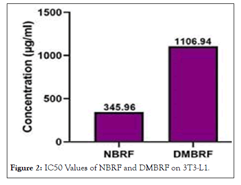
In our study, the IC50 Value of NBRF and DMBRF on 3T3- L1 cell line were found to be 345.96 μg/mL and 1106.94 μg/mL respectively. Therefore, according to the NCI, the NBRF had weak cytotoxicity and DMBRF is a non-toxic (Figure 2).

Figure 2: IC50 Values of NBRF and DMBRF on 3T3-L1.
The phase contrast microscopic displays the different concentrations of NBRF and DMBRF treated normal cells. The results revealed that there is no morphological changes occurred in treated and untreated 3T3-L1 cells
Both native and dual modified black rice flour exhibited cytotoxicity and proved to be beneficial as anti-cancer agent. The observations strongly suggests that the when compared with dual modified black rice flour, native black rice flour may have possible therapeutic potential against Human colon cancer cells .This may be due effect of methods applied in dual modification had impact on antioxidant content and properties. These findings provide important information to improve human health, especially prevention of cancer by encouraging the consumption of black rice and its use in food product development.
The authors express sincere thanks for the Management and Principal of Cauvery College for Women (Autonomous), Trichy -620 018, Tamil Nadu, India.
[CrossRef] [Google Scholar] [PubMed]
[CrossRef] [Google Scholar] [PubMed]
[CrossRef] [Google Scholar] [PubMed]
[CrossRef] [Google Scholar] [PubMed]
[CrossRef] [Google Scholar] [PubMed]
[CrossRef] [Google Scholar] [PubMed]
[CrossRef] [Google Scholar] [PubMed]
[CrossRef] [Google Scholar] [PubMed]
[CrossRef] [Google Scholar] [PubMed]
[CrossRef] [Google Scholar] [PubMed]
[CrossRef] [Google Scholar] [PubMed]
[CrossRef] [Google Scholar] [PubMed]
Citation: Webster R (2022) In Vitro Anticancer Activity of Native and Modified Black Rice Flour against Colon Cancer Cell Line. Intern Med. 12:359.
Received: 03-Jan-2022, Manuscript No. IME-21-359; Editor assigned: 05-Jan-2022, Pre QC No. IME-21-359 (PQ); Reviewed: 19-Jan-2022, QC No. IME-21-359; Revised: 24-Jan-2022, Manuscript No. IME-21-359 (R); Published: 31-Jan-2022 , DOI: 10.35248/2165-8048.22.12.359
Copyright: © 2022 Webster R. This is an open-access article distributed under the terms of the Creative Commons Attribution License, which permits unrestricted use, distribution, and reproduction in any medium, provided the original author and source are credited.