Pancreatic Disorders & Therapy
Open Access
ISSN: 2165-7092
ISSN: 2165-7092
Review Article - (2015) Volume 5, Issue 2
Disconnected duct syndrome (DDS) is defined by a complete discontinuity of the pancreatic duct, such that the secretions of pancreas distal to the discontinuity fails to drain into the duodenum. It usually follows acute necrostising pancreatitis. This duct disruption occurs predominately in the pancreatic neck region, which represents a watershed area that is vulnerable to perfusion abnormalities. Failure of the disconnected duct to drain its secretions leads to serious complications including fistula, peripancreatic collections, sepsis, pancreatic ascites and chronic disability inlcuding diabetes mellitus, malabsorption and portal hypertension. How do we manage DDS and mend this bridge to nowhere. This article reviews the etiology, presentation, investigation, management options and complications of this syndrome.
<Keywords: Disconnected duct syndrome, Acute necrotizing pancreatitis, Pancreatic pseudo cyst, Pancreatic fistula
Disconnected duct syndrome (DDS) is defined by a complete discontinuity of the pancreatic duct, such that a viable left side of the pancreas does not drain downstream into the duodenum, through an intact duct that traverses the length of the pancreas [1-4]. This syndrome occurs predominately in the pancreatic neck region, which represents a watershed area that is vulnerable to perfusion abnormalities [3,5]. DDS is a recognized complication of acute necrotizing pancreatitis and is reported to occur in 30 to 50% of them [5-8]. Infrequently, DDS occurs in chronic pancreatitis and following abdominal trauma [3,7,8]. How do we manage DDS and mend this bridge to nowhere?
Patients with DDS represent an extremely difficult therapeutic dilemma. A multidisciplinary approach is mandatory to optimize the outcome [1-5]. Despite this, patient may encounter frequent interventions (surgical, radiological or endoscopic) in addition to the potential risk of developing diabetes mellitus and chronic disability [3-5,7]. In DDS, the downstream pancreas may continue to drain via papilla in a physiological manner, although frequently leaks may occur in retrograde fashion [2-5,7]. However, the viable pancreatic tissue upstream beyond the transection continues to secrete pancreatic juice, through the point of disruption, without access to the duodenum. Enzyme rich pancreatic secretions may accumulate intra or retroperitonealy from these leaks, to produce pseudocysts, persistent fistulae, psuedoaneurysm, pancreatic ascites, and pancreaticopleural fistula and pancreatic/ peripancreatic necrosis [3,9,10]. This sequence of abnormal drainage will essentially continue until such time as the drainage is redirected endoscopically or surgically or the disconnected pancreas is resected or undergoes atrophy [3-5,7]. DDS has been characterized as type 111 ductal injuries, which is further classified into type 111A (when duct is occluded) or type 111B when it drains freely with resultant pancreatic fistula [11].
Several investigation modalities may facilitate in diagnosing DDS [12-14]. DDS may be diagnosed by cross sectional imaging. This is usually by contrast enhanced CT, which demonstrates a non-perfused pancreatic neck, body or tail [12,13]. Endoscopic retrograde cholangiopancreatography (ERCP) is often recommended to demonstrate the finding of pancreatic duct cut-off or extravasation of contrast (Figure 1) [15] however the concern is that of risk of infection following ERCP, of an otherwise sterile pancreatic necrosis [16] and the accompanying 5 fold increase in the mortality [17].
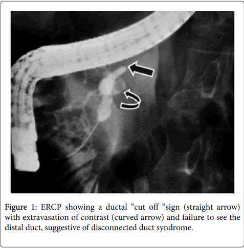
Figure 1: ERCP showing a ductal “cut off “sign (straight arrow) with extravasation of contrast (curved arrow) and failure to see the distal duct, suggestive of disconnected duct syndrome.
The criteria for diagnosing DDS include:
Evidence on ERCP of main pancreatic duct cut-off or discontinuity, with the inability of accessing or cannulating the upstream pancreatic duct (Figure 1).
2) Computed tomography (CT) evidence of viable pancreatic tissue upstream beyond the pancreatic duct cut-off or discontinuity and
3) A non-healing pancreatic fistula, pseudo cyst or fluid collection despite a course of conservative medical management [12].
Other diagnostic criteria proposed are:
1) Necrosis of at least 2 cm of the pancreas;
2) Viable pancreatic tissue upstream i.e. toward the pancreatic tail and
3) Extravasation of contrast material injected into the main pancreatic duct at pancreatography [14]. An accurate preoperative diagnosis may require both cross-sectional imaging (CT or MRI) and pancreatography (Figure 2) [3,12-14].
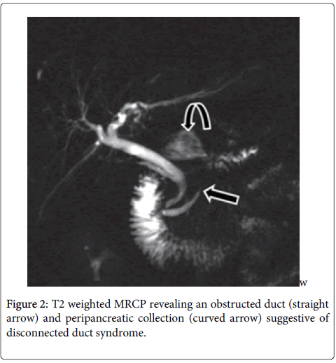
Figure 2: T2 weighted MRCP revealing an obstructed duct (straight arrow) and peripancreatic collection (curved arrow) suggestive of disconnected duct syndrome./p>
Presently secretin enhanced MR cholangiopancreatography has been proposed as an alternative to ERCP in the diagnosis of DDS and external fistula [14]. However its sensitivity in demonstrating a ductal leak at the site of ductal disconnection is lower than ERCP. Features that predict DDS in the early stage of acute pancreatitis (at the time of necrosis) include, a large intra-pancreatic collection or necrosis of a section of the pancreatic head, neck or body, combined with a viable segment of the distal body or tail. When visible, the duct in the pancreatic tail enters the collection at an angle of approximately 90 degree [14]. Despite these multimodal-imaging techniques that readily identifies the disconnected pancreatic duct; the diagnosis is often missed or delayed [3]. Moreover, it may be difficult to ascertain a pseudo cyst that arises from complete discontinuity of the pancreatic duct from one that arises from the more common incomplete duct disruption [3]. Hence DDS should be in the differential diagnosis of all patients with a pancreatic pseudo cyst [3-5].
Management of DDS requires multidisciplinary approach and could involve radiological, endoscopic or surgical intervention [2-5]. Although the initial treatment of DDS is conservative, interventions would be required at an optimum time of 4-6 weeks as most of the conservative measures fail [2,18]. Interventional radiologists can offer minimally invasive surgery sparing techniques by blocking the disrupted duct with cyanoacrylate or other glues [19,20]. This is achieved by advancing guidewire into the main pancreatic duct that is isolated. Subsequently a microcatheter is advanced over the wire and glue is injected to completely fill the pancreatic duct and all its branches within this segment of the pancreas. The ideal situation would be a short 3-4 cm of disconnected pancreas [19]. The procedure however may be associated with mild pancreatitis in 50% of the patients [19,20].
Endoscopic procedures achieve drainage by developing a tract between the pseudocyst and stomach or duodenum. The decision to choose the transgastric or transduodenal approach is based on the site of fistula/ collection and its relationship to the stomach or the duodenum. If the collection is amenable to either, then transduodenal drainage is preferred because of its similarity to the natural path of drainage of pancreatic secretion and a possible greater patency of the tract [2]. Internal drainage of pseudocysts, associated with DDS, is reported to compare favorably with initial success of 76%, in resolution of the pseudocyst [2,7]. However, the high risk of recurrence is a concern as the viable upstream pancreas continues to secrete into the pseudocyst without an intact drainage system to drain into the duodenum [7]. Recurrent pseudocyst has been reported in 50% of patients post drainage [7]. A combination of transmural and transpapillary endoscopic drainage is often performed with transpapillary stent placement [3,7,21]. Prolonged placement of stent in DDS has a role in preventing fluid reaccumulation [19]. Some have used prolonged stent placement (up to 1 to 2 years) thus contributing significantly to their long term success [7,22]. Similar success has also been reported with long-term transmural drainage in randomized controlled trial [23]. Extended stent placement with a resultant pancreaticoenteric fistula, could possibly prevent pancreatic fluid reaccumulation until there is atrophy of the disconnected portion of the gland [21-24]. In univariate analysis, early stent retrieval is a predominant cause of recurrence of fistula [23]. Interestingly, stent migration did not predispose to increased recurrence and is believed to be due to fusion of the cyst edge with gut mucosa, maintaining the lumen of the tract, which may not be achieved if the stent is retrieved early [23]. Removal of the stent, 2 to 6 weeks after endoscopic therapy is at a risk of developing recurrence, despite a reassuring repeat US finding [7,23]. Long-term placement of stent however may predispose to stent occlusion, migration and infection and secondary changes in the duct and its branches [2,3,5,7,23]. Leaving the stent permanently in place could prevent recurrence by creating a permanent fistula between the main pancreatic duct and the gastrointestinal tract, which remains patent even in case of stent occlusion [2]. The stent is believed to act a s a wick, keeping the fistula open, independent of the patency of the stent itself [2]. Permanent plastic stents may however predispose to complications, including recurrence of peripancreatic fluid accumulation due to stent occlusion, superinfection, stent migration leading to intestinal obstruction and stent fracture. Importantly, there are studies to suggest that such complications though potential, do not often occur despite the stent being in place for over two years [7]. As expected, there others who question the role of long term stent placement and raise the concerns of its associated complications and failure to resolve the pseudocyst or fistula, as there are no long term data to support the patency of this approach [3].
In these patients where nonsurgical measures fail, surgical intervention may be warranted and has been considered as optimal therapy for the disconnected portion of the pancreas tail by some [2,3,7,12]. In one of the study, an initial success rate of 73%, 70% and 100% was reported following drainage through ERCP facilitated transmural, transpapillary approach and surgical drainage respectively. However disappointingly, a recurrence rate of 45%, 40% and 60% respectively was observed during the follow up [2,7]. Surgical intervention for recurrence was required in 63% of these patients [7]. Surgical intervention may involve debridement, resection or drainage procedure [2,3,5,7]. The caution however is that surgical intervention may be associated with technical difficulties due to the presence of left sided portal hypertension, ongoing inflammation and adhesions, due to previous debridement [3,7]. Some have reduced the risk of bleeding during resection and the blood transfusion requirement by preoperative splenic artery embolization [3,25]. Unfortunately, recurrence of pancreaticocutaneous fistula has been reported in 23% of patients despite surgical intervention [5]. Those who advocate surgical intervention over other approach believe that the long term benefit claimed of preserving endocrine and exocrine function by nonsurgical approach, in the presence of clearly damaged pancreas, is not often realized [3,7]. An alternative surgical approach to resection is cystojejunostomy or lateral pancreaticojejunostomy in patients with a large remnant and dilated pancreatic duct [2,3,7]
DDS may vary both in terms of its time of presentation and its leading cause [3]. In a recent report by Fischer et al., DDS has been identified to occur in three discrete groups of patients with abdominal symptoms related to nonenteric pancreatic juice secretion [3]. These include concurrent DDS, delayed DDS and chronic pancreatitis associated DDS. The presentation, treatment and outcomes are reported to differ markedly between these three groups [3]. They have suggested that the various subcategories of DDS would require unique operative approaches that balance the objective of preserving pancreatic parenchyma vs. the risk of an ongoing pancreatic fistula. Among the 50 patients with DDS that they treated, 28(56%) had concurrent DDS, 15(30%) had delayed pseudocyst presentation (Delayed DDS) and 7(14%) were associated with chronic pancreatitis3. Concurrent DDS was treated with necrosectomy including body/tail resection within 60 days of onset of pancreatitis and was complicated by grade B/C fistula in 36%. Delayed DDS required distal pancreatectomy in 440 days after the diagnosis, with 7% fistula rate. Chronic pancreatitis DDS was treated with lateral pancreaticojejunostomy at 417 days with no associated complication of fistula [3]. The location of DDS in the neck and body was 50% & 50% in concurrent DDS, 47% and 53% in delayed DDS and 71%:29% in chronic pancreatitis respectively [3].
DDS have been associated with long-term complications [3,5,7,8]. A significant number of them develop complications including left sided portal hypertension, diabetes and exocrine deficiency [3,5,7]. This is reflected in studies reporting the incidence of left sided portal hypertension (53%) and diabetes (50%) during a mean follow up of 38 months [7]. The diabetes unfortunately is not transient and most of them would require medications. In a study of 50 cases of DDS, the incidence of exocrine deficiency in concurrent, delayed and chronic pancreatitis associated DDS was 36%, 60% and 57% and the incidence of postoperative diabetes was 57%, 73% and 14% respectively [3].
Early recognition of DDS and its effective management is important for a better outcome. Disconnection of the duct usually as a consequence of acute necrotizing pancreatitis leads to a bridge that leads to nowhere specific and unfortunately causes serious complications, including pseudocysts, fistula, pancreatic/ peripancreatic necrosis and pseudoaneurysms. While contrast enhanced CT scan are often used to establish the diagnosis, ERCP may be needed both for diagnostic and therapeutic purpose. Secretin- MRCP off late plays a major diagnostic role. Once confirmed, the initial management is non-surgical intervention using endoscopic placement of transpapillary or transcystic stents to drain pseudocyst and the proximal duct or a radiological approach to block the duct with glues when feasible. Unfortunately, there is a significant risk of recurrence warranting surgical intervention. Surgical approach could involve debridement; resection or anastomosis based on whether the DDS is concurrent or develops several months after acute pancreatitis or chronic pancreatitis. Hence, the bridge to nowhere in DDS needs multidisciplinary approaches to mend it and can be challenging and time consuming.