Journal of Fertilization: In Vitro - IVF-Worldwide, Reproductive Medicine, Genetics & Stem Cell Biol
Open Access
ISSN: 2375-4508
ISSN: 2375-4508
Research Article - (2014) Volume 2, Issue 4
Objective : To develop an animal model of thin endometrium, and to evaluate the histologic effect of 95%
ethanol in the uterine cavity.
Design: Experimental prospective.
Setting: Teaching hospital affiliated with Central South University, Changsha.
Patient(s): Twenty and five female adult rats.
Intervention(s): Animals were divided into three groups: control group were submitted to injection of physiological saline in uteri horn, 5 minutes group received 95% ethanol with 5 minutes of retention, 10 minutes group received 95% ethanol with 10 minutes of retention. Endometrial morphology was analyzed by hematoxylin-eosin staining (HE), and the growth of the epithelial, stromal and vascular cells were evaluated by immunohistochemistry with cytokeratin, vimentin and VEGF.
Main outcome measure(s): Histologic effects.
Result(s): Fourteen uterine endometria thinned in 5 minutes group and the endometrial layers, even all uterine layers of 16 uteri were necrosed in 10 minutes group. Compared with control group, the cytokeratin area per unit endometrium area and the vimentin area per unit endometrium stromal were less and the expression of VEGF decreased in model group. All endometria were characterized by poor regrowth in model group.
Conclusion(s): The study developed an experimental rat model of thin endometrium with 5 minutes retention of 95% ethanol, and the success rate was 70%.
Keywords: Thin endometrium, Ethanol, Rat model, Poor regrowth
Endometrial thickness is defined as the minimal distance between the echogenic interfaces of myometrium and endometrium. An adequate thickness of endometrium is indispensable for embryo implantation [1-4]. The minimum endometrial thickness that may cause a successful implantation is called “threshold thickness”. The endometrium that is thinner than “threshold thickness” is called “thin endometrium” [1,5].
The causative factors [6-8] for thin endometrium include Asherman’s syndrome, using oral contraceptive, clomiphene citrate, endometrial tuberculosis and other unknown factors. Various recent modalities proposed for the treatment of thin endometrium seem to be useless and inefficient. At present, stem cell therapeutics may be a promising treatment for thin endometrium. So development of an animal model of thin endometrium is particularly essential for further research.
Destruction of the endometrium has been tried with many agents such as fluoride, electromagnetic wave, 5-amino acid, 80°C water, but the procedures are either ineffective or complicated [9,10]. So we need to find a simple, effective method to develop an animal model. 95% ethanol has dehydration and protein denaturation efficacy. Many studies found aspiration and sclerotherapy with 95% ethanol is an effective treatment of ovarian endometriomas [11,12]. So we proposed the experimental use of 95% ethanol for developing animal model of thin endometrium. The objective of the present study is to develop an animal model of thin endometrium, and to evaluate the histologic and histomorphometric effects of 95% ethanol.
Animals
Central South University Institutional Review Board approved the study protocol. A total of 25 female adult Sprague Dawley (SD) rats weighing 200-250 g were maintained in temperature-controlled (24°C) quarters with free access to food and water. A 14-hour light and 10- hour dark cycle was maintained.
Groups
The rats were grouped as follows:
(i) Control group (5 rats with 10 uteri) were submitted to injection of physiological saline in uteri horn;
(ii) 5 minutes group (10 rats with 20 uteri) received 95% ethanol with 5 minutes of retention;
(iii) 10 minutes group (10 rats with 20 uteri) received 95% ethanol with 10 minutes of retention.
Two estrous cycles after operation, rats in the experimental group with thinner endometrium can be seen as model group, otherwise be considered as modeling fail.
Surgical techniques
The rats were anesthetized with 10% Chloral Hydrate (3 ml/kg; Xiangya hospital; Changsha; China); they were allowed to breathe spontaneously during the procedure. Then the rats were placed on the operating table in a supine position, and the inferior abdomen was sterilized and shaved. An incision of approximately 2.5 cm was made into the inferior abdomen through the skin and underlying layers. After uterus was exposed, clipped each uterine horn with vascular clip and placed sterile gauze around them. And injected the chemical agent (0.3 mL) into the uterine horn with a 1 mL syringe with a 16-gauge needle. 2 minutes later another 0.1 mL was injected and repeated twice. So about 0.5 mL physiological saline kept in the cavity for 5 minutes in the control group and about 0.5 mL and 0.8 mL 95% ethanol was injected in 5 minutes group and 10 minutes group, respectively. The abdomen was then closed, and rats were allowed to recover from the anesthetic.
Specimen collection
All rats were sacrificed in the estrus phase after recovery of 2 normal estrus cycles. The phases of estrous cycle were determined by observing the vaginal smear as described by Zarrow et al. [13]. A 2 cm segment from uterine horns was removed and placed into 10% buffered formalin, embedded in paraffin wax, sectioned transversely at 4 μm, and waiting for further research.
Histology and immunohistochemistry
All the specimens were stained with hematoxylin and eosin (HE). The thickness of each endometrial layer, glands and vessel and the morphology of glandular epithelium cells were evaluated and compared among groups. The growth of epithelium cells, cells in stroma and vascular cells was analyzed via immunohistochemistry with cytokeratin, vimentin and VEGF
Statistical analysis
We compared all groups on the categorical variables using the student’s t test. Statistical analysis was performed with SPSS version 16.0 (SPSS Institute, Inc. Cary, NC). A value of P<0.05 was considered statistically significant.
Observation of the specimen
The uteri demonstrated normal morphology in control group. In 5 minutes group, one rat died and four uteri became normal, and the other fourteen uteri were with moderate abnormal morphology and thinner endometrium in 5 minutes group, which were referred as model group. In 10 minutes group, two rats died and the other eight rats with extensive necrosis had no viable material to be analyzed.
Histopathological observations
In control group, the endometrium was of normal histological appearance and consisted of columnar epithelium and a lamina propria. The lamina propria was intact with a rich glandular component and highly cellular stroma.
In model group, there were moderate changes in the endometrium when compared with the control group. All layers of uterine were thinner in model group, especially in the endometrial layer. The number of the glands and vessels were moderately decreased.
In 10 minutes group, the uterine presented extensive coagulative necrosis in all layers of the endometrium and myometrium, with thin or absence of glandular and luminal epithelium, extensive stromal edema, thrombosis in musclar layers.
The Regrowth of epithelial cells
The regrowth of epithelium cells was evaluated by the cytokeratin area per unit endometrial area. Cytokeratin staining was observed in endometrial epithelium cytoplasm. The cytokeratin area per unit endometrial area in model group was less than that in control group (P < 0.05) (Figure 1).
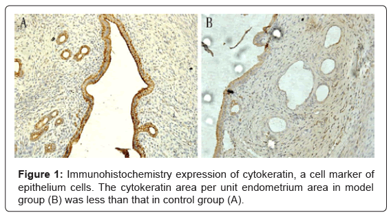
Figure 1: Immunohistochemistry expression of cytokeratin, a cell marker of epithelium cells. The cytokeratin area per unit endometrium area in model group (B) was less than that in control group (A).
The Regrowth of cells in stroma
The regrowth of epithelium cells was evaluated by the vimentin area per unit stromal area. Vimentin staining was positive in the cytoplasm of cells in stroma (stromal cells and a small amount of endothelial cells). The vimentin area per unit endometrial area in model group was less than that in control group (P < 0.05) (Figure 2).
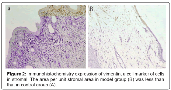
Figure 2: Immunohistochemistry expression of vimentin, a cell marker of cells in stromal. The area per unit stromal area in model group (B) was less than that in control group (A).
VEGF expression
VEGF mainly expressed in the cytoplasm of endometrial epithelial and a few of stromal cells. The average optical density of VEGF in the model group was lower than that in control group (Table 1) (P < 0.05).
| groups | uteri | AOD |
| model group | 14 | 0.033 ± 0.0053 |
| control group | 10 | 0.054 ± 0.0084 |
Note: There was statistical significance between the two groups. P < 0.05
Table 1: Comparison of the average optical density (AOD) of VEGF in endometria between the control group and model group
Thin endometrium can result in infertility. There is lots of debate on the administration of estrogen, low-dose aspirin, vaginal sildenafil citrate, pentoxifylline, vitamin E for the management of thin endometrium [14]. Stem cell therapeutics may be a promising treatment [15]. So development of an animal model for thin endometrium is urgently needed. In this study, we developed a rat model of thin endometrium with 95% ethanol for the first time.
Sclerotherapy was originally used to treat tuberculosis pneumonitis and is currently used by medical oncologists to treat malignant pleural effusion. Noma and Yoshida [11] reported that ethanol instillation into the cyst cavity for >10 minutes was most effective at reducing the recurrence rate.
Considering the thickness of the rat endometrium layer, which is much thinner than the human endometrium, we shorten the retention time. The effect of 95% ethanol over the endometrium and myometrium visualized in the 5 minutes and 10 minutes groups denoted its powerful capacity for tissue destruction. In this study, our result show that most rats survived the procedure, allowing histologic evaluation in the following day, thus validating our model of thin endometrium.
95% ethanol could reach the deepest endometrium layers. All layers of uterine were thinner, and the number of the glands and vessels were moderately decreased in model group compared with controls, which represented poor endometrial growth were consistent with the changes of Asherman’s syndrome [16].
The uterine endometrium mainly consists of epithelial cells, stromal cells and vascular cells, which completely renewed in each monthly menstrual cycle [17]. Most studies have shown that VEGF is expressed in the human endometrium and regulates vascularization in the endometrium. A team led by Miwa et al. [18] has shown that thin endometrium is characterized by poor growth of glandular epithelium, high uterine blood flow impedance, decreased VEGF expression, and poor vascular development. High blood flow impedance as a trigger impairs the growth of the glandular epithelium and results in a decrease in VEGF level in the endometrium. Low VEGF level causes poor vascular development, which in turn further decreases blood flow in the endometrium. The vicious circle leads to a thin endometrium that is related to impaired endometrial receptivity. In our study, we analyzed the regrowth of endometrium via evaluating the immunohistochemical expression of cytokeratin, vimentin and VEGF. The result of our study showed the immunohistochemical expression of Cytokeratin, Vimentin, and VEGF were decreased in model group than that in the control group, which were consistent with the proposal by Miwa et al. [18] .
In conclusion, the result of the study confirmed that we successfully developed a rat model of thin endometrium when 0.5 mL 95% ethanol injected with 5 minutes of retention, and the success rate was 70%. Further studies are needed to perfect the procedure to get a higher success rate, such as adjusting the volume and retention time of 95% ethanol, and search for endometrial stem cell markers to evaluate the model.