Journal of Pollution Effects & Control
Open Access
ISSN: 2375-4397
ISSN: 2375-4397
Research Article - (2016) Volume 4, Issue 1
In this study antibiotic resistant Escherichia coli from household water collected in Hafr AlBatin, Saudi Arabia, were characterized by prevalence of extended spectrum betalactamase (ESBL). Samples were collected from drinking and washing water at 12 locations. From the 144 samples obtained, Millipore membrane filters incubated on trypton soya agar plates produced colonies that yielded 34 isolates of E. coli as verified by biochemical tests. Isolates suspected ESBL producing were tested by using MicroScan analysis and disc diffusion test as ESBL phenotypic confirmatory methods. Phenotypically confirmed ESBL isolates were examined for antimicrobial susceptibility against 30 antibiotics and amplification of blaVEM, blaCTX, blaTEM, blaGES and blaSHV genes by polymerase chain reaction. Out of 34 E. coli isolates, only 6 (17.6%) were positive for ESBL producing according to MicroScan analysis. Disk diffusion as a confirmatory test indicates sensitivity of MicroScan system. PCR results indicated that the VEB was the most prevalent (83.3%) followed by CTX gene (16.6%) between these isolates. The ESBL-producing E. coli isolates was fully susceptible to pip/tazo (100%) and fully resistance to ampicillin, cefazolin and pepracillin (100%). This study showed that ESBL-producing E. coli are multidrug-resistant and existent in Hafr Al Batin’s water. Also, data indicated that wastewater maybe contributes as a source and reservoir of antibiotic resistance.
Keywords: Antimicrobial resistance; ESBL; E. coli; Water
One of the major ways of diffusion of pathogenic and/or antibiotic resistant bacteria, through water, soil and air. Multidrug resistant bacteria have been revealed in different water sources including rivers, lakes, groundwater and drinking water [1-4]. Water consumption, can command to habitation of the gastrointestinal tract of humans and animals by bacteria containing resistance genes and barter genes with bacteria previously present in the intestinal tract [5,6].
Some bacteria can make beta-lactamase enzymes that cleave the beta-lactam ring and then disrupt the antimicrobial action [7]. An extended spectrum beta-lactamase (ESBL)-producing bacteria is capable to disrupt the third generation cephalosporins and monobactum [8]. Also, shown to be able to combat quinolones [9]. ESBL-producing bacteria especially of Enterobacteriaceae, foremost E. coli and Klebsiella pneumonia [10]. ESBL-producing E. coli have 3 various resistance mechanisms. The most widespread is production of beta-lactamase which hydrolyzes the beta-lactam ring in penicillins and cephalosporins. The second is mutation which decrease betalactam uptake [11]. The third resistance mechanism is existence of efflux pumps, which exports antibiotics outside of the cell.
Penicillins and concerned antibiotics have been utilized extensively for the control and treatment of bacterial infections. Efficiency of this group of antimicrobial agents has been amended, because of the evolution of multidrug-resistant strains of bacteria [13]. Through the years, incalculable penicillin derivatives [14] have been designed and tested, and a variety of new β-lactam ring systems have been introduced such as cephalosporins, cephamycins, oxacepems, clavulanic acid and carbapenems.
Saudi Arabia with an area of 2.15 million km2 is a desert and water deficit country, with limited fresh water-supplies. It is also recognized by low annual rainfall and privation perennial rivers. The water resources in the Saudi Arabia are surface and underground deposits. Water collected through rainfall (surface water).
In Hafr Al Batin city about 100% of water supply comes from groundwater. Water transported by car to the house where stored in the wells dedicated to it, which may be located next to the sewage tanks. No available information about extended-spectrum beta-lactamases (ESBLs) producing bacteria in water Hafr Al Batin. The present study was to gain insight into the prevalence of AMR E. coli in washing and drinking water, and to check the possible role of sewage tanks as AMR contamination source of water.
Water sampling
Water samples were taken from 12 locations in the Hafr Al-Batin city, Saudi Arabia (Figure 1). Three sampling sites were located on each location. Two of which were from washing water that taken from house tanks in most of residential neighborhoods and one from drinking water from charity refrigerators. Sampling was done twice, the first on January (winter) and the second on July (summer). Two samples were collected each time at each collection site at two different times, and we used three replicates from each sample for tests. Collected samples were transported by standard methods as mentioned in APHA, 1989 [15].

Figure 1: Location map of the study area of Hafr Al-Batin city, Saudi Arabia.
Isolation of E. coli
100 mL water sample is filtered through 0.45 μm pore size membrane filters (Millipore, the Netherlands). Filters were incubated on tryptone soya agar (TSA) at 36 ± 2°C during 4-5 hours, and subsequently transferred to tryptone bile agar (TBA) then incubated for 19-20 hours at 44 ± 0.5°C as described by Sing et al. [16]. E. coli identified using the TSA/TBA method (i.e. indole-positive), were supplementary confirmed as E. coli by testing for ß-glucuronidase activity on Brilliance E. coli/coliform agar. Beta-glucuronidase-positive colonies identified using tryptone bile x-glucuronide agar (TBX) was extra confirmed by MicroScan [17] for identification and antibiotic susceptibility.
Analysis of antimicrobial resistance
Generally, 99 E. coli isolates (91 from washing water and 8 from drinking water) were obtained. These were screened for confirmation of identification and its susceptibility to a panel of antimicrobials of human and veterinary clinical relevance, using MicroScan. MicroScan® instrumentation (auto SCAN®-4 and WalkAway® System) (Siemens Healthcare Diagnostics Inc, USA) was used. Panels used were MicroScan Dried Gram Positive MIC/Combo, Dried Gram Positive Breakpoint Combo and Dried Gram Positive ID Type 2 or 3. Also, MicroScan Dried Gram Negative MIC/Combo panels and Dried Gram Negative Breakpoint Combo Panels were used. MicroScan panels were designed for use in determining agent susceptibility and/or identification to the species level of rapidly growing aerobic and facultative Gram positive cocci or aerobic and facultatively anaerobic Gram negative bacilli. MICs obtained for ceftazidime and cefotaxime with CA were compared with those obtained with the same drugs without CA. Subsequently, strains were considered as ESBL-positive or -negative in accordance with CLSI recommendations (CLSI, 2010). The tests were performed as recommended by supplier guidelines [17]. Susceptible, intermediate and resistant isolates were arranged according to antibiogram results. Multi-drug resistance was defined as resistance to 3 or more different classes of antimicrobials according to CLSI, 2010 [18].
Confirmation of ESBL-producing E. coli
Suspected ESBL-E. coli isolates (after MicroScan analysis) were confirmed for ESBL-production by double disc synergy test (DDST) according to CLSI guidelines (CLSI, 2010) [18]. DDS test results analysis. Susceptibility testing was performed (McFarland 0.5 standard) on Mueller–Hinton agar (MHA) by placing discs on the agar surface containing 30 mg cefotaxime or ceftazidime, with and without 10 mg CA. Plates were incubated at 37°C for 24 h. According to CLSI guidelines, strains were considered positive for ESBL production whenever zone diameters increased by ¢5 mm for cefotaxime or ceftazidime when tested in combination with CA. This method was considered the gold standard for method comparison.
DNA isolation and genotyping
A single colony from each ESBL-producing isolate was transferred into 100 μL of sterile distilled water and the bacterial DNA was extracted by using boiling method included microwave pre-heating according to Ahmed et al. [19]. PCR screening for presence of different beta-lactamase genes was performed. PCR was carried out and specific primers (Table 1) were used for VEB, CTX, TEM and GES genes. PCR mixtures were prepared by using 5 μL template DNA (about 500 pg of DNA), 12.5μL PCR master mix; 1 × PCR buffer [Tris-Cl, KCl, (NH4)2SO4, 1.5mM MgCl2] (pH 8.7), 200 μM dNTP, and 1 μL of each 10 pM primer and 0.5U Taq DNA polymerase in a final volume of 25 μL. The amplification reaction was carried out in a Thermal Cycler (Eppendorf master cycler®, MA) with an initial denaturation (94°C for five minutes) followed by 30 cycles of denaturation (94°C for 25 seconds) annealing (52°C for 40 seconds), and extension (72°C for 50 seconds) and a single final extension at 72°C for six minutes [20]. The amplified products were electrophoresed on 2% agarose gel and visualized on a gel document system after staining with ethidium bromide (0.5 mg/mL). A non-ESBLproducing strain (E. coli ATCC 25922) was used as a negative control.
| Genes | Primer used (5'-3') | PCR product size |
| VEB | CGACTTCCATTTCCCGATGC GGACTCTGCAACAAATACGC |
585 bp |
| CTX | CAAAGAGAGTGCAACGGATG ATTGGAAAGCGTTCATCACC |
205 bp |
| TEM | TAATCAGTGAGGCACCTATCTC GAGTATTCAACATTTCCGTGTC |
800 bp |
| GES | ATGCGCTTCATTCACGCAC CTATTTGTCCGTGCTCAGG |
846 bp |
| SHV | GGTTATGCGTTATATTCGCC TTAGCGTTGCCAGTGCTC |
867 bp |
Table 1: The sequences of primers used in PCR amplification of beta-lactamase genes.
Water borne infections still ravage the global community and are responsible for millions of deaths per year. Water that looks clear and pure may be sufficiently contaminated with pathogenic microorganisms to be a health hazard.
ESBL-producing E. coli
Ninety-nine E. coli isolates were detected and isolated from the water samples using the TSA/TBA method. Only 34 isolates were confirmed as E. coli by MicroScan. ESBL-producing strains (6⁄34) were found in all the samples analyzed by MicroScan system (Table 2). Five ESBL producing E. coli were detected from the washing water samples at five locations (Al-Khalediyah, Al-Rabwah, Al-Rawdhah, Al-Nayefiyah and Al-Nakheel) at summer season only, no winter was found. On the other hand, ESBL-producing E. coli was detected and isolated from drinking water in one location (Al-Baladiyah) out of 12.
| Location | Season | Washing water isolates | Drinking water isolates | Total/ confirmed (%) | ||
| Total | + | Total | + | |||
| Abu-Musa al-Asha'ari | Winter Summer |
1 4 |
0 0 |
0 0 |
0 0 |
1/0 (0.0) 4/0 (0.0) |
| Al-Aziziah | Winter Summer |
0 2 |
0 1 |
0 0 |
0 0 |
0/0 (0.0) 2/1 (50.0) |
| Al-Khalediyah | Winter Summer |
1 2 |
0 0 |
0 0 |
0 0 |
1/0 (0.0) 2/0 (0.0) |
| Al-Rabwah | Winter Summer |
0 1 |
0 1 |
0 1 |
0 0 |
0/0 (0.0) 2/1 (50.0) |
| Al-Muhammadiyah | Winter Summer |
0 3 |
0 0 |
0 0 |
0 0 |
0/0 (0.0) 3/0 (0.0) |
| Al-Baladiyah | Winter Summer |
1 3 |
0 0 |
0 2 |
0 1 |
1/0 (0.0) 5/1 (20.0) |
| Al-Rawdhah | Winter Summer |
0 4 |
0 1 |
0 0 |
0 0 |
0/0 (0.0) 4/1 (25.0) |
| Al-Nayefiyah | Winter Summer |
0 1 |
0 1 |
0 0 |
0 0 |
0/0 (0.0) 1/1 (100.0) |
| Al-Sulaimaniyah | Winter Summer |
0 0 |
0 0 |
0 0 |
0 0 |
0/0 (0.0 0/0 (0.0) |
| Al-Faisaliyah | Winter Summer |
0 1 |
0 0 |
0 0 |
0 0 |
0/0 (0.0) 1/0 (0.0) |
| Al-Masyef | Winter Summer |
0 4 |
0 0 |
0 0 |
0 0 |
0/0 (0.0) 4/0 (0.0) |
| Al-Nakheel | Winter Summer |
0 2 |
0 1 |
0 1 |
0 0 |
0/0 (0.0) 3/1 (33.3) |
| Total | 30 | 5 | 4 | 1 | 34/6 (17.6) | |
Table 2: Total number and ESBL-E. coli isolates, which confirmed (+) by MicroScan system from 13 locations.
The prevalence of ESBL-producing E. coli varied between waters that differed with regard to region, type of water, and time of the year at sampling. Perhaps, prevalence may vary with the number of faecal dirtiness sources in the neighborhood of sampling sites and factors affecting when and how often releasing takes place (climate or season). Our results agreed with Blaak et al. [21]; Adnan et al. [22].
All ESBL-producing isolates also showed resistance to other antimicrobials: 100% to ampicillin, cefazolin, cefepime, cfuroxime, mezlocillin, piperacillin, trimethoprime, trimethoprime⁄sulfamethoxazole; 83.4% to ciprofloxacin, gentamicin, levofloxacin, norfioxacin, tetracycline, tobramycin; 50% to cefoxitin; 33.3% to ertapenem and 16.6% to fosfomycin, imipenem, meropenem, nitrofurantain, tigecycline (Table 3). The study by Babypadmini and Appalaraju [23] reported 74% resistance to trimethoprim/ sulfamethoxazole in ESBL-producing E. coli pathogens by disk diffusion method, which is lower than our results. This difference may be due to use of different methods of evaluation for determining the susceptibility. We determined the antimicrobial resistance by the MicroScan method which is more sensitive than disk diffusion method.
| Antibiotics | Washing water-E. coliisolates | Drinking water-E. coliisolate | ||||||||||
| 1 | 2 | 3 | 4 | 5 | MIC | Interps | ||||||
| MIC | Interps | MIC | Interps | MIC | Interps | MIC | Interps | MIC | Interps | |||
| Amikacin | ≤16 | S | >32 | R | ≤16 | S | ≤16 | S | ≤16 | S | >32 | R |
| Amox/K Clav | >16/8 | R | >16/8 | R | ≤8/4 | S | 16/8 | I | 16/8 | I | >16/8 | R |
| Ampicillin | >16 | R | >16 | R | >16 | R | >16 | R | >16 | R | >16 | R |
| Cefazolin | >16 | R | >16 | R | >16 | R | >16 | R | >16 | R | >16 | R |
| Cefepime | >16 | R | >16 | R | >16 | R | >16 | R | >16 | R | >16 | R |
| Cefotaxime | >32 | ESBL | >32 | ESBL | >32 | ESBL | >32 | ESBL | >32 | ESBL | >32 | ESBL |
| Cefotaxime/K Clav. | ≤0.5 | ≤0.5 | ≤0.5 | ≤0.5 | ≤0.5 | ≤0.5 | ||||||
| Cefoxitin | ≤8 | S | >8 | R | ≤8 | S | ≤8 | S | >8 | R | >8 | R |
| Ceftazidime | >16 | ESBL | >16 | ESBL | >16 | ESBL | >16 | ESBL | >16 | ESBL | >16 | ESBL |
| Ceftazidime/k clav. | 2 | 2 | ≤0.25 | ≤0.25 | 2 | |||||||
| Cefuroxime | >16 | R | >16 | R | >16 | R | >16 | R | >16 | R | >16 | R |
| Ciprofloxacin | >2 | R | >2 | R | ≤1 | S | >2 | R | >2 | R | >2 | R |
| Colistin | ≤2 | S | ≤2 | S | ≤2 | S | ≤2 | S | ≤2 | S | >4 | R |
| Ertapenem | ≤2 | S | >4 | R | ≤2 | S | ≤2 | S | ≤2 | S | >4 | R |
| Fosfomycin | ≤32 | S | ≤32 | S | ≤32 | S | ≤32 | S | ≤32 | S | >32 | R |
| Gentamicin | >8 | R | >8 | R | ≤4 | S | >8 | R | >8 | R | >8 | R |
| Imipenem | ≤4 | S | ≤4 | S | ≤4 | S | ≤4 | S | ≤4 | S | >8 | R |
| Levofloxacin | >4 | R | >4 | R | ≤2 | S | >4 | R | >4 | R | >4 | R |
| Meropenem | ≤1 | S | ≤1 | S | ≤1 | S | ≤1 | S | ≤1 | S | >8 | R |
| Mezlocillin | >64 | R | >64 | R | >64 | R | >64 | R | >64 | R | >64 | R |
| Moxifloxacin | >1 | R | >1 | R | 1 | I | >1 | R | >1 | R | >1 | R |
| Nitrofurantoin | ≤32 | S | ≤32 | S | ≤32 | S | ≤32 | S | ≤32 | S | >64 | R |
| Norfioxacin | >8 | R | >8 | R | >4 | S | >8 | R | >8 | R | >8 | R |
| Pip/tazo | 64 | I | ≤16 | S | ≤16 | S | ≤16 | S | ≤16 | S | ≤16 | S |
| Piperacillin | >64 | R | >64 | R | >64 | R | >64 | R | >64 | R | >64 | R |
| Tetracycline | >8 | R | >8 | R | ≤ | S | >8 | R | >8 | R | >8 | R |
| Tigecycline | ≤1 | S | ≤1 | S | ≤1 | S | ≤1 | S | ≤1 | S | >2 | R |
| Tobramycin | >8 | R | >8 | R | ≤4 | S | >8 | R | >8 | R | >8 | R |
| Trimeth/sulfa | >2/38 | R | >2/38 | R | >2/38 | R | >2/38 | R | >2/38 | R | >2/38 | R |
| Trimethoprim | >8 | R | >8 | R | >8 | R | >8 | R | >8 | R | >8 | R |
Table 3: Antibiotic susceptibility pattern among ESBL-E. coli resistant isolates.
Validation of ESBL-producing isolates
ESBL confirmation test was carried out by disk diffusion method. For validity testing of 6 E. coli ESBL- producing isolates, no errors were showed between the two test methods, MicroScan and disk diffusion test.
ESBL genes
VEB and CTX-M genes were detected in 5 and 1 ESBL-producing E. coli isolates from washing and drinking water samples, respectively. The results of ESBL genotyping showed that VEB gene was the most prevalent (83.3%) followed by CTX gene (16.6%), TEM (0.0%), GES (0.0%) and SHV (0.0%). ESBL-producing E. coli isolate from drinking water was carried CTX gene (Figures 2 and 3). In washing water, only VEB gene was predominant. VEB was the most frequent resistant gene in ESBL-producing E. coli isolates in this study. The study by Rezai et al. [24] reported that TEM resistant gene was most prevalent. TEM, GES and SHV resistant genes were not found in ESBL-producing E. coli isolates which is in line with the low frequency of this gene in ESBLproducing E. coli strains [25].
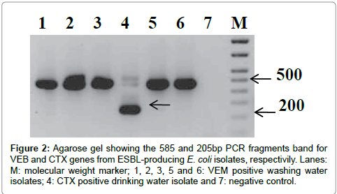
Figure 2: Agarose gel showing the 585 and 205bp PCR fragments band for VEB and CTX genes from ESBL-producing E. coli isolates, respectivily. Lanes: M: molecular weight marker; 1, 2, 3, 5 and 6: VEM positive washing water isolates; 4: CTX positive drinking water isolate and 7: negative control.
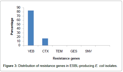
Figure 3: Distribution of resistance genes in ESBL-producing E. coli isolates.
Fecal contamination of tank water in Hafr Al Batin city by sewage may be occurs through discharge of untreated sewage through sewage leaking or during heavy rainfall. Water distribution and storage systems in Hafr Al-Batin city could serve as an incubator for growth of certain ARB populations and as an important reservoir for the spread of antibiotic resistance to opportunistic pathogens. Out of six ESBL-producing E. coli isolates, five were obtained from washing water tanks adjacent to sewage tanks as shown in proposed pathway of contamination transport (Figure 4). Leaks happen when a pipe isn’t sealed in a specific spot. Most old septic tanks didn’t have any sealants applied to the mating joints and troubleshooting difficult. Also, heavy vehicles can crush septic system drain lines causing leakage and toxic smells and odors. The septic cover can cause leaks because they may not be well-secured. From here, we recommend that the system of quantitative and qualitative microbial risk estimate must be applied on houses water tanks at Hafr Al-batin city. This process was successfully implemented for estimate exposure and infection hazard of bacterial and other pathogens from consumption of drinking water and washing water. Water-borne transmission has been demonstrated to be a relevant route of transmission for faecal bacterial species, including Salmonella and E. coli [26,27]. To evaluate risks of human exposure, the system of quantitative microbial risk assessment could be used [28]. This process was successfully implemented for estimate exposure and infection hazard of bacterial and other pathogens from consumption of drinking water and recreational water [29,30].
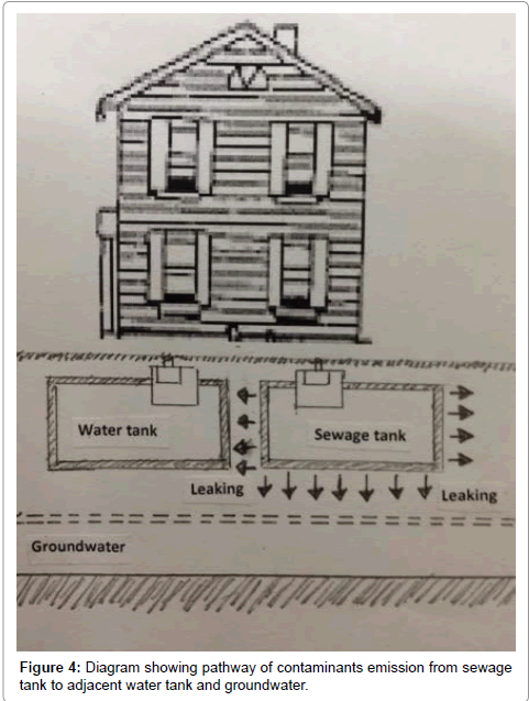
Figure 4: Diagram showing pathway of contaminants emission from sewage tank to adjacent water tank and groundwater.
Our results suggested the possible emissions of the ESBL-producing E. coli from sewage tanks to the environment. Water distribution systems in Hafr Al-Batin city could serve as an incubator for growth of certain ARB populations and as an important reservoir for the spread of antibiotic resistance to opportunistic pathogens.
The authors extend their appreciation to the Deanship of Scientific Research at University of Dammam for funding this work through research project no. 2014027.