International Journal of Physical Medicine & Rehabilitation
Open Access
ISSN: 2329-9096
ISSN: 2329-9096
Research Article - (2023)Volume 11, Issue 5
Study design: Experimental study.
Background: In degenerative disc-radiculopathies, medical treatment provides only temporary relief and the pathology may progress to paralysis if the spinal disc impingement is not resolved. Given the long-term harmful effects of Pharmacotherapy, the WHO recommends management by non-pharmacological therapeutic measures. Addressing this concern that is why we conducted this study on neurovertebral decompression using a non-invasive, safe and effective method.
Objective: To determine the deformation threshold of the anatomo-histological structures of the cervical spine of the goat subjected to a traction force, with a view to the transposition of results in humans for safe and effective cervical traction in degenerative neck pain.
Methods: This experimental in vitro study, carried out on 12 goats, divided into two groups of 6 each, the first of which included goats subjected to cervical traction with muscle mass and neck skin in place and the second, goats stripped of muscle mass and skin. During the period from February 2020 to March 2021.
Results: For progressively increasing tensile forces from 0 to 100 kgf at a rate of 10 kgf per sequence, the maximum duration beyond which no elongation was observed was 5 minutes. All parameters remaining constant (duration of the sequence, tensile load), the elongations observed in the center of the cervical spine (were far superior to those observed at the periphery in a ratio of 1 of 8. The progression of the elongation at the periphery was very low, evolving from 0 to 2 mm compared to that of the center, which evolves from 0 to 17 mm. All the parameters remaining constant (duration of the sequence, tensile load), the elongations observed at the center of the cervical spine (in the goats were by far superior to those of the periphery according to a calculated ratio of 1 of 8. Progression of elongation in periphery was very weak, evolving from 0 to 3 mm compared to that of the center which evolved from 0 to 25 mm. Peripheral elongations in group II goats is not as pronounced as that of group I, ratio:1 of 2. We note a series of deformation of the cervical spine: intervertebral dislocation and ligament cracking, with spinal cord exposure, focused on C2, C3 on all 6 goats from a tensile force of 50 kgf to 80 kgf. Comparison of means between the lengthening observed in periphery and in center in intra-group I is clearly significant (test t: p˂0.001). The elongation in center is greater than that of periphery. The same is true for the elongation observed in intragroup II (t test: p˂0.001), the elongation observed in the center is greater than that observed in periphery. By comparing the average lengthening observed between the two groups in periphery, we note a significant difference with predominance in group I (t test: p=0.001). Between groups I and II, the comparison of average lengthening of the center is as significant difference (test t: p˂0.001).
Conclusion: The anatomo-histological structures of the cervical spine of the goat subjected to a traction force are deformable from 50 kgf of traction force up to 80 kgf. For a constant force, the center of the spine lengthens more than the periphery. The elongation resulting from traction at 20 kgs is beneficial for application in humans in the clinic. This information is essential to us when performing cervical traction in humans, for safe and effective cervical traction.
Cervical traction; In vitro experiment on goats; cervicarthrosis.
“Ut tensio sic vis, such extension such force”. This law introduced by Robert Hooke, in 1678, linking the deformation of a spring to the force exerted on it, is still very useful to us in the experimentation of the cervical traction in animals [1]. The in vitro experimental study of the spine is essential for the understanding of its physiological functioning and to justify the various interventions carried out in this segment both for the purpose of evaluation and for various therapeutic treatments and especially in our case, non-invasive treatments involving intervene eccentric mechanical forces during degenerative pathologies at the base of disco-radicular conflict. This is one of the forms of so-called common neck pain and in the vast majority of cases it is this neck pain that is frequently observed in a clinical setting [2,3].
This neck pain, due to a degenerative deterioration (cervicarthrosis) and/or a functional musculo-ligament disorder of the cervical region. It is extremely frequent (more than 50% of individuals after forty) and it increases with age [4]. The evolution of common neck pain is generally favourable but often with repeated acute attacks. And, sometimes, chronic neck pain [5,6]. In this regard, postures, repetitive movements at work, can contribute to the occurrence of neck pain in the context of musculoskeletal disorders [7,8]. The genesis of these pains is complex and their management must integrate a coherent, effective and lasting prevention of musculoskeletal disorders [9,10]. The problems which arise and which are at the center of our concern are at the level of the choice of non-surgical methods which can lead either to neurovertebral decompression in the event of chronic common rachialgia and or recurrent cervical spinal pain on disco-radicular impingement. Indeed, by observing the different techniques or methods of care in the Physiotherapy departments of our various hospitals and rehabilitation centers, we have observed:
That the cervical traction devices used to date are not calibrated and the forces mobilized are either insufficient or exaggerated in relation to the patient’s weight, contrary to the therapist’s expectations [11]. That the manual neurovertebral decompression techniques applied to date often have limitations: even well-executed passive self-stretching does not always provide the maximum force required for better neurovertebral decompression; muscle toning and other means can initially bring relief but very often the recurrence is early, the reason is that these measures will not be able to remove the pressures exerted on the nerve roots or the backbone, which pressures irritate the nerves and weaken nerve impulses. Therefore, freeing the damaged nerve roots by means of a non-invasive technique is therefore the goal of short-term relief. Mechanical cervical traction is therefore the best technique for lasting relief [12]. It would have been desirable for the experiment to be carried out in a bipedal animal, in this case the chimpanzee. But since it is a protected species and would be enormously expensive, we chose the goat as an experimental animal, because of its accessibility, the similarity of its anatomo-histological structures of the neck and the transposition of its results to man [13].
Theoretical framework of the study and physiological basis of traction
We must first know on the one hand that the cervical segment is composed of vertebrae that we can consider as Hooke’s solid, which can be deformable even in a minimal way because enjoying the viscoelasticity of biological tissues and that on the other hand a body subject to external forces (gravity, muscle action and resistance from the air or liquid environment) and internal forces (resistance of the material) is in a state of equilibrium, therefore these two forces cancel each other out. On the other hand, all stresses, that is to say any force brought back to the surface on which it is exerted, whether it is a matter of traction, compression, shearing, torsion, or bending, is likely to cause a deformation, which deformation results in the modification of the length, either the elongation or the shortening of the stressed tissue. Biological tissues subjected to deformation can react in different ways: By elasticity, i.e. they return to their initial shape when the force has been removed; either by plasticity, which corresponds to the property that a body has of retaining its shape obtained once the force has been removed.This in vitro experimental study, carried out in goats, relating to the determination of the deformation threshold of the anatomo-histological structures of the neck, was carried out during the period from February 2020 to March 2021. The study population was composed of all the mature goats ranging in age from 3 to 5 years old, available at the freedom market in the commune of Masina. Our sample included 12 goats, mature horns in good health and without visible defects or malformations. Having, in the opinion of specialists, met the following standards.
Study framework
A well-appointed makeshift laboratory sheltered from the public, in an enclosure, served as the setting for our experiment near a slaughter point for goat sellers.
Inclusion criteria
Mature goats, horned, with no visible physical malformation, living shortly before the start of the experiment. Criteria for non-inclusion, goat with a visible physical malformation, not horned, immature and slaughtered more than 5 minutes before the experiment.
Material description
1. Twelve goats weighing an average of 25 kg each, divided into two groups, whose average age is estimated at 4 years, their throats cut 5 minutes before the start of the experiment, installed one after the other on the traction table described below.
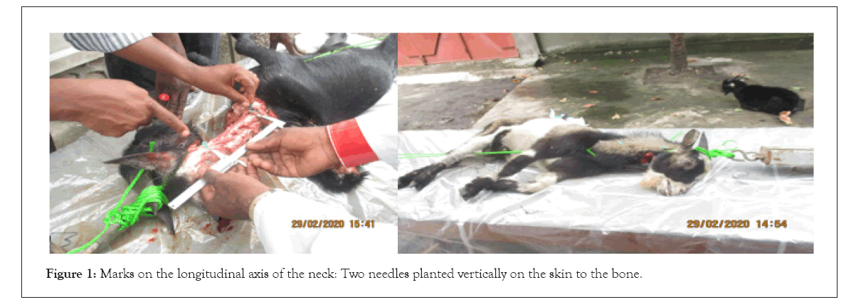
2. A table, 2 meters long, 50 cm wide and 1 meter high. Comprising two attachment points, one at the base of the goat’s skull, connected to the cervical traction device, the other at the base of the neck fixed on the traction table (Figure 1).

Figure 1: Marks on the longitudinal axis of the neck: Two needles planted vertically on the skin to the bone.
3. A dynamometer inserted between the cervical traction device and the strap attached to the base of the goat’s skull.
4. A calibrated cervical traction device, operating as an inter- support lever, with a maximum load set at 100 kg.
5. The calliper is the measuring instrument used when taking successive measurements as the tensile load increases from 0 to 100 kg following an arithmetic progression at the rate of 10 kgf at each sequence.
6. A clock was used in the collection of the time that a traction sequence lasts for each goat in order to determine the necessary useful time that a cervical traction can last.
7. The landmarks: two 5 cm long needles, planted vertically in the muscle, up to the level of the vertebra (Figure 1), over a distance of 14 cm from each other, along the longitudinal axis of the goat’s cervical spine, served as a benchmark, allowing measurement of spinal elongation during vertebral traction, both centrally and peripherally. According to the formula: Δl=l-l0 (Figure 1).
In which:
l0: this is the initial length of 14 cm as a reference or benchmark length, along the longitudinal axis of the goat’s neck; l: it is the length reached after each sequence of the traction; Δl=l-l0: this is the deviation or elongation obtained in relation to the initial length during traction.
Experimentation procedure
The activities took place according to the schedule below. Slaughtered shortly before the experiment, each goat was placed in lateral decubitus on the traction table, tied at the base of the neck and fixed at the base of the skull. The progressive traction starting from zero load, the progressive deviations corresponding to the progressive increase in the traction load following an arithmetic progression of 10 kg per sequence, are observed in terms of neck elongation and deformation of the anatomo- histological structures, up to the maximum load (set at 100 kg). During the experiment on group II, all the muscle mass and the skin around the neck over a distance of 20 cm were removed, leaving bare the vertebrae, cervical ligaments and some residues of the fleshy mass of the neck. The two needle markers, placed longitudinally along the axis of the neck, over a distance of 14 cm.
To minimize observation errors, two people were responsible for recording the data displayed during the experiment.
Variables of study
They were summarized in: Tensile loads in kg, Sequential duration of traction in minutes and Elongations observed at each sequence in millimetres (Table 1).
| Group: I | Group: II |
|---|---|
| D1: February 15. 2020: 2:00. to 4 pm | D1: February 15. 2020: 2:00. to 4 pm |
| D2: March 26. 2021: 7:30 to 9:00 | D1: March 31. 2021: 8:20 to 9:45 |
| D3: March 27. 2021 : 8 :00 to 9 am | D2: April 2. 2021: 8:17 to 9:40 |
| D4: March 28. 2021 : 8:25 to 9:00 | D3: April 3. 2021 8:12 to 9:45 |
| D5: March 29. 2021 : 8:00 to 9:00 | D4: April 09. 2021 : 8:45 to 10:00 |
| D6: March 30. 2021 : 9:30 to 10:00 | D5: April 10. 2021: 8:52 to 9:45 |
Table 1: Execution schedule of the experiment.
Statistical analysis
The Shapiro-Wilk normality test was used to determine the normality of the distribution of the main variables, and these variables were normally distributed (p ≥ 0.05). Therefore, Student’s parametric differential t-statistic test was used for comparison of independent means on discrete data.
Movement of the markers (needles) during traction
The two needles pressed vertically, moved clockwise during the pulling sequences, i.e. tilting to the right and not vertically (Table 2). For progressively increasing tensile forces from 0 to 100 kgf at a rate of 10 kgf per sequence, the maximum duration beyond which no elongation was observed was 5 minutes, during an experimental sequence. All the parameters remaining constant (duration of the sequence, tensile load), the elongations observed in the center of the cervical spine (Δ) (goats I, II, III, IV, V, VI), are far superior to those observed at the periphery (Δ’) in a ratio of 1 to 8. The progression of the elongation in the periphery is very weak evolving from 0 to 2 mm compared to that of the hundred (Table 3). For progressively increasing forces from 0 to 100 kgf at a rate of 10 kgf per sequence, no elongation is observed beyond 5 minutes during traction. All parameters remaining constant (duration of the sequence, tensile load), the elongations observed at the center of the cervical spine (Δ’) in goats (I, II, III, IV, V, VI) are far superior to those of the periphery (Δ) (ratio of 1 in 25). The progression of elongation in the periphery is very weak, evolving from 0 to 3 mm compared to that of the center which evolves from 0 to 25 mm. Peripheral elongation in Group II goats is not as pronounced as in Group I, ratio:1 of 2. We note a series of deformation of the cervical spine in the form of intervertebral dislocation, centred on C2, C3 on all 6 goats from 50 kgf to 80 kgf (Table 4).
| Duration of pull sequence In minutes (t) | Différences observed in mm in the 1st goat | Differences observed in mm in the 2nd goat | Différences observed in mm in the 3th goat | Différences observed in mm in the 4th goat | Différences observed in mm in the 5th goat | the deviations observed in the 6th goat | Déformations observed by sequ | Aérage of the déviations between the periphery and the center of group I | ||||||||||
|---|---|---|---|---|---|---|---|---|---|---|---|---|---|---|---|---|---|---|
| ∆l | ∆l’ | ∆ | ∆l’ | ∆ | ∆l’ | ∆ | ∆l’ | ∆ | ∆l’ | ∆ | ∆l’ | ∆ | ∆l’ | ∆ | ∆l’ | |||
| 0 | 0 | 0 | 0 | 0 | 0 | 0 | 0 | 0 | 0 | 0 | 0 | 0 | 0 | 0 | 0 | 0 | ||
| 3 | 0 | 11 | 1.8 | 8.8 | 0 | 4 | 0.6 | 2 | 1.2 | 2.2 | 0 | 1 | 0 | 0 | 0.6 | 4.8 | ||
| 5 | 1 | 12 | 1.2 | 14 | 0 | 5 | 1 | 6 | 1.4 | 5 | 1.8 | 1 | 0 | 0 | 0.9 | 7.3 | ||
| 3 | 3.5 | 11.6 | 1.6 | 14 | 2.5 | 9 | 1.6 | 6 | 1.5 | 9 | 1.2 | 2 | 0 | 0 | 1.9 | 8.6 | ||
| 4 | 4 | 12.5 | 1.7 | 16 | 2.5 | 12 | 1.8 | 8 | 1.4 | 11 | 1.6 | 2 | 0 | 0 | 2 | 8.9 | ||
| 5 | 4 | 12.5 | 1.7 | 16 | 3.8 | 14 | 1.8 | 10 | 1.5 | 15 | 1.7 | 3 | 0 | 0 | 2.6 | 10 | ||
| 5 | 4 | 12.5 | 1.7 | 17 | 3.8 | 16 | 2 | 12 | 1.4 | 16 | 1.7 | 4 | 0 | 0 | 2.4 | 14.2 | ||
| 5 | 4 | 13 | 1.7 | 19 | 4.5 | 16 | 2.2 | 16 | 1.4 | 16 | 1.7 | 5 | 0 | 0 | 2.4 | 13.2 | ||
| 5 | 4 | 14 | 1.7 | 20 | 3.6 | 17 | 2.2 | 17 | 1.4 | 17 | 1.7 | 11 | 0 | 0 | 2.4 | 16 | ||
| 5 | 5 | 14 | 1.7 | 21 | 3.6 | 19 | 2.6 | 19 | 1.8 | 19 | 1.7 | 11 | 0 | 0 | 2.7 | 17.1 | ||
| 4 | 5 | 15 | 1.7 | 21 | 3.6 | 19 | 2 | 20 | 1.8 | 22 | 1.7 | 21 | 0 | 0 | 2.3 | 17.3 | ||
| Total | 18.4 | 94.0 | ||||||||||||||||
Table 2: The variations of intervertebral spaces observed during traction in Millimetres (mm). According to the formula: ∆l=l-l0; on the goats of group I (with muscle Mass. ligaments and skin of the nec).
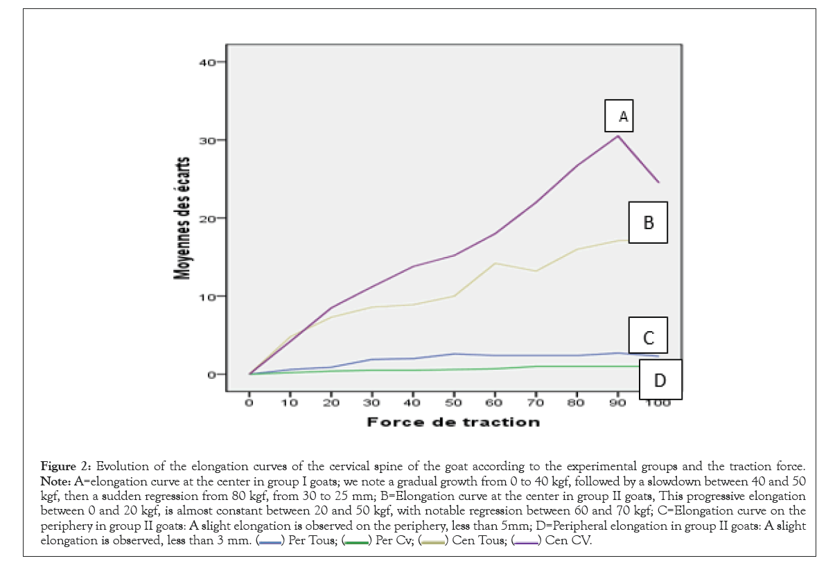
The comparison of the means between the elongation observed in the periphery and in the center within the same group I is clearly significant (p˂0.001). The elongation in the center is greater than that of the periphery. The same is true for the elongation observed in goats of group II (p˂0.001), the elongation observed in the center is greater than that observed in periphery. By comparing the average lengthening’s observed between the two groups in the periphery, we note a significant difference with a predominance in group I (p=0.001). In between groups, the comparison of the average lengthening of the center is as significant as in the periphery (p˂0.001) (Figure 2). The present experimental study based on the determination of deformation thresholds of the anatomo-histological structures of the cervical spine as well as its operating mechanism starting from the model observed in the goat, has made it possible to draw important information essential in the improvement of the grip in charge of degenerative pathologies with disco-radicular conflict.

Figure 2: Evolution of the elongation curves of the cervical spine of the goat according to the experimental groups and the traction force. Note: A=elongation curve at the center in group I goats; we note a gradual growth from 0 to 40 kgf, followed by a slowdown between 40 and 50 kgf, then a sudden regression from 80 kgf, from 30 to 25 mm; B=Elongation curve at the center in group II goats, This progressive elongation between 0 and 20 kgf, is almost constant between 20 and 50 kgf, with notable regression between 60 and 70 kgf; C=Elongation curve on the periphery in group II goats: A slight elongation is observed on the periphery, less than 5mm; D=Peripheral elongation in group II goats: A slight elongation is observed, less than 3 mm. 
The duration of the traction
The maximum sequential duration of experimental traction (useful time) for each of the 12 goats was 5 minutes with an average of 4.4 minutes (Tables 1 and 2), the time during which we observed maximum elongation, and beyond, which no column elongation was observed. This fact could either be justified by the fact that under the viscoelasticity of biological tissues according to Hooke’s laws, the elongation of a structure subjected to a tensile force is done in three important phases: The first is the phase elastic during which the structure elongates and as soon as the maximum load is reached, the plastic phase occurs, during which no elongation will be observed. Beyond this phase, rupture occurs during the overload [13].
| Tensile load in kg | Sequence duration in minutes | Différences observed in the 1th goat | Différences observed in the 2th goat | Différences observed in the 3th goat | Différences observed in the 4th goat | Différences observed in the 5th goat | Differences observed in the 6th goat | Synthesis the average of elongations observed | |||||||
|---|---|---|---|---|---|---|---|---|---|---|---|---|---|---|---|
| 0 | 0 | 14 | 14 | 14 | 14 | 14 | 14 | ||||||||
| ∆l | ∆l’ | ∆l | ∆l’ | ∆l | ∆l’ | ∆l | ∆l’ | ∆l | ∆l’ | ∆l | ∆l’ | ∆l | ∆l’ | ||
| 10 | 3 | 0.2 | 4 | 0 | 4 | 1 | 4 | 0 | 4 | 0 | 5 | 0 | 4 | 0.2 | 4.2 |
| 20 | 3 | 0.9 | 9 | 0 | 9 | 1.4 | 10 | 0.2 | 7 | 0 | 8 | 0 | 9 | 0.4 | 8.5 |
| 30 | 3 | 1.3 | 11 | 0 | 11 | 1.3 | 13 | 0.2 | 11 | 0 | 10 | 0 | 11 | 0.5 | 11.2 |
| 40 | 4 | 1.4 | 14 | 0 | 14 | 1.4 | 16 | 0.4 | 12 | 0 | 12 | 0 | 14 | 0.5 | 13.8 |
| 50 | 3 | 1.4 | 14 | 0.3 | 14 | 1.5 | 16 | 0.3 | 17 | 0.5 | 15 | 0.3 | 14 | 0.6 | 15.2 |
| 60 | 5 | 1.4 | 17 | 0.2 | 17 | 1.5 | 18 | 0.5 | 20 | 0.8 | 17 | 0.2 | 17 | 0.7 | 18 |
| 70 | 3 | 1.8 | 20 | 0.5 | 20 | 2 | 22 | 0.5 | 22 | 0.5 | 17 | 0.5 | 20 | 1 | 22 |
| 80 | 5 | 1.8 | 21 | 0.6 | 21 | 2 | 27 | 0.6 | 26 | 0.5 | 17 | 0.6 | 21 | 1 | 26.7 |
| 90 | 5 | 1.9 | 24 | 0.6 | 24 | 2 | 28 | 0.6 | 25 | 0.5 | 17 | 0.6 | 24 | 1 | 30.5 |
| 100 | 5 | 1.9 | 26 | 0.6 | 26 | 2 | 28 | 0.6 | 25 | 0.5 | 17 | 0.6 | 26 | 1 | 24.5 |
Table 3: The variations of intervertebral spaces observed during traction in Millimetres (mm). According to the formula: ∆l=l-l0; on the goats of group I (without muscle Mass. ligaments and skin of the neck).
Revision to reducing the necessary duration of a calibrated pull sequence, which is currently 15 minutes on average, is possible. But already other authors recognize that this time can be reduced to 3 minutes, maintaining that beyond this duration the traction no longer acts because the force is already reduced compared to the initial force [14]. The experiment on the stretching of certain muscles has shown that the traction force decreases after a few minutes of stretching (For example it decreases by 22 Newton after 5 minutes for the triceps and by 15 to 20 Newton after 4 to 5 stretching of quadriceps). The traction must be done for a determined time because it loses its effectiveness after too long a time [15]. On the other hand speaks of a continuous traction which can last 1 to 2 hours. The results of our second study of this thesis showed that cervical traction exercised according to the protocol defined in this work, only for an optimal force whose intensity is twice the weight of the head for a maximum duration of 15 minutes, already brought relief in 90% of cases [12]. This leads us to conclude that the minimum duration observed through the present in vitro experiment is close to reality. It remains that a study be conducted in the future to retain this value as obvious and applicable with satisfactory results.
| Groups | Means ± standard déviation | p value | |
|---|---|---|---|
| Intragroup GI | In periphery | 1.8 ± 0.9 | 0.000 |
| In the center | 10.7 ± 5.5 | ||
| Intragroup GII | In periphery | 0.6 ± 0.4 | 0.000 |
| In the center | 15.9 ± 9.6 | ||
| Intergroup in periphery | GI | 1.8 ± 0.9 | 0.001 |
| GII | 0.6 ± 0.4 | ||
| Intergroup in the center | GI | 10.7 ± 5.5 | 0.000 |
| GII | 15.9 ± 9.6 | ||
Table 4: Means of cervical spine elongation within the same group and between two groups.
| ∆ | ∆’ | ∆ | ∆’ | ∆ | ∆’ | ∆ | ∆’ | ∆ | ∆’ | ∆ | ∆’ | ∆ | ∆’ | ∆ | ∆’ | ||
|---|---|---|---|---|---|---|---|---|---|---|---|---|---|---|---|---|---|
| 0 | 0 | 0 | 0 | 0 | 0 | 0 | 0 | 0 | 0 | 0 | 0 | 0 | 0 | 0 | 0 | 0 | 0 |
| 10kg | 3 | 0 | 11 | 1.8 | 8.8 | 0 | 4 | 0.6 | 2 | 1.2 | 2.2 | 0 | 1 | 0 | 0 | 0.6 mm | 4.8 mm |
| 20kg | 5 | 1 | 12 | 1.2 | 14 | 0 | 5 | 1 | 6 | 1.4 | 5 | 1.8 | 1 | 0 | 0 | 0.9 mm | 7.3 mm |
Table 5: Extract from table 1 the average elongation of two 3 last columns.
The shift in deformation of the cervical spine
We observed during the experiment, significant differences between the elongation in the periphery and in the center. As an indication, for a total elongation of 13.6 mm in the center against 3.3 mm in the periphery in the goats of group II. And for a total lengthening of 94 mm in the center against 18 mm at the periphery in the goats of group I. We can conclude from these data that during cervical traction, the lengthening is done according to the principle of a shock absorber coiled, or it is the interior that oscillates more and the exterior is almost static, as observed during the movement of the markers during the pulling sequences. This fact is particularly important because, coupled with cervical lordosis, justifies the damping of gravity stresses in the preservation of the intervertebral discs of this segment of the body and therefore against premature wear.
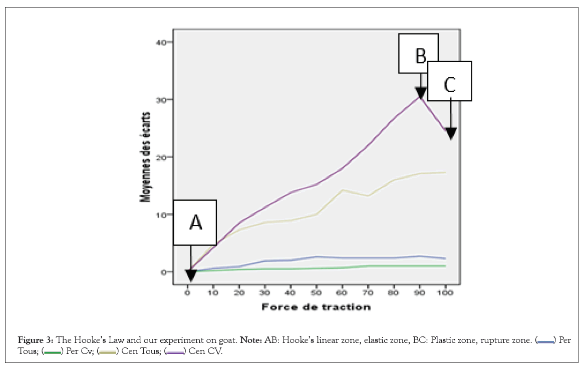
The application that we intend to make of this discovery is that during cervical traction, the elongation obtained cannot be measured on the basis of the extension of the external wall of the neck, this can remain identical both before and during the traction, the real elongation is reflected in the center of the neck along its vertical axial axis and is reflected in the shift displayed on the dynamometer incorporated in the traction device. Under these conditions, the determining factor for the effectiveness of the traction is rather the traction load mobilized by the device as well as the duration of this load and not the length of the external mass of the neck, because it is almost unnoticed. As we can see through this curve (graphic 1), the elongation at the center (axial) in group I goats is progressively increasing from 0 to 40 kg, it slows down between 40 and 50 kg and resumes progression for regress suddenly from 80 kgf of traction. Logically the points of modification of this curve correspond to the breaking points of the means of union of the vertebral column from 50 kgf, with a sudden drop from 80 kgf, corresponding to disjunction of C1 and C2. These lesions are not visualized due to the presence of muscle mass around the cervical spine. However, in accordance with Hooke’s law mentioned above, the three successive phases of the graph of a solid subjected to a tensile force are evident through our graph and reflect the deformation or vertebral disjunction (Figure 3).

Figure 3: The Hooke’s Law and our experiment on goat. Note: AB: Hooke’s linear zone, elastic zone, BC: Plastic zone, rupture zone. 

Physiological and anatomical deformations
As we can see in the images the proximal parts of the column are the most influenced during traction because the intensity is much more pronounced there, because they are close to the point of application of the force. Concerning the elongation in the center in group II, this progressive elongation between 0 and 20 kgf, is almost constant, a sign of structural integrity. Disturbances in this progression are observed between 20 and 50 kgf, but always in progression, with notorious regression between 60 and 80 kgf. These changes in the curve during progressive traction reflect the lesions observed on the bare column; these lesions vary from cracking to dislocation or joint dislocation after deformation of the ligaments: Regarding the elongation observed at the periphery in group I, as shown by curve C of graphic I, a slight elongation is observed at the periphery, less than 5 mm compared to the initial length. The same applies to the elongation observed on the periphery in group II, which is less than 3 mm. If we consider the two elongations during head oscillation movements during walking, we can assimilate the functioning of the cervical spine to a coil shock absorber, only the central axis is more stressed during oscillations. Thus, during cervical traction, the outer wall cannot be considered as a reference for the gain obtained, because it remains almost static with respect to the center. This disposition of the cervical spine, in addition to its physiological lordosis, largely contributes to the damping of the gravitational constraints of the weight of the head on the cervical spine.
The measurement of elongations during traction (extract from table 1 below)
The observation of the first two lines, shows us at the level of the last 2 columns the average of the elongations observed during the first two sequences: 0.6 mm and 0.9 mm on the periphery for 10 kg and 20 kg against 4.8 mm and 7, 3 mm in the center for the same loads. We consider its elongations compatible with the practice of cervical traction on degenerative disc disease. Considering that 4.8 and 7.3 are respectively the elongation observed over the entire cervical spine, the 7 intervertebral spaces would be elongated by at least 1 mm on average. Given that the height of the normal cervical intervertebral disc which is (5 to 6 mm and 10 to 12 mm for the lumbar region). This intensity is therefore normal and tolerable to reconstitute the pinched intervertebral space and allow the reconstitution by imbibition of the disc intervertebral if it is still viable (Table 5). The analysis of this figure illustrating the main sites of deformation of solids subjected to constraint forces according to Hooke’s law, allows us to identify 2 main zones: A elastic zone corresponding to the linear zone of Hooke’s law, B Plastic zone, corresponding to the maximum stress, It is the zone of rupture which corresponds to us with the deflection of the curve. In accordance with Hooke’s law mentioned in our introduction, the size of the deformation is proportional to the applied load “such extension such force” and that the mechanical stress/deformation curve of materials such as the skin, the ligaments and the intervertebral discs, exhibited a linear region at the onset of load application. It is that observed in figure, curve AB and the rupture zone. BC appears from 80 kg load for group I goats and from 50 kg for group II goats.
This experimental study on the threshold of deformation of the anatomical and histological structures of the cervical spine in the goat allowed us to focus on major questions relating to spinal manipulation, in particular cervical traction: The duration of a sequence of traction can be limited to less time than it takes to date, saving time for other impatient patients in the waiting room; lengthening of the neck during traction cannot in any case constitute a reference for better traction because the true elongation is unnoticed and is axial and this is tolerable. Finally, the breaking thresholds are from 50 kg for the cervical spine, this reassures us because our tractions remain below this threshold in the clinic. During cervical traction, the intensity is all the higher as one approaches the junction point with the traction force, therefore the proximal intervertebral spaces are more increased than those of the distal part. Cervical traction certainly leads to a release of the intervertebral space likely to release a nerve root in pain, this space, however small, allows the intervertebral disc to redo itself if necessary. We therefore recommend the use of this non-pharmacological and non-invasive technique, but able to relieve many patients suffering from common neck pain.
The deformations observed in group II goats, those caused during the experiment in group I goats could only be observed under imaging. Fortunately, in accordance with the above-mentioned stress/strain law, observation of this graph enabled us to detect the strain thresholds in the two groups.
The authors declare that they have no conflict of interest.
[Crossref] [Google Scholar] [PubMed]
[Crossref] [Google Scholar] [PubMed]
[Crossref] [Google Scholar] [PubMed]
[Crossref] [Google Scholar] [PubMed]
[Crossref] [Google Scholar] [PubMed]
[Crossref] [Google Scholar] [PubMed]
[Crossref] [Google Scholar] [PubMed]
[Crossref] [Google Scholar] [PubMed]
[Crossref] [Google Scholar] [PubMed]
Citation: Kiala GM (2023) Deformation Threshold of Cervical Spine Structures Subjected to Tensile Stress: In Vitro Experiment on Goats. Int J Phys Med Rehabil. 11:673.
Received: 31-Mar-2023, Manuscript No. JPMR-23-22739; Editor assigned: 30-Apr-2023, Pre QC No. JPMR-23-22739 (PQ); Reviewed: 18-Apr-2023, QC No. JPMR-23-22739; Revised: 26-Apr-2023, Manuscript No. JPMR-23-22739 (R); Published: 04-May-2023 , DOI: 10.35248/2329-9096.23.11.673
Copyright: © 2023 Kiala GM. This is an open-access article distributed under the terms of the Creative Commons Attribution License, which permits unrestricted use, distribution, and reproduction in any medium, provided the original author and source are credited.