Journal of Antivirals & Antiretrovirals
Open Access
ISSN: 1948-5964
ISSN: 1948-5964
Research - (2019)Volume 11, Issue 3
Background: Full length Ebolavirus glycoprotein (GP) intersperses the outer most lipid membrane to form spikes, where it mediates virus-host cell interaction. A secretory form of GP (sGP) is also produced by all 5 known Ebolavirus species. These attributes make GP an ideal target for research and development (R and D) of Ebolavirus and possibly pan-filovirus targeted Rapid Diagnostic Tests (RDTs), bio-therapeutics and vaccines. Prior cloning of recombinant Zaire ebolavirus (EBOV) GP has majorly used insect (baculovirus ) expression systems. We report the cloning, expression and purification of the full length and extracellular domain (ECD) forms of recombinant EBOV GP in mammalian cell-lines.
Methods and results: 2034 and 1956 base-pair (bp) coding DNA sequences corresponding to the 669 and 643 amino acids (aa) residues of full length and ECD forms of EBOV GP were sub-cloned into the pTGE plasmids. Recombinant pTGE-plasmids were used to transfect 293-6E HEK mammalian cells grown in serum-free FreeStyleTM 293 Expression Medium. Cell lysates and or culture supernatants were used to obtain purified protein, followed by analysis on SDS-PAGE and Western blot. Purified full length GP was detected as membrane bound protein in cell lysates with estimated molecular weight of ~100 kDa (Cal.M.W.~71.67 kDa) on Western blot; and 0.02 mg GP (Concentration: 0.2 mg/mL, Purity: ~50%) derived. On the contrary, ECD GP was detected in supernatants of cell culture broth with estimated molecular weights of ~116 kDa based on SDS-PAGE and Western blot; and 1.6 mg (Concentration: 0.4 mg/ml, Purity: ~70%) of GP_ECD was obtained.
Conclusion: Within mammalian cells, recombinant full length EBOV GP is predominantly expressed as transmembrane protein (tGP), while ECD GP is eluted into the culture medium. Both recombinant forms of GP are critical for the R and D of rapid diagnostic tests (RDTs).
Ebola virus ; Glycoprotein; RDTs; Therapeutics; Vaccines
Ebolavirus is one of two common genera of the enveloped, nonsegmented single-stranded RNA virus family Filoviridae, order Mononegavirales [1]. Filoviruses are the cause of rare but highly fatal viral hemmorhagic fever (VHFs) in remote villages of equatorial Africa [1,2]. Between Dec 2013 through to April 2016, the most widespread outbreak of ebola virus disease(EVD) in history occured in the West African countries of Guniea, Liberia and Sierra Leone [3]. WHO documents a total of 28, 616 cases, with 11, 310 deaths [4]. The occurence of several cases involving travellers to Europe and America underscored EVD as a public health emergency of international concern (PHEIC) [5]. As of today, there is an on-going outbreak of EVD in 3 eastern provinces of the Democratic Republic of Congo-DRC (North Kivu, Ituri and Goma) that has intermitently spilled over to Uganda and continues to threaten the health-security of other neighbouring countries and populations.
Species members of the genera Ebola virus and its relative Marburgvirus are structurally similar [2,3,6]. Only one species of Marburgvirus (Marburg Marburgvirus-MARV) is known. On the other hand, there are five known species of Ebola virus, namely Bundibugyo Ebola virus (BDBV), Zaire Ebola virus (EBOV), Reston Ebola virus (RESTV), Sudan Ebola virus (SUDV), and Tai Forest Ebola virus (TAFV). At the deoxyribose acids (DNA) level, both filovirus genomes have seven open reading frames. These encode the virion envelope glycoprotein (GP), nucleoprotein (NP), matrix proteins VP24 and VP40, nucleocapsid associated proteins VP30 and VP35, and the viral polymerase [6].
The EBOV GP gene—through transcriptional editing, encodes two glycoproteins. The major product is a soluble, dimeric glycoprotein (sGP) that is secreted abundantly. A minor product, resulting from transcriptional editing, is the transmembrane-anchored, trimeric viral surface glycoprotein (tGP). tGP is responsible for cell targeting and virus entry by mediating receptor binding and membrane fusion [7-10]. Both these GP forms primarily comprise of two disulfide-linked subunits (denoted GP1 and GP2) that are generated by proteolytic cleavage of the GP precursor (Gp1, Gp2) effected by the cellular subtilisin-like protease furin [11,12]. EBOV and MARV GP1, Gp2 pre-proteins share 31% identity in amino acid sequence—with most variabilities seen in the middle region, and conservation of the N- and C-terminal regions [13-17]. Surface GP is a trimeric type I transmembrane protein (tGP) that forms structural spikes at the surface of infected cells and virions [15]. Naturally, MARV only exhibits the transmembrane type of GP (tGP), while EBOV also manifests a secretory form of GP (sGP). Contrary to EBOV that requires transcriptional editing to express tGP, the GP gene of MARV is organized in a way that transcription results in a single sub-genomic RNA species used for the synthesis of the full-length envelope glycoprotein [13,14]. As a result, MARV does not express the sGP that is synthesized from the non-edited mRNA species during EBOV infection and secreted into the culture medium [14]. Expression of tGP by EBOV is limited during virus replication, since most GP gene-specific mRNAs (80 %) is directed towards synthesis of the small secreted non-structural glycoprotein (sGP) [13]. In addition, significant amounts of tGP are shed from the surface of infected cells due to cleavage by the cellular metalloprotease tumour necrosis factor alpha-converting enzyme (TACE) [14].
Our team has previous rigorously explored the biophysical profiles of filovirus GP with an interest in devising pan-filovirus rapid diagnostic tests (RDTs) [18,19]. Current EBOV antigen targeted RDTs detect the matrix protein antigen-VP40 [20,21]. By virtue of its surface localization and secretory form, GP is an ideal target for research and development (R andD) of filovirus specific rapid diagnostic tests (RDTs), bio-therapeutics and vaccines [20-28]. Most previous studies expressing recombinant EBOV GP have exploited an insect (baculovirus) expression system. Such systems might, however, not generate recombinant GP with close to natural post-translational modifications seen during human EBOV infection [10-20]. Here, we report the cloning, expression and purification of the full length and extracellular domain (ECD) forms of EBOV GP in mammalian cell-lines.
Cloning of the full length and ECD forms of EBOV GP cDNA into pTGE plasmids
The 2034 nucleotide base-pair (bp) coding DNA sequences corresponding to the 669 amino acids (aa) residues of full length EBOV GP were obtained from National Center of Biotechnology Information (NCBI) Genbank (ID: ALT19603.1 and accession #HALT19603) and respective cDNA synthezed (Figure 1). Through target-site mutation (truncation), the same cDNA was modified to generate the 1956 nucleotide bp coding DNA sequences corresponding to the 643 amino acids (aa) long residues of the extracellular domain (ECD) variant of EBOV GP (Figure 2). These were then sub-cloned into the pTGE plasmids.
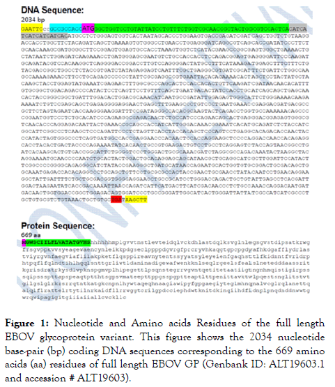
Figure 1. Nucleotide and Amino acids Residues of the full length EBOV glycoprotein variant. This figure shows the 2034 nucleotide base-pair (bp) coding DNA sequences corresponding to the 669 amino acids (aa) residues of full length EBOV GP (Genbank ID: ALT19603.1 and accession # ALT19603).
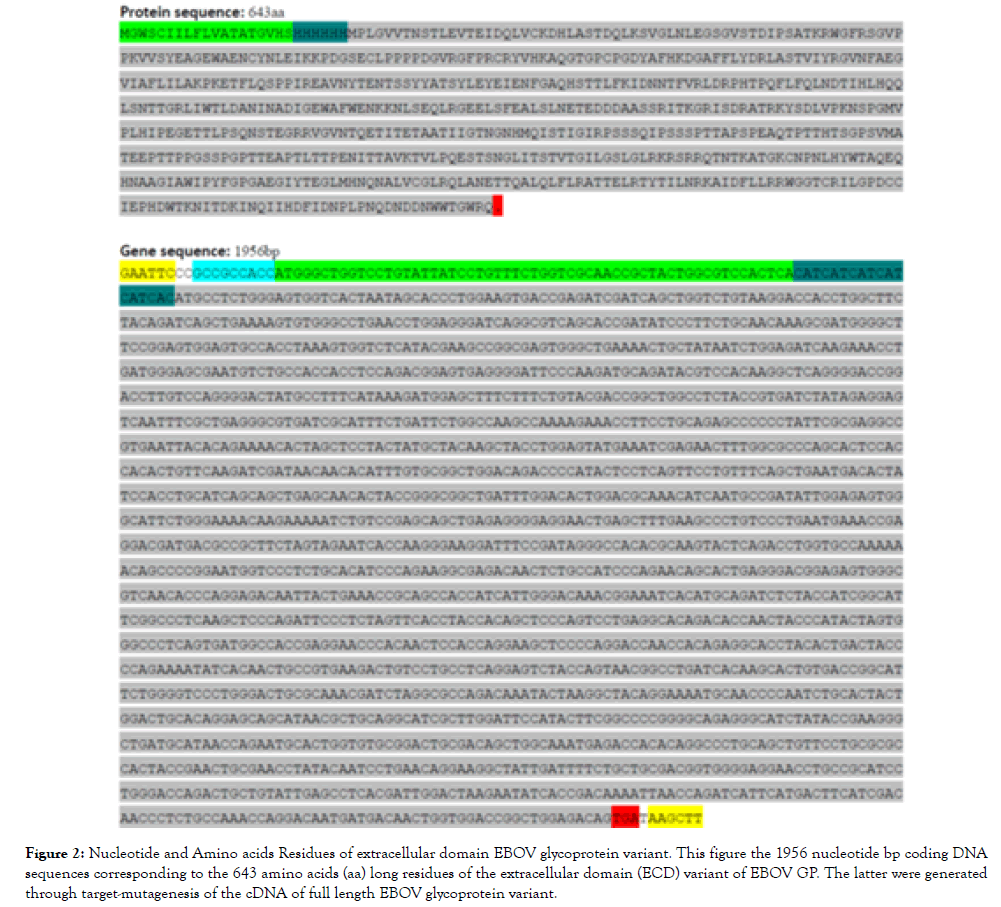
Figure 2. Nucleotide and Amino acids Residues of extracellular domain EBOV glycoprotein variant. This figure the 1956 nucleotide bp coding DNA sequences corresponding to the 643 amino acids (aa) long residues of the extracellular domain (ECD) variant of EBOV GP. The latter were generated through target-mutagenesis of the cDNA of full length EBOV glycoprotein variant.
On one hand, (a) for full length EBOV GP—a Kozak sequence, ATG, an Artificial Signal Peptide, and 6xHis were inserted upstream of the cDNA followed by a stop codon. The artificial signal peptide is required to drive protein expression within the defined environment, and various proteins and expression systems demand different artificial signals [8]. The resulting artificial gene construct was placed between the EcoRI and HindIII restriction sites at the 5' and 3' primer ends, respectively (Figure 3).

Figure 3. Construct of the pTGE plasmid vector for full length EBOV glycprotein-variant expression. This figure shows the construct of the pTGE plasmid vector for full length EBOV glycprotein expression. Specifically, a Kozak sequence, ATG, an Artificial Signal Peptide, and 6xHis were inserted upstream of the cDNA of full length zEBOV GP followed by a stop codon. The issuing construct was placed between the EcoRI and HindIII restriction sites at the 5' and 3' primer ends, respectively.
On the other hand, (b) For the ECD of EBOV GP, a Kozak sequence, an Artificial signal peptide, and 6xHis tag were placed upstream of the cDNA of EBOV ECD-GP (33-671 aa optimized for HEK293) followed by a stop condon. As the case above, the resulting artificial gene construct was flanked by the EcoRI and HindIII restriction sites at the 5' and 3' primer ends, respectively (Figure 4).

Figure 4. Construct of the pTGE plasmid vector for extracellular domain EBOV glycprotein-variant expression. This figure shows the construct of the pTGE plasmid vector for extracellular domain EBOV glycprotein-variant expression. A Kozak sequence, an Artificial signal peptide, and 6xHis tag were placed upstream of the cDNA of zEBOV ECD-GP (33‐671 aa optimized for HEK293) followed by a stop codon. As the case above, the issuing construct was flanked by the EcoRI and HindIII restriction sites at the 5' and 3' primer ends, respectively.
Cell cultures and transfection
A freshly thawed innoculum of 293-6E cells was seeded and grown in serum-free FreeStyleTM 293 Expression Medium (Life Technologies, Carlsbad, CA, USA). The cells were maintained in Erlenmeyer Flasks (Corning Inc., Acton, MA) at 37°C with 5% CO2 on an orbital shaker (VWR Scientific, Chester, PA). One day before transfection, the cells were seeded at an appropriate density in Corning Erlenmeyer Flasks. On the day of transfection, DNA and PEI transfection reagent (Polysciences, Eppelheim, Germany) were mixed in the ratio of 1:4 (2 μg plasmid DNA for 8 μL Transporter™) and then added into the flask with cells ready for transfection. The plasmid DNA encoding GP was transiently transfected into 100 mL suspension 293-6E cells.
Purification and analysis
The purification of recombinant full length protein and extracellular domain took different patterns. (a) The the initial attempt to purify full length EBOV Gp using culture supernatants failed to yield substantial amounts of the protein, possibly because of the trans membrane localization of full length GP that restricts it localization to the cell membranes. Specifically, centrifugation supernatants of cells collected at day 4 post-transfection were purified by loading onto Ni Sepharose 6 FF 0.3 mL resins. The samples from purification step were analyzed by SDS-PAGE and Western blot analysis as shown in Figure 5.
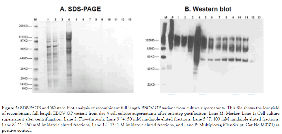
Figure 5. SDS-PAGE and Western blot analysis of recombinant full length EBOV GP variant from culture supernatants. This file shows the low yield of recombinant full length EBOV GP variant from day 4 cell culture supernatants after one-step purification. Lane M: Marker, Lane 1: Cell culture supernatant after centrifugation, Lane 2: Flow-through, Lane 3~4: 50 mM imidazole eluted fractions, Lane 5~7: 100 mM imidazole eluted fractions, Lane 8~11: 250 mM imidazole eluted fractions, Lane 12~13: 1 M imidazole eluted fractions, and Lane P: Multiple-tag (GenScript, Cat.No.M0101) as positive control.
This attempt to purify the target full length GP from cell culture supernatant was unsuccessful as very little protein could be detected in eluted fractions. This shows that the protein is not well expressed in suspension 293-6E cells; and that attempts to purify it from supernatants yield only minimal protein that is not bound well onto the resin. The existing literature denoting full length GP as a transmembrane protein led us to optimize strategies for purifying the same protein from cell-lysates rather than supernatants. Cell lysate collected from day 1 to day 4 posttransfection were prepared and analyzed by SDS-PAGE and Western blot as shown in Figure 6. The cells were harvested at day 4 post-transfection, lysed to collect the target protein. The extracted membrane bound proteins were loaded onto Ni Sepharose 6 FF 0.3 mL resin to capture target protein, and analyzed by SDS-PAGE and Western blot as shown in Figure 7. The fractions containing purified GP were collected and desalted to PBS, with 0.1% DDM, pH 7.2. The final products were analyzed by SDS-PAGE and Western blot analysis as shown in Figure 8.
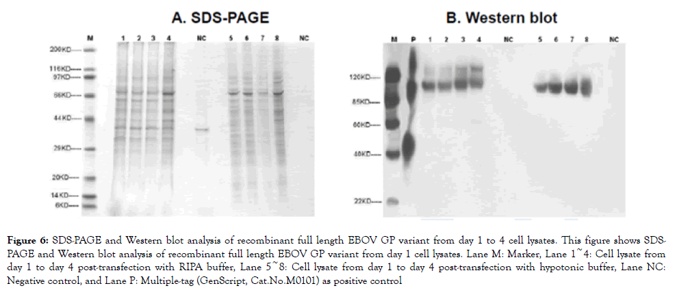
Figure 6. SDS-PAGE and Western blot analysis of recombinant full length EBOV GP variant from day 1 to 4 cell lysates. This figure shows SDSPAGE and Western blot analysis of recombinant full length EBOV GP variant from day 1 cell lysates. Lane M: Marker, Lane 1~4: Cell lysate from day 1 to day 4 post-transfection with RIPA buffer, Lane 5~8: Cell lysate from day 1 to day 4 post-transfection with hypotonic buffer, Lane NC: Negative control, and Lane P: Multiple-tag (GenScript, Cat.No.M0101) as positive control
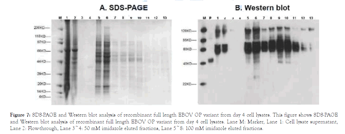
Figure 7. SDS-PAGE and Western blot analysis of recombinant full length EBOV GP variant from day 4 cell lysates. This figure shows SDS-PAGE and Western blot analysis of recombinant full length EBOV GP variant from day 4 cell lysates. Lane M: Marker, Lane 1: Cell lysate supernatant, Lane 2: Flow-through, Lane 3~4: 50 mM imidazole eluted fractions, Lane 5~8: 100 mM imidazole eluted fractions.
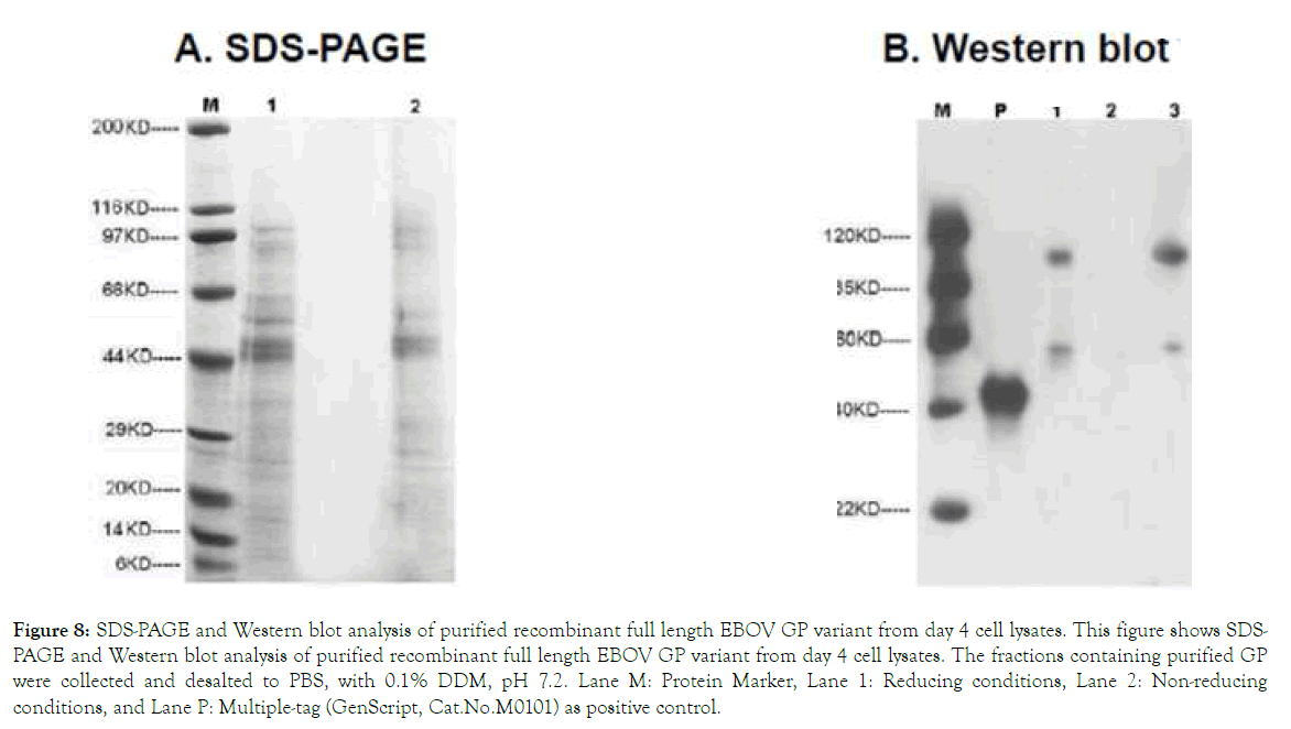
Figure 8. SDS-PAGE and Western blot analysis of purified recombinant full length EBOV GP variant from day 4 cell lysates. This figure shows SDSPAGE and Western blot analysis of purified recombinant full length EBOV GP variant from day 4 cell lysates. The fractions containing purified GP were collected and desalted to PBS, with 0.1% DDM, pH 7.2. Lane M: Protein Marker, Lane 1: Reducing conditions, Lane 2: Non-reducing conditions, and Lane P: Multiple-tag (GenScript, Cat.No.M0101) as positive control.
On the other hand, (b) The purification of ECD-GP was done in a similar way as full length GP, except that after centrifugation, the filtered supernatant was loaded onto HisTrapTM FF Crude 5 ml (GE, Cat.No.17-5286-01) at 3.0 ml/ min. After washing and elution with appropriate buffer, the eluted fractions were pooled and buffer exchanged to PBS, pH 7.2. The purified protein was analyzed by SDS-PAGE and Western blot by using standard protocols for molecular weight, yield and purity measurements. The primary antibody for Western blot was Mouse-anti-his mAb (GenScript, Cat.No.A00186). The target protein GP_ECD was detected with estimated molecular weights of ~116 kDa based on SDS-PAGE and Western blot analysis as shown in Figure 9. 1.6 mg (Concentration: 0.4 mg/ml, Purity: ~70%) of GP_ECD was obtained from 400 ml of cells at a density of 2–3 × 10⁶ cells ml−1.
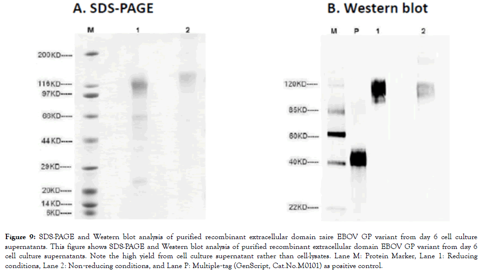
Figure 9. SDS-PAGE and Western blot analysis of purified recombinant extracellular domain zaire EBOV GP variant from day 6 cell culture supernatants. This figure shows SDS-PAGE and Western blot analysis of purified recombinant extracellular domain EBOV GP variant from day 6 cell culture supernatants. Note the high yield from cell culture supernatant rather than cell-lysates. Lane M: Protein Marker, Lane 1: Reducing conditions, Lane 2: Non‐reducing conditions, and Lane P: Multiple‐tag (GenScript, Cat.No.M0101) as positive control.
We report the cloning, expression and purification of the full length and extracellular domain forms of EBOV GP within 293-6E HEK mammaliian cell-lines. Whereas majority prior attempts to aimed to express recombinant GP have exploited insect (baculovirus) expression systems, mammalian cell expressed recombinant forms of GP might be more representative of GP proteolytic and or post-translational modifications that occur in vivo, thereby making them more appropriate for R and D of EBOV targeted RDTs, biotherapeutics and vaccines.
The expression of both EBOV GP forms was achieved by sub-cloning their respective coding DNAs into pTGE plasmids (Figures 1-4). Consistent with the natural expression of the transmembrane form of GP (tGP), full length GP was only recovered from cell-lysates. Specifically, purified full length GP was detected as membrane bound protein in cell lysates with estimated molecular weight of ~100 kDa (Cal.MW. ~71.67kDa) on Western blot; and 0.02 mg GP (Concentration: 0.2 mg/mL, Purity: ~50%) derived (Figures 5-8). On the other hand, extracellular domain (ECD) GP was expressed in a secretory manner similar to what is observed during viral expression of the sGP variant of EBOV. This was detected in supernatants of cell culture broth with estimated molecular weights of ~116 kDa based on SDS-PAGE and Western blot; and 1.6 mg (Concentration: 0.4 mg/ml, Purity: ~70%) of GP_ECD was obtained (Figure 9). Thus, similar to its behavior on the EBOV virions, recombinant full length GP is predominantly expressed as transmembrane protein (tGP) within mammalian cell-lines [7-20]. On the other hand, ECD-GP is eluted into the culture medium (presumably following similar expression pattern of sGP), thereby being captured in the supernatants. This makes the expression of recombinant ECD-GP in mammalian cells similar to that of sGP. Overall, these data point to differences in expression-patterns of the two recombinant forms of EBOV GP in mammalian cells. No drastic differences were noted between the measured and theoretically (in silico) predicted in biophysical profiles of both forms of GP [19].
In conclusion, recombinant full length EBOV GP is predominantly expressed in mammalian cells as transmembrane protein (tGP) while ECD-GP is eluted into the culture medium. Both these recombinant GP forms have proven relevant towards the R and D of rapid diagnostic tests (RDTs), just as they may for biotherapeutics and or vaccines.
Ethics approval and consent to participate
This study obtained ethical approval and clearance from the School of Biomedical Sciences Institutional Review and Ethics Committee (IREC), College of Health Sciences, Makerere University, Kampala, UG (Protocol Approval #s : SBS 138 and 228); as well as the Uganda National Council of Science and Technology (UNCST; Protocol Clearance # HR 1521). Specific waiver approval to use 293-6E HEK mammalian cell-lines was also obatined.
Consent to publish
All authors read and approved of this draft of the manuscript for publication.
Availability of data and materials
All data and material required to replicate this work is supplied in the manuscript. Gene sequences of the cloned zaire Ebola virus glycoprotein are accessible at the National Center for Biotechnology Information: Genbank id: ALT19603.1 accession # ALT19603
Competing interests
MW has filed a PCT for conserved B cell epitopes of filovirus GP and derivative PAbs and MAbs (see: Wayengera M. Conserved B cell epitopes of filovirus glycoprotein and their use as either biomarkers or therapeutics and sub-unit vaccines for Ebola virus and Marburg virus International Application #:PCT/IB2014/066251; Pub #:WO/2016/079572; Date: 26.05.2016; Filing Date:21.11.2014). MW is Chief Scientific Officer at Restrizymes Biotherapeutics (U) LTD—a Ugandan start-up biotech.
Funding
This work was made possible by two grants to MW from Grand Challenges Canada and the Government of Canada i.e, Rising Stars in Global Health award # S4_0280-01; and a Transition to Scale award # TTS-0709-05. MW currently holds a European Developing Country Clinical Trails (EDCTP) Career Fellowship # TMA2016CDF-1545. MW is recipent of emergency Ebola funding under the EDCTP Mobilization of research funds in case of Public Health Emergencies, grant # RIA2018EF-2081.
Author’s contribution
MW conceived the idea for this study. PB, CM, SK, JJL and MW conducted the experiments. RD, JJL, EM, JW, JP, MLJ and MW provided technical guidance and materials and reagents. MW wrote the initial draft of the manuscript.
Acknowledgements
The authors thank Ms. Geraldine Nalwadde and Ms. Joanitta B Birungi who carried out the administrative work for these projects. The staff of the Immunology Laboratory, Department of Immunology and Molecular Biology at the School of Biomedical Sciences, MakCHS, Kampala, UG and Arbovirology and CDC-Filovirology Laboratories at the Uganda Virus Research Institute-UVRI, Entebbe, UG offered indispensable support. We thank Mr. Ken Simiyu and Mr. Andrew Taylor who acted as program officers at Grand Challenges Canada.
Citation: Wayengera M, Babirye P, Musubika C, Kirimunda S, Joloba ML (2019) Cloning, Expression and Purification of recombinant forms of Full Length and Extracellular Domain EBOV Glycoprotein within Mammalian Cell-Lines. J Antivir Antiretrovir. 11:187. doi: 10.35248/1948-5964.19.11.187
Received: 12-Sep-2019 Accepted: 26-Sep-2019 Published: 03-Oct-2019 , DOI: 10.35248/1948-5964.19.11.187
Copyright: © 2019 Wayengera M, et al. This is an open-access article distributed under the terms of the Creative Commons Attribution License, which permits unrestricted use, distribution, and reproduction in any medium, provided the original author and source are credited.
Sources of funding : This work was made possible by two grants to MW from Grand Challenges Canada and the Government of Canada i.e, Rising Stars in Global Health award # S4_0280-01; and a Transition to Scale award # TTS-0709-05. MW currently holds a European Developing Country Clinical Trails (EDCTP) Career Fellowship # TMA2016CDF-1545. MW is recipent of emergency Ebola funding under the EDCTP Mobilization of research funds in case of Public Health Emergencies, grant # RIA2018EF-2081.