Journal of Medical & Surgical Pathology
Open Access
ISSN: 2472-4971
ISSN: 2472-4971
Research Article - (2020)Volume 5, Issue 2
Ewing’s sarcoma is the second most common childhood bone neoplasm, which is considered rare in African population. This is the first study in Sudan. The aim of the study is to demonstrate the clinical and histo-morphological findings of these tumours and to compare the results with similar studies. A retrospective cross-sectional study was done, conducted over 6 years’ period on 82 cases diagnosed as ES/PNET at Histo-Centre located in Khartoum state. Twenty-six of them were found to have CD99 immune-stain and ten cases have PAS special stain, data were collected, H and E, PAS and CD99 stained slides were examined by light microscope. Data analysis showed that the peak incidence of ES/PNET occurs between 5 to <18 years (58.5%) with a Mean age: 16.9 years. It shows obvious male predominance accounting for 64.6% of cases. The majority of cases are intra osseous tumour, with only 11% of cases are extra osseous, the lower limb is the most common site accounting for 54.9% of the cases, followed by the flat bones of the pelvis 13.4% with the vertebra and the skull being the least common sites accounting for 1.2% each. In clinical presentation 74.4% presented with localized swelling, 34% as localized pain, Fever and pathological fractures were the presentation in 2.4% and 3.7% respectively and none of the study population were presented as metastatic disease. Microscopic examinations of the H and E stained slides showed that 57.3% of the cases are ES devoid of neural differentiation while the rest are PNET showing some degree neural differentiation devoid of neural differentiation, among PNET cases, Homer Wright rosette were the most common rosettes in about 70% of cases. Focal areas of atypical features were found in 15.9% of cases with the pleomorphic large cells and spindle cells being the most commonly encountered features. Necrosis is present in 52.4% of cases. PAS was positive in 9 out of the 10 cases. While all the 26 cases with CD99 stained slides were positive, only 61.5% showed diffuse strong membranous positivity. In Conclusion although the demographic features and clinical presentations of ES/PNET in the study population were found to be similar to other global and regional studies finding 82 cases/6 years in single centre makes its rarity among African nations doubtful.
Ewing’s sarcoma; Primitive neuroectodermal tumor; CD99 immune stain; Sudan
Ewing’s sarcoma families of tumours are group of aggressive malignant neoplasms. They are the second most common childhood bone tumours after osteosarcoma accounting for 5%- 10% of paediatric bone tumours [1], in Sudan there is no data registry for Ewing’s sarcoma and more studies and documentation are required in this field. A common feature shared among these group of sarcomas is the presence of chimeric transcript, resulting from fusion of the EWS gene with genes that encode for structurally related transcription factors, usually FLI1 or ERG2 [2] .They show a morphologic features of undifferentiated small round blue cell tumour [3] with varying degree of neuroendocrine differentiation. Ewing’s sarcoma family of tumours is in general called ES/PNET and include: Ewing sarcoma of bone, extra-osseous Ewing’s sarcoma and peripheral primitive neuroendocrine tumour which can be osseous or extra-osseFous, and called Askin tumour when it occurs in the chest wall [4]. The distention between Ewing sarcoma and PNET is based on the degree of neural differentiation, ES is poorly differentiated tumour where’s PNET is showing some evidence of neural differentiation [5]. ES/PNET typically occurs as skeletal mass in the second decade of life with strong male predilection and well known racial variation [6,7]. Clinically patients with ES/PNET present with pain located at the site of the tumour, some present with palpable swelling and one third of patients relate the onset of the symptoms to previous minor trauma [8]. However it’s not uncommon for ES/PNET to present as fever and leukocytosis mimicking osteomyelitis [9,10]. Histologically ES/PNET displays a spectrum of morphologic variations and variable degrees of differentiation making it sometimes difficult to reach final diagnosis without ancillary tests which include special stains and IHC stains panel and sometimes the final diagnosis cannot be reached without molecular studies which are not available in our country, so this study was focused on analyzing the available tests aiming to improve the diagnostic yield for this aggressive neoplasm.
This is a cross-sectional retrospective laboratory based study conducted in Histo Centre which is located in Khartoum state. The study covered all the cases diagnosed as Ewing sarcoma during the period from January 2013 till November 2018 which is 82 cases, 26 of them have CD99 immune-stain and 9 have PAS special stain. The clinical information of the patients was obtained from the patient records and the slides were examined under the light microscope.
The study results show that the majority of the patients (58.5%) are within the age group 5 to <18 years with the mean age 16.9 years. 3.7% below 5 years and 4.9% above 40 years (Table 1). Distribution of sex shows that 64.6% (53/82) are males and 35.4% (29/82) of cases are females. Eighty-nine percent cases are intra osseous tumour and 11% are extra osseous with the long bone of the lower limbs being the most common site accounting for 54.9% (45/82) followed by the flat bones of the pelvis 13.4% (11/82), long bones of the upper limbs 6.1% (5/82), ribs, clavicle and sternum all together 4.9% (4/82), small bones of the hands and feet 3.7%, skull and vertebrae 1.2% (1/82) each (Figure 1). Regarding the clinical presentation of the patients as mentioned by the treating clinicians 74.4% presented with localized swelling, followed by pain then pathological fractures and 2.4% presented with fever. The specimens sent for diagnosis was biopsies in 81.7% (67/82) of cases and excision or amputation in 18.3% (15/82). H and E stained slides demonstrates that 57.3% (47/82) of cases are small round blue cell tumour without neural differentiation (ES) and 42.7% (35/82) are showing some degree of neural differentiation in the form of rosettes (PNET), the majority of the rosettes are Homer Wright rosettes (Figures 2-5). Necrosis was found in 52.4% (43/82) of cases which was prominent in 25.6% (21/82). Focal areas of atypical features were found in 15.9% of cases with the pleomorphic large cells and spindle cells being the most commonly encountered features. PAS stained slides showed positivity in 9 out of the 10 cases. CD99 is positive in 100% of cases (26/26) with different patterns of positivity, strong membranes (3+) in 61.5%, strong focal (2+) in 23.1% and faint patchy (1+) in 15.4% of cases (Figures 6-8).
| Frequency | Percent | |
|---|---|---|
| <5 | 3 | 3.7 |
| 5 to <18 | 48 | 58.5 |
| 18 to < 40 | 27 | 32.9 |
| Above 40 | 4 | 4.9 |
| Total | 82 | 100 |
Table 1: Patients with age.
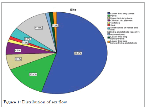
Figure 1: Distribution of sex flow.
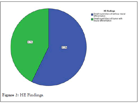
Figure 2: HE Findings
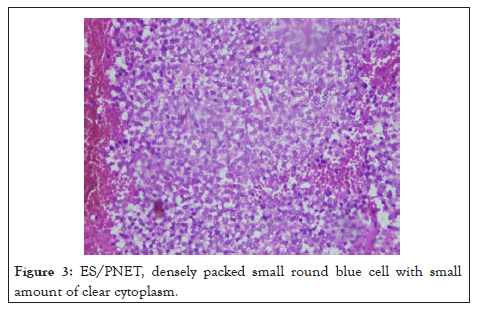
Figure 3: ES/PNET, densely packed small round blue cell with small amount of clear cytoplasm.
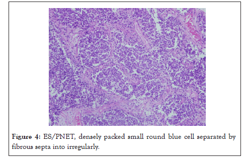
Figure 4: ES/PNET, densely packed small round blue cell separated by fibrous septa into irregularly.
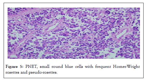
Figure 5: PNET, small round blue cells with frequent Homer-Wright rosettes and pseudo-rosettes.
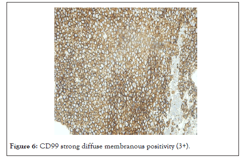
Figure 6: CD99 strong diffuse membranous positivity (3+).
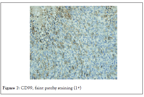
Figure 7: CD99, faint patchy staining (1+)
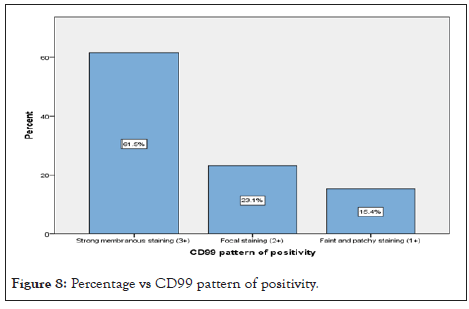
Figure 8: Percentage vs CD99 pattern of positivity.
ES/PNET is an aggressive small round blue cell tumour with significant racial variability, stated to be rare among patients from African descents like Sudanese’s population. In such a developing country, the diagnosis of ES/PNET is a challenge due to the limited resources and the complete absence of important diagnostic tools like molecular analysis which is now considered to be essential for the diagnosis of such tumors. Complete panel of immune-histochemical stains are costly and not available most of the time. Thus the diagnosis of ES/PNET mostly depends on careful interpretation of clinical, histo-morphological findings and the minimal number of IHC stains. In this study we reviewed 82 cases of ES/PNET, twenty-six of them were found to have CD99 immune-stain. The study population was found to have a wide age range from less than 5 years to 47 years. The great majority (58.5%) are within the age group 5 to <18 years the neoplasm was found to be rare below 5 and above 40 years. Our findings are consistent with the global figures and studies among non-African populations [11,12]. The mean age of 16.9 years in our patients is much lower compared to other studies [13,14] reflecting a narrower age range in the study population? The majority of patients were males showing obvious male gender predilection in concordance with other studies [15]. Regarding the site of the tumour, more than 85% are intra osseous while extra osseous sites account for only 11%. The lower limbs were the most common site representing 52.2% of the cases followed by the flat bone of the pelvis 19.5% supporting similar results in some studies [7-13] despite a much higher percentage in our study for the lower limbs as primary site, and opposed to other small study that shows truncal condensation (63.6%) [16]. Most of the patients presented with localized swelling 74.4% and 34% presented with localized pain, a minority related the compliant to previous trauma and other small fraction reported nocturnal pain. Fever and pathological fractures were the presentation in 2.4% and 3.7% respectively. Those presentations are similar to that mentioned in literature [8,9], but unlike some studies that found presentation as metastatic disease in about 30% of cases [17] none of our patients presented as metastatic disease. Microscopic examinations of the H&E sections showed densely packed small round blue cell tumour, 57.3% of them were ES devoid of any neural differentiation while the rest 42.7% were PNET with some degree of neural differentiation, making ES more common in our population like other study which shows predominance of Ewing sarcoma [15]. Ewing sarcoma highest incidence is in the age group between 5 to <18 years with only 8.3% of PNET occurring in this age group. PNET is defined by the presence of neural differentiation in the form of rosettes; three types of rosettes were encountered. Homer Wright rosette were the most commonly found type of rosettes, a finding that is similar to what is stated in the literature followed by pseudo rosettes and the least ones were the Flexner-Winterstainer rosettes. Necrosis was found in 50% of cases, in 25.6% of them the necrosis was prominent involving more than 50% of the tumour. Apart from the small round blue cells about 15.3% of cases found to have focal areas of atypical features which is slightly lower compared with the 20% found in two other studies [18,19]. Putting this in mind is important when interpreting small biopsies taken from the areas with atypical features. Large pleomorphic cells and spindle cells were the most commonly encountered features. PAS stain was positive in 9 out of the 10 cases with cytoplasmic positivity supporting the diagnosis of ES/PNET but it is neither sensitive nor specific finding and can be used in classic cases only to support the diagnosis. After reviewing the CD99 stained slide which were 26 cases, it’s found that all of them (100%) were positive with varying degree of positivity, but only 68% of the cases are showing strong diffuse membranous positivity (3+) a figure that is much lower than other studies where they showed a strong membranous positivity in more than 90% of cases [14- 20] this could be due to the variation in the specimen fixation time or the use of manual staining procedure in the lab more comparative studies is required in this area. The focal and faint patchy positivity should be interpreted with caution and supported by other immune-stains to rule out the other differentials. This highlights the importance of using CD99 as part of small round blue cell tumour panel to increase its specificity. Comparing the distribution of atypical features and CD99 expression between Ewing sarcoma and PNET there were no obvious variations unlike necrosis which is found to occur more frequently in PNET (60%) when compared with Ewing sarcoma (40%). The limitations of the study were many, the top one was the lack of financial fund to do CD99 to the 82 specimen, also the study was challenged by the of the lack of confirmatory molecular testing and relayed only on morphology. Other limitation was the deficient cancer registry in Sudan which is needed as reference to compare with it.
• In Sudan as African country, finding 82 cases of ES/PNET in 6 years’ period in a single center makes its rarity doubtful.
• The demographic features (age, sex and anatomical site) and clinical presentations of ES/PNET in this study population are found to be similar to other global and regional studies. The only differences demonstrated in the study population, are a much younger mean age of patients and a more predominance of the lower limbs as primary site of presentation.
• Histomorphologicaly, PNET is predominant over Ewing sarcoma and the atypical features are found in both groups with no obvious variation between them, unlike necrosis which is more frequent in PNET cases.
• PAS is strongly positive in about 90% of the cases in which it was done.
• CD99 is positive in all the cases in which it was done, with the majority of cases expressing strong diffuse membranous pattern of positivity.
Citation: Elidrisi A, Abdelraheem S, Abdelsatir AA, Satir AA. (2020) Clinicopathologic Study of Ewing’s Sarcoma in 82 Sudanese Patients Diagnosed at Histo Centre/ Khartoum (2013-2018). J Med Surg Pathol. 5:177
Received: 10-Jul-2020 Accepted: 24-Jul-2020 Published: 31-Aug-2020 , DOI: 10.35248/2472-4971.20.5.177
Copyright: © 2020 Elidrisi A, et al. This is an open-access article distributed under the terms of the Creative Commons Attribution License, which permits unrestricted use, distribution, and reproduction in any medium, provided the original author and source are credited.